Click image to see more details
-

-
-
-
-
+2
Product Info Summary
| SKU: | M00254-11 |
|---|---|
| Size: | 100 μl |
| Host: | Rabbit |
| Application: | IF, IHC, ICC |
Customers Who Bought This Also Bought

Product info
Product Name
Anti-Ki67/MKI67 Rabbit Monoclonal Antibody
SKU/Catalog Number
M00254-11
Size
100 μl
Form
Liquid
Description
Boster Bio Anti-Ki67/MKI67 Rabbit Monoclonal Antibody catalog # M00254-11. Tested in IHC, IF, ICC/IF applications. This antibody reacts with Human, Mouse, Rat.
Storage & Handling
Store at -20°C for one year. For short term storage and frequent use, store at 4°C for up to one month. Avoid repeated freeze-thaw cycles.
Cite This Product
Anti-Ki67/MKI67 Rabbit Monoclonal Antibody (Boster Biological Technology, Pleasanton CA, USA, Catalog # M00254-11)
Host
Rabbit
Contents
Rabbit IgG in stabilizing components, phosphate buffered saline, pH 7.4, 150mM NaCl, 0.02% sodium azide and 50% glycerol.
*This antibody is supplied in a stabilized formulation.
Compatibility with conjugation reactions depends on the chemistry of the conjugation method used.
For conjugation methods that are not compatible with the stabilizing components present in this formulation, a carrier-free antibody format is required.
Clonality
Monoclonal
Isotype
Rabbit IgG
Immunogen
Recombinant protein with human Ki67 (Position: K2860-I3256).
Reactive Species
M00254-11 is reactive to MKI67 in
Calculated molecular weight
358.7 kDa
Antibody Validation
Boster validates all antibodies on WB, IHC, ICC, Immunofluorescence, and ELISA with known positive control and negative samples to ensure specificity and high affinity, including thorough antibody incubations.
Application & Images
Applications
M00254-11 is guaranteed for IF, IHC, ICC Boster Guarantee
Recommend Dilution
IHC 1:50-1:200
IF 1:50-1:200
ICC/IF 1:50-1:200
Tested application
Use TE buffer pH 9.0 for antigen retrieval; (*) citrate buffer pH 6.0 is an alternative.
Validation Images & Assay Conditions
Click image to see more details
IHC analysis of Ki67/MKI67 using anti-Ki67/MKI67 antibody (M00254-11).
Ki67/MKI67 was detected in a paraffin-embedded section of human colon cancer tissue. Heat mediated antigen retrieval was performed in EDTA buffer (pH 8.0, epitope retrieval solution). The tissue section was blocked with 10% goat serum. The tissue section was then incubated with 1:50 rabbit anti-Ki67/MKI67 Antibody (M00254-11) overnight at 4°C. Peroxidase Conjugated Goat Anti-rabbit IgG was used as secondary antibody and incubated for 30 minutes at 37°C. The tissue section was developed using HRP Conjugated Rabbit IgG Super Vision Assay Kit (Catalog # SV0002) with DAB as the chromogen.

Click image to see more details
Histological morphological features. (A) H&E staining for pathological changes of rats’ prostate tissues (left panel), and the prostate thickness of rats (right panel). (B) The distribution and expression of Ki67 in rat prostate tissues (left panel), and the proportion of Ki67 positive cells positive (%) (right panel). Red arrow: prostatic epithelium; Blue arrow: prostatic stroma. Each bar in the graph represents the mean ± S.D. Scale bar = 50 µm, n = 3. * p < 0.05, ** p < 0.01, ns, not significant; when compared with the Con group. Download full-size image DOI:
Index in PubMed under a CC BY license. PMID: 41112764

Click image to see more details
IHC analysis of Ki67/MKI67 using anti-Ki67/MKI67 antibody (M00254-11).
Ki67/MKI67 was detected in a paraffin-embedded section of mouse spleen tissue. Heat mediated antigen retrieval was performed in EDTA buffer (pH 8.0, epitope retrieval solution). The tissue section was blocked with 10% goat serum. The tissue section was then incubated with 1:50 rabbit anti-Ki67/MKI67 Antibody (M00254-11) overnight at 4°C. Peroxidase Conjugated Goat Anti-rabbit IgG was used as secondary antibody and incubated for 30 minutes at 37°C. The tissue section was developed using HRP Conjugated Rabbit IgG Super Vision Assay Kit (Catalog # SV0002) with DAB as the chromogen.

Click image to see more details
IHC analysis of Ki67/MKI67 using anti-Ki67/MKI67 antibody (M00254-11).
Ki67/MKI67 was detected in a paraffin-embedded section of rat spleen tissue. Heat mediated antigen retrieval was performed in EDTA buffer (pH 8.0, epitope retrieval solution). The tissue section was blocked with 10% goat serum. The tissue section was then incubated with 1:50 rabbit anti-Ki67/MKI67 Antibody (M00254-11) overnight at 4°C. Peroxidase Conjugated Goat Anti-rabbit IgG was used as secondary antibody and incubated for 30 minutes at 37°C. The tissue section was developed using HRP Conjugated Rabbit IgG Super Vision Assay Kit (Catalog # SV0002) with DAB as the chromogen.

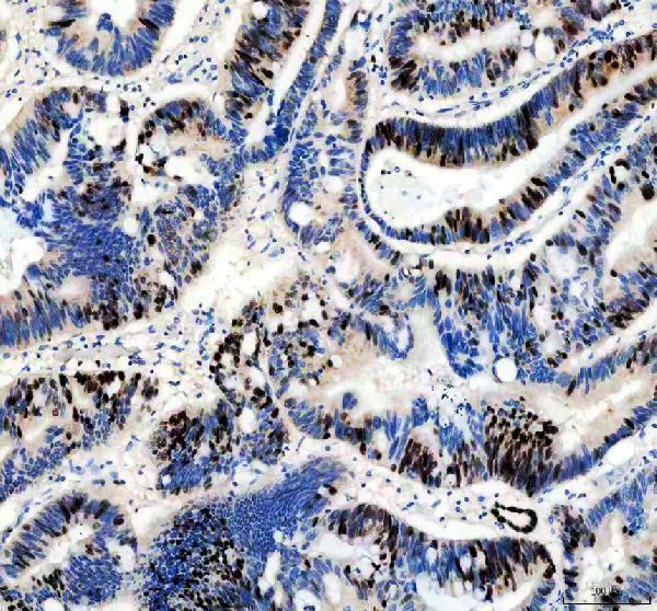
Click image to see more details
IF analysis of Ki67/MKI67 using anti-Ki67/MKI67 antibody (M00254-11).
Ki67/MKI67 was detected in a paraffin-embedded section of human intestine cancer tissue. Heat mediated antigen retrieval was performed in EDTA buffer (pH 8.0, epitope retrieval solution). The tissue section was blocked with 10% goat serum. The tissue section was then incubated at 1:50 rabbit anti-Ki67/MKI67 Antibody (M00254-11) overnight at 4°C. DyLight®488 Conjugated Goat Anti-Rabbit IgG (BA1127) was used as secondary antibody at 1:500 dilution and incubated for 30 minutes at 37°C. Visualize using a fluorescence microscope and filter sets appropriate for the label used.

Click image to see more details
IF analysis of Ki67/MKI67 using anti-Ki67/MKI67 antibody (M00254-11).
Ki67/MKI67 was detected in an immunocytochemical section of A549 cells. Enzyme antigen retrieval was performed using IHC enzyme antigen retrieval reagent (AR0022) for 15 mins. The cells were blocked with 10% goat serum. And then incubated at 1:50 rabbit anti-Ki67/MKI67 Antibody (M00254-11) overnight at 4°C. DyLight488 Conjugated Goat Anti-Rabbit IgG (BA1127) was used as secondary antibody at 1:500 dilution and incubated for 30 minutes at 37°C. Visualize using a fluorescence microscope and filter sets appropriate for the label used.
Specific Publications For Anti-Ki67/MKI67 Rabbit Monoclonal Antibody (M00254-11)
Loading publications
Recommended Resources
Here are featured tools and databases that you might find useful.
- Boster's Pathways Library
- Protein Databases
- Bioscience Research Protocol Resources
- Data Processing & Analysis Software
- Photo Editing Software
- Scientific Literature Resources
- Research Paper Management Tools
- Molecular Biology Software
- Primer Design Tools
- Bioinformatics Tools
- Phylogenetic Tree Analysis
Customer Reviews
Have you used Anti-Ki67/MKI67 Rabbit Monoclonal Antibody?
Share your experimental results or join a short interview to earn up to $1,000 in product credits or other rewards.
0 Reviews For Anti-Ki67/MKI67 Rabbit Monoclonal Antibody
Customer Q&As
Have a question?
Find answers in Q&As, reviews.
Can't find your answer?
Submit your question
