Click image to see more details
-

-
-
-
-
+5
Product Info Summary
| SKU: | M01524 |
|---|---|
| Size: | 100 μl |
| Reactive Species: | Human, Mouse, Rat |
| Host: | Rabbit |
| Application: | IP, IF, ICC, WB |
Customers Who Bought This Also Bought

Product info
Product Name
Anti-LC3B MAP1LC3B Rabbit Monoclonal Antibody
SKU/Catalog Number
M01524
BM4827 is an alternative SKU for this antibody, used in previous lots.
Size
100 μl
Form
Liquid
Description
Boster Bio Anti-LC3B MAP1LC3B Rabbit Monoclonal Antibody catalog # M01524. Tested in WB, ICC/IF, IP applications. This antibody reacts with Human, Mouse, Rat.
Storage & Handling
Store at -20°C for one year. For short term storage and frequent use, store at 4°C for up to one month. Avoid repeated freeze-thaw cycles.
Cite This Product
Anti-LC3B MAP1LC3B Rabbit Monoclonal Antibody (Boster Biological Technology, Pleasanton CA, USA, Catalog # M01524)
Host
Rabbit
Contents
Rabbit IgG in stabilizing components, phosphate buffered saline, pH 7.4, 150mM NaCl, 0.02% sodium azide and 50% glycerol.
*This antibody is supplied in a stabilized formulation.
Compatibility with conjugation reactions depends on the chemistry of the conjugation method used.
For conjugation methods that are not compatible with the stabilizing components present in this formulation, a carrier-free antibody format is required.
Clonality
Monoclonal
Clone Number
IEC-13
Isotype
Rabbit IgG
Immunogen
A synthesized peptide derived from human LC3B
Reactive Species
M01524 is reactive to MAP1LC3B in Human, Mouse, Rat
Observed Molecular Weight
15, 18 kDa
Calculated molecular weight
14.7 kDa
Antibody Validation
Boster validates all antibodies on WB, IHC, ICC, Immunofluorescence, and ELISA with known positive control and negative samples to ensure specificity and high affinity, including thorough antibody incubations.
Application & Images
Applications
M01524 is guaranteed for IP, IF, ICC, WB Boster Guarantee
Recommend Dilution
WB 1:500-2000
IHC 1:50-200
ICC/IF 1:50-200
IP 1:50
Tested application
Suggested blocking solution with 5% non-fat milk or BSA; (*)Recommended protein loading: 20-40 µg per lane
Use TE buffer pH 9.0 for antigen retrieval; (*) citrate buffer pH 6.0 is an alternative.
Validation Images & Assay Conditions
Click image to see more details
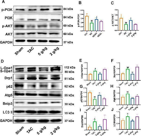
AEA improved CHF via PI3K/AKT/Bnip3 axis. (A) Representative images of PI3K/AKT axis. (B, C) The phosphorylation level of PI3K and AKT. (D) Representative images of Opa1, Drp1, Bnip3, p62, Atg5 and LC3II. (E–J) The expression level of Opa1, Drp1, Bnip3, p62, Atg5 and LC3II. (n = 3).
Index in PubMed under a CC BY license. PMID: 40206063

Click image to see more details
AEA improved NE-induced injuries via PI3K/AKT/Bnip3 axis. (A) Representative images of PI3K/AKT axis in H9c2 cells. (B, C) The phosphorylation level of PI3K and AKT. (D) Representative images of Opa1, Drp1, TrxR2, Bnip3, p62, Atg5 and LC3II in cells. (E–K) The expression level of Opa1, Drp1, TrxR2, Bnip3, p62, Atg5 and LC3II in H9c2 cells. (n = 3).
Index in PubMed under a CC BY license. PMID: 40206063

Click image to see more details
TMEM232 functions in autophagy in testis. A Western blotting analysis for the comparison of ARMC3 protein levels between Tmem232 −/− ( n = 2) and Tmem232 +/+ ( n = 2) testes and sperms at 2-months-old. α-Tubulin served as a loading control. The corresponding optical density readings for each image are shown. The representative image of biological duplicates is shown. B Western blotting analysis revealed the protein levels of four subunits (VPS15, VPS34, ATG14, and Beclin1) of PIK3C3-C1 complex in the testes of 2-month-old Tmem232 +/+ ( n = 2) and Tmem232 −/− ( n = 2) male mice. α-Tubulin served as a loading control. The representative image of biological duplicates is shown. C Co-immunoprecipitation assay results revealed the interaction between the TMEM232 and ATG14 protein in cells. Anti-Flag beads were used for immunoprecipitation. Anti-Flag and anti-GFP antibodies were used for western blotting analysis. The results shown are representative of three independent experiments. The representative image of biological duplicates is shown. D TMEM232-GFP was partly colocalized with LC3 in HeLa cells. Subcellular localization of target proteins (red) was probed with an anti-GOLGI97 antibody (upper panel, marker of Golgi apparatus), an anti-LAMP1 antibody (middle panel, marker of lysosome), and an anti-LC3 antibody (lower panel, marker of autophagosome). Cell nuclei were counterstained with DAPI. Scale bars, 10 μm. E The absence of TMEM232 in mice resulted in the accumulation of P62 and LC3. Tmem232 +/+ ( n = 2) and Tmem232 −/− ( n = 2) testes were separated and prepared for western blotting analysis. α-Tubulin served as a loading control. The corresponding optical density readings for each image are shown. The representative image of biological duplicates is shown. F Immunofluorescence staining of the P62 protein performed on frozen sections of Tmem232 +/+ ( n = 2) and Tmem232 −/− ( n = 2) testes. Blue, DAPI; green, P62. Scale bars, 20 μm. G Western blotting analysis of LC3B protein levels in HeLa cells after Flag-TMEM232 overexpression treatment with or without bafilomycin A1 (BafA1, 50 nM) treatment. HeLa cells were pretreated with Baf-A1 for 1 h and transfected with p-TMEM232×3FLAG-Myc-CMV-24 plasmid for 24 h. α-Tubulin served as a loading control. The corresponding optical density readings for each image are shown below. The results shown are representative of three independent experiments. The representative image of biological duplicates is shown. H A proposed model for the assumption that TMEM232 is involved in autophagy to regulate sperm formation in mice.
Index in PubMed under a CC BY license. PMID: 39516485

Click image to see more details
Western blot analysis of LC3B using anti-LC3B antibody (M01524).
Electrophoresis was performed on a 12% SDS-PAGE gel at 80V (Stacking gel) / 120V (Resolving gel) for 2 hours. The sample well of each lane was loaded with 30 ug of sample under reducing conditions.
Lane 1: human Hela whole cell lysates,
Lane 2: human MCF-7 whole cell lysates,
Lane 3: human A549 whole cell lysates,
Lane 4: human U2OS whole cell lysates,
Lane 5: rat brain tissue lysates,
Lane 6: rat C6 whole cell lysates,
Lane 7: mouse brain tissue lysates,
Lane 8: mouse Neurao-2a whole cell lysates.
After electrophoresis, proteins were transferred to a nitrocellulose membrane at 150 mA for 50-90 minutes. Blocked the membrane with 5% non-fat milk/TBS for 1.5 hour at RT. The membrane was incubated with rabbit anti-LC3B antigen affinity purified monoclonal antibody (Catalog # M01524) at 1:500 overnight at 4°C, then washed with TBS-0.1%Tween 3 times with 5 minutes each and probed with a goat anti-rabbit IgG-HRP secondary antibody at a dilution of 1:500 for 1.5 hour at RT. The signal is developed using an ECL Plus Western Blotting Substrate (Catalog # AR1196-200) with Tanon 5200 system. A specific band was detected for LC3B at approximately 15, 18 kDa. The expected band size for LC3B is at 15 kDa.

Click image to see more details
IHC analysis of LC3B using anti-LC3B antibody (M01524).
LC3B was detected in a paraffin-embedded section of human glioma tissue. Heat mediated antigen retrieval was performed in EDTA buffer (pH 8.0, epitope retrieval solution). The tissue section was blocked with 10% goat serum. The tissue section was then incubated with 1:50 rabbit anti-LC3B Antibody (M01524) overnight at 4°C. Peroxidase Conjugated Goat Anti-rabbit IgG was used as secondary antibody and incubated for 30 minutes at 37°C. The tissue section was developed using HRP Conjugated Rabbit IgG Super Vision Assay Kit (Catalog # SV0002) with DAB as the chromogen.

Click image to see more details
IHC analysis of LC3B using anti-LC3B antibody (M01524).
LC3B was detected in a paraffin-embedded section of mouse brain tissue. Heat mediated antigen retrieval was performed in EDTA buffer (pH 8.0, epitope retrieval solution). The tissue section was blocked with 10% goat serum. The tissue section was then incubated with 1:50 rabbit anti-LC3B Antibody (M01524) overnight at 4°C. Peroxidase Conjugated Goat Anti-rabbit IgG was used as secondary antibody and incubated for 30 minutes at 37°C. The tissue section was developed using HRP Conjugated Rabbit IgG Super Vision Assay Kit (Catalog # SV0002) with DAB as the chromogen.

Click image to see more details
IHC analysis of LC3B using anti-LC3B antibody (M01524).
LC3B was detected in a paraffin-embedded section of rat brain tissue. Heat mediated antigen retrieval was performed in EDTA buffer (pH 8.0, epitope retrieval solution). The tissue section was blocked with 10% goat serum. The tissue section was then incubated with 1:50 rabbit anti-LC3B Antibody (M01524) overnight at 4°C. Peroxidase Conjugated Goat Anti-rabbit IgG was used as secondary antibody and incubated for 30 minutes at 37°C. The tissue section was developed using HRP Conjugated Rabbit IgG Super Vision Assay Kit (Catalog # SV0002) with DAB as the chromogen.

Click image to see more details
Immunofluorescent analysis using the Antibody at 1:150 dilution.

Click image to see more details
Immunofluorescent analysis of Hela cells treated with choroquine, using LC3B Antibody.
Specific Publications For Anti-LC3B MAP1LC3B Rabbit Monoclonal Antibody (M01524)
Loading publications
Recommended Resources
Here are featured tools and databases that you might find useful.
- Boster's Pathways Library
- Protein Databases
- Bioscience Research Protocol Resources
- Data Processing & Analysis Software
- Photo Editing Software
- Scientific Literature Resources
- Research Paper Management Tools
- Molecular Biology Software
- Primer Design Tools
- Bioinformatics Tools
- Phylogenetic Tree Analysis
Customer Reviews
Have you used Anti-LC3B MAP1LC3B Rabbit Monoclonal Antibody?
Share your experimental results or join a short interview to earn up to $1,000 in product credits or other rewards.
0 Reviews For Anti-LC3B MAP1LC3B Rabbit Monoclonal Antibody
Customer Q&As
Have a question?
Find answers in Q&As, reviews.
Can't find your answer?
Submit your question
