Click image to see more details
-

-
-
-
-
+1
Product Info Summary
| SKU: | A01524-2 |
|---|---|
| Size: | 100 μl/vial |
| Reactive Species: | Human, Mouse, Rat |
| Host: | Rabbit |
| Application: | ELISA, IF, IHC, ICC, WB |
Customers Who Bought This Also Bought

Product info
Product Name
Anti-LC3B/MAP1LC3B Antibody
SKU/Catalog Number
A01524-2
Size
100 μl/vial
Form
Liquid
Description
Boster Bio Anti-LC3B/MAP1LC3B Antibody catalog # A01524-2. Tested in WB, IF, IHC, ICC, ELISA applications. This antibody reacts with Human, Mouse, Rat.
Storage & Handling
12 months from date of receipt,-20℃ as supplied. 6 months 2 to 8℃ after reconstitution. Avoid repeated freezing and thawing.
Cite This Product
Anti-LC3B/MAP1LC3B Antibody (Boster Biological Technology, Pleasanton CA, USA, Catalog # A01524-2)
Host
Rabbit
Contents
500 μg/ml antibody with PBS, 0.02% NaN3, 1 mg stabilizing protein and 50% glycerol
*This antibody is supplied in a stabilized formulation.
Compatibility with conjugation reactions depends on the chemistry of the conjugation method used.
For conjugation methods that are not compatible with the stabilizing components present in this formulation, a carrier-free antibody format is required.
Clonality
Polyclonal
Immunogen
Peptide
Reactive Species
A01524-2 is reactive to MAP1LC3B in Human, Mouse, Rat
Calculated molecular weight
14.7 kDa
Background of MAP1LC3B
LC3B, also named as MAP1LC3B, MAP1A/1BLC3, belongs to the MAP1 LC3 family. It is a subunit of neuronal microtubule-associated MAP1A and MAP1B proteins, which are involved in microtubule assembly and important for neurogenesis. In cell biology, autophagy, or autophagocytosis, is a catabolic process involving the degradation of a cell's own components through the lysosomalmachinery. It is a major mechanism by which a starving cell reallocates nutrients from unnecessary processes to more-essential processes. Two forms of LC3, called LC3-I (17-19kd) and -II(14-16kd), were produced post-translationally in various cells. LC3-I is cytosolic, whereas LC3-II is membrane bound. The precursor molecule is cleaved by APG4B/ATG4B to form the cytosolic form, LC3-I. This is activated by APG7L/ATG7, transferred to ATG3 and conjugated to phospholipid to form the membrane-bound form, LC3-II. The amount of LC3-II is correlated with the extent of autophagosome formation. LC3-II is the first mammalian protein identified that specifically associates with autophagosome membranes. MAP1LC3 has 3 isoforms MAP1LC3A, MAP1LC3B and MAP1LC3C. MAP1LC3A and MAP1LC3C are produced by the proteolytic cleavage after the conserved C-terminal Gly residue, like their rat counterpart, MAP1LC3B does not undergo C-terminal cleavage and exists in a single modified form. This antibody is specific to LC3B.
Antibody Validation
Boster validates all antibodies on WB, IHC, ICC, Immunofluorescence, and ELISA with known positive control and negative samples to ensure specificity and high affinity, including thorough antibody incubations.
Application & Images
Applications
A01524-2 is guaranteed for ELISA, IF, IHC, ICC, WB Boster Guarantee
Assay Dilutions Recommendation
The recommendations below provide a starting point for assay optimization. The actual working concentration varies and should be decided by the user.
Western blot, 1:500-2000
Immunohistochemistry, 1:50-400
Immunocytochemistry/Immunofluorescence, 1:50-400
ELISA, 1:100-1000
Validation Images & Assay Conditions
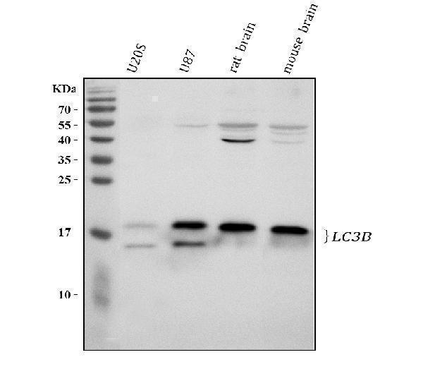
Click image to see more details
Western blot analysis of LC3B/MAP1LC3B using anti-LC3B/MAP1LC3B antibody (A01524-2).
Electrophoresis was performed on a 10% SDS-PAGE gel at 80V (Stacking gel) / 120V (Resolving gel) for 2 hours. The sample well of each lane was loaded with 30 ug of sample under reducing conditions.
Lane 1: human U2OS whole cell lysates,
Lane 2: human U87 whole cell lysates,
Lane 3: rat brain tissue lysates,
Lane 4: mouse brain tissue lysates.
After electrophoresis, proteins were transferred to a nitrocellulose membrane at 150 mA for 50-90 minutes. Blocked the membrane with 5% non-fat milk/TBS for 1.5 hour at RT. The membrane was incubated with rabbit anti-LC3B/MAP1LC3B antigen affinity purified polyclonal antibody (A01524-2) at 1:1000 overnight at 4°C, then washed with TBS-0.1%Tween 3 times with 5 minutes each and probed with a goat anti-rabbit IgG-HRP secondary antibody at a dilution of 1:5000 for 1.5 hour at RT. The signal is developed using an ECL Plus Western Blotting Substrate (Catalog # AR1196-200) with Tanon 5200 system. A specific band was detected for LC3B/MAP1LC3B at approximately 15,18 kDa. The expected band size for LC3B/MAP1LC3B is at 15 kDa.

Click image to see more details
IHC analysis of LC3B/MAP1LC3B using anti-LC3B/MAP1LC3B antibody (A01524-2).
LC3B/MAP1LC3B was detected in a paraffin-embedded section of human glioma tissue. Heat mediated antigen retrieval was performed in EDTA buffer (pH 8.0, epitope retrieval solution). The tissue section was blocked with 10% goat serum. The tissue section was then incubated with 1:100 rabbit anti-LC3B/MAP1LC3B Antibody (A01524-2) overnight at 4°C. Peroxidase Conjugated Goat Anti-rabbit IgG was used as secondary antibody and incubated for 30 minutes at 37°C. The tissue section was developed using HRP Conjugated Rabbit IgG Super Vision Assay Kit (Catalog # SV0002) with DAB as the chromogen.

Click image to see more details
IHC analysis of LC3B/MAP1LC3B using anti-LC3B/MAP1LC3B antibody (A01524-2).
LC3B/MAP1LC3B was detected in a paraffin-embedded section of mouse brain tissue. Heat mediated antigen retrieval was performed in EDTA buffer (pH 8.0, epitope retrieval solution). The tissue section was blocked with 10% goat serum. The tissue section was then incubated with 1:100 rabbit anti-LC3B/MAP1LC3B Antibody (A01524-2) overnight at 4°C. Peroxidase Conjugated Goat Anti-rabbit IgG was used as secondary antibody and incubated for 30 minutes at 37°C. The tissue section was developed using HRP Conjugated Rabbit IgG Super Vision Assay Kit (Catalog # SV0002) with DAB as the chromogen.

Click image to see more details
IHC analysis of LC3B/MAP1LC3B using anti-LC3B/MAP1LC3B antibody (A01524-2).
LC3B/MAP1LC3B was detected in a paraffin-embedded section of rat brain tissue. Heat mediated antigen retrieval was performed in EDTA buffer (pH 8.0, epitope retrieval solution). The tissue section was blocked with 10% goat serum. The tissue section was then incubated with 1:100 rabbit anti-LC3B/MAP1LC3B Antibody (A01524-2) overnight at 4°C. Peroxidase Conjugated Goat Anti-rabbit IgG was used as secondary antibody and incubated for 30 minutes at 37°C. The tissue section was developed using HRP Conjugated Rabbit IgG Super Vision Assay Kit (Catalog # SV0002) with DAB as the chromogen.

Click image to see more details
PBM alleviates abnormal mitochondrial autophagy and promotes mitochondrial energy metabolism in mice with AD. A RT-PCR was used to detect autophagy-related protein (Beclin1, LC3II.) and glycolysis-related proteins (TSPO and HK2) in mouse brain tissues. B WB was used to detect the autophagy protein LC3II, and the oxidative phosphorylation-related proteins PGC-1α and NRF-1. C Immunohistochemical staining of mouse brains using an anti-HK2 antibody (scale bars 100 μm and 20 μm, respectively) with brown plaques of HK2. D , E WB was used to detect the glycolysis-related proteins GLUT1, PKM2, and HK2. Experimental data are presented as the mean ± standard deviation. Compared with the CON group, * p < 0.05, ** p < 0.01, *** p < 0.001; Compared with the AD group, # p < 0.05, ## p < 0.01
Index in PubMed under a CC BY license. PMID: 40188044
Specific Publications For Anti-LC3B/MAP1LC3B Antibody (A01524-2)
Loading publications
Recommended Resources
Here are featured tools and databases that you might find useful.
- Boster's Pathways Library
- Protein Databases
- Bioscience Research Protocol Resources
- Data Processing & Analysis Software
- Photo Editing Software
- Scientific Literature Resources
- Research Paper Management Tools
- Molecular Biology Software
- Primer Design Tools
- Bioinformatics Tools
- Phylogenetic Tree Analysis
Customer Reviews
Have you used Anti-LC3B/MAP1LC3B Antibody?
Share your experimental results or join a short interview to earn up to $1,000 in product credits or other rewards.
0 Reviews For Anti-LC3B/MAP1LC3B Antibody
Customer Q&As
Have a question?
Find answers in Q&As, reviews.
Can't find your answer?
Submit your question
