Click image to see more details
-

-
-
-
-
+8
Product Info Summary
| SKU: | A01758-2 |
|---|---|
| Size: | 100 μg/vial |
| Reactive Species: | Human, Mouse, Rat |
| Host: | Rabbit |
| Application: | ELISA, Flow Cytometry, IF, IHC, ICC, WB |
Customers Who Bought This Also Bought

Product info
Product Name
Anti-METTL3 Antibody Picoband®
SKU/Catalog Number
A01758-2
Size
100 μg/vial
Form
Lyophilized
Description
Boster Bio Anti-METTL3 Antibody Picoband® catalog # A01758-2. Tested in ELISA, Flow Cytometry, IF, IHC, ICC, WB applications. This antibody reacts with Human, Mouse, Rat. The brand Picoband indicates this is a premium antibody that guarantees superior quality, high affinity, and strong signals with minimal background in Western blot applications. Only our best-performing antibodies are designated as Picoband, ensuring unmatched performance.
Storage & Handling
Store at -20˚C for one year from date of receipt. After reconstitution, at 4˚C for one month. It can also be aliquotted and stored frozen at -20˚C for six months. Avoid repeated freeze-thaw cycles.
Cite This Product
Anti-METTL3 Antibody Picoband® (Boster Biological Technology, Pleasanton CA, USA, Catalog # A01758-2)
Host
Rabbit
Contents
Each vial contains 4mg Trehalose, 0.9mg NaCl, 0.2mg Na2HPO4, 0.005mg NaN3.
Clonality
Polyclonal
Isotype
Rabbit IgG
Immunogen
E.coli-derived human METTL3 recombinant protein (Position: E71-L580).
Cross-reactivity
No cross-reactivity with other proteins.
Reactive Species
A01758-2 is reactive to METTL3 in Human, Mouse, Rat
Observed Molecular Weight
64 kDa
Calculated molecular weight
64.5 kDa
Background of METTL3
N6-adenosine-methyltransferase 70 kDa subunit (METTL3) is an enzyme that in humans is encoded by the METTL3 gene. It is mapped to 14q11.2. This gene encodes the 70 kDa subunit of MT-A which is part of N6-adenosine-methyltransferase. This enzyme is involved in the posttranscriptional methylation of internal adenosine residues in eukaryotic mRNAs, forming N6-methyladenosine.
Antibody Validation
Boster validates all antibodies on WB, IHC, ICC, Immunofluorescence, and ELISA with known positive control and negative samples to ensure specificity and high affinity, including thorough antibody incubations.
Application & Images
Applications
A01758-2 is guaranteed for ELISA, Flow Cytometry, IF, IHC, ICC, WB Boster Guarantee
Recommend Dilution
| Application | Dilution | Species |
|---|---|---|
| Western blot | 0.25-0.5μg/ml | Human, Mouse, Rat |
| Immunohistochemistry (Paraffin-embedded Section) | 2-5μg/ml | Human, Mouse, Rat |
| Immunocytochemistry/Immunofluorescence | 5μg/ml | Human |
| Flow Cytometry (Fixed) | 1-3μg/1x106 cells | Human |
| ELISA | 0.1-0.5μg/ml | - |
Tested application
Suggested blocking solution with 5% non-fat milk or BSA; (*)Recommended protein loading: 20-40 µg per lane
Use TE buffer pH 9.0 for antigen retrieval; (*) citrate buffer pH 6.0 is an alternative.
Validation Images & Assay Conditions
Click image to see more details
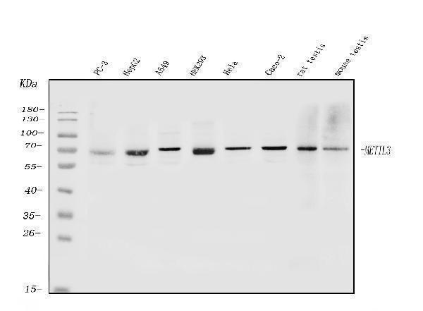
Western blot analysis of METTL3 using anti-METTL3 antibody (A01758-2).
Electrophoresis was performed on a 5-20% SDS-PAGE gel at 70V (Stacking gel) / 90V (Resolving gel) for 2-3 hours. The sample well of each lane was loaded with 50ug of sample under reducing conditions.
Lane 1: human PC-3 whole cell lysates,
Lane 2: human HEPG2 whole cell lysates,
Lane 3: human A549 whole cell lysates,
Lane 4: human HEK293 whole cell lysates,
Lane 5: human HELA whole cell lysates,
Lane 6: human CACO-2 whole cell lysates,
Lane 7: rat testis tissue lysates,
Lane 8: mouse testis tissue lysates.
After Electrophoresis, proteins were transferred to a Nitrocellulose membrane at 150mA for 50-90 minutes. Blocked the membrane with 5% Non-fat Milk/ TBS for 1.5 hour at RT. The membrane was incubated with rabbit anti-METTL3 antigen affinity purified polyclonal antibody (Catalog # A01758-21) at 0.5 μg/mL overnight at 4°C, then washed with TBS-0.1%Tween 3 times with 5 minutes each and probed with a goat anti-rabbit IgG-HRP secondary antibody at a dilution of 1:5000 for 1.5 hour at RT. The signal is developed using an Enhanced Chemiluminescent detection (ECL) kit (Catalog # EK1002) with Tanon 5200 system. A specific band was detected for METTL3 at approximately 64KD. The expected band size for METTL3 is at 64KD.

Click image to see more details
SOX2OT-dependent m6A modification and stabilization of GLI1 mRNA by METTL3/14 and IGF2BP2. A Proteins interacted with both GLI1 mRNA and SOX2OT were predicted using StarBase. B RIP assay demonstrated that SOX2OT and GLI1 mRNA were enriched for m6A modification and interacted with METTL3/14/IGF2BP2. C RIP-PCR validated that METTL3/14/IGF2BP2 overexpression promoted m6A modification of GLI1 mRNA. D METTL3/14/IGF2BP2 upregulation promoted GLI1 expression detected via immunoblotting. E RNA stability assay revealed that METTL3/14/IGF2BP2 overexpression increased GLI1 mRNA stability compared to the pENTER backbone. F Expression of GLI1 mRNA upon METTL3/14/IGF2BP2 overexpression or co-transfection with SOX2OT shRNA. G Expression of GLI1 at the protein level upon METTL3/14/IGF2BP2 overexpression or co-transfection with SOX2OT shRNA.
Index in PubMed under a CC BY license. PMID: 37149646

Click image to see more details
METTL3/14/IGF2BP2-mediated proliferation of NSCLC stem cells upon GLI1 repression. A Proliferation assay of NSCLC cells transfected with METTL3/14/IGF2BP2 and/or GLI1 shRNA. B and C Spheroid formation assay and histogram of NSCLC cells transfected with METTL3/14/IGF2BP2 and/or GLI1 shRNA. D and E Clonal formation assay and histogram of NSCLC cells transfected with METTL3/14/IGF2BP2 and/or GLI1 shRNA. Magnification = ×100, scale bar = 200 μm.
Index in PubMed under a CC BY license. PMID: 37149646

Click image to see more details
GLI1 positively regulates the expression of METTL3/14/IGF2BP2 via SOX2OT. A , B METTL3/14/IGF2BP2 expression at the mRNA and protein level in NSCLC cells upon SOX2OT knockdown, detected using RT-qPCR and immunoblotting, respectively. C , D Upregulation of METTL3/14/IGF2BP2 promoted by GLI1 transfection was blocked by SOX2OT suppression measured via RT-qPCR and immunoblotting. E , F miR-186-5p inhibitor promoted the expression of METTL3/14/IGF2BP2 and counteracted their downregulation upon SOX2OT interference detected via RT-qPCR and immunoblotting.
Index in PubMed under a CC BY license. PMID: 37149646

Click image to see more details
Pharmacological intervention of GLI1 and METTL3 attenuates lung cancer tumor growth in vivo. A Tumor bulk in mice subcutaneously implanted with A549. B Tumor growth curves in groups treated with GANT58, SAH, or their combination. The control group was treated with saline. C IHC staining of Ki67, GLI1, METTL3/14, and IGF2BP2 in treatment and control groups. Scale bar = 100 µM.
Index in PubMed under a CC BY license. PMID: 37149646

Click image to see more details
Positive correlation of the GLI1/SOX2OT loop and METTL3/14/IGF2BP2 expression at the tissue level. A Representative immunohistochemistry images of the GLI1/SOX2OT loop and METTL3/14/IGF2BP2. Magnification = ×200, Scale bar = 100 μm. B Expression level of the GLI1/SOX2OT loop and METTL3/14/IGF2BP2 in paired NSCLC specimens and adjacent normal tissues. C Correlation analysis between the expression levels of GLI1 with that of METTL3, METTL14, IGF2BP2, and SOX2OT in lung cancer specimens. D Diagram illustrating the postulated molecular mechanisms of the GLI1/SOX2OT loop m6A modification in NSCLC cells.
Index in PubMed under a CC BY license. PMID: 37149646

Click image to see more details
IHC analysis of METTL3 using anti-METTL3 antibody (A01758-2).
METTL3 was detected in paraffin-embedded section of human esophageal squamous carcinoma tissue. Heat mediated antigen retrieval was performed in EDTA buffer (pH8.0, epitope retrieval solution). The tissue section was blocked with 10% goat serum. The tissue section was then incubated with 2μg/ml rabbit anti-METTL3 Antibody (A01758-2) overnight at 4°C. Biotinylated goat anti-rabbit IgG was used as secondary antibody and incubated for 30 minutes at 37°C. The tissue section was developed using Strepavidin-Biotin-Complex (SABC) (Catalog # SA1022) with DAB as the chromogen.

Click image to see more details
Figure 3 IHC analysis of METTL3 using anti-METTL3 antibody (A01758-2).
METTL3 was detected in paraffin-embedded section of human tonsil tissue. Heat mediated antigen retrieval was performed in EDTA buffer (pH8.0, epitope retrieval solution). The tissue section was blocked with 10% goat serum. The tissue section was then incubated with 2μg/ml rabbit anti-METTL3 Antibody (A01758-2) overnight at 4°C. Biotinylated goat anti-rabbit IgG was used as secondary antibody and incubated for 30 minutes at 37°C. The tissue section was developed using Strepavidin-Biotin-Complex (SABC) (Catalog # SA1022) with DAB as the chromogen.

Click image to see more details
Figure 4 IHC analysis of METTL3 using anti-METTL3 antibody (A01758-2).
METTL3 was detected in paraffin-embedded section of human placenta tissue. Heat mediated antigen retrieval was performed in EDTA buffer (pH8.0, epitope retrieval solution). The tissue section was blocked with 10% goat serum. The tissue section was then incubated with 2μg/ml rabbit anti-METTL3 Antibody (A01758-2) overnight at 4°C. Biotinylated goat anti-rabbit IgG was used as secondary antibody and incubated for 30 minutes at 37°C. The tissue section was developed using Strepavidin-Biotin-Complex (SABC) (Catalog # SA1022) with DAB as the chromogen.

Click image to see more details
IF analysis of METTL3 using anti-METTL3 antibody (A01758-2).
METTL3 was detected in immunocytochemical section of PC-3 cells. Enzyme antigen retrieval was performed using IHC enzyme antigen retrieval reagent (AR0022) for 15 mins. The cells were blocked with 10% goat serum. And then incubated with 5μg/mL rabbit anti-METTL3 Antibody (A01758-2) overnight at 4°C. DyLight®488 Conjugated Goat Anti-Rabbit IgG (BA1127) was used as secondary antibody at 1:100 dilution and incubated for 30 minutes at 37°C. The section was counterstained with DAPI. Visualize using a fluorescence microscope and filter sets appropriate for the label used.

Click image to see more details
Flow Cytometry analysis of HL-60 cells using anti-METTL3 antibody (A01758-2).
Overlay histogram showing HL-60 cells stained with A01758-2 (Blue line). To facilitate intracellular staining, cells were fixed with 4% paraformaldehyde and permeabilized with permeabilization buffer. The cells were blocked with 10% normal goat serum. And then incubated with rabbit anti-METTL3 Antibody (A01758-2, 1μg/1x106 cells) for 30 min at 20°C. DyLight®488 conjugated goat anti-rabbit IgG (BA1127, 5-10μg/1x106 cells) was used as secondary antibody for 30 minutes at 20°C. Isotype control antibody (Green line) was rabbit IgG (1μg/1x106) used under the same conditions. Unlabelled sample without incubation with primary antibody and secondary antibody (Red line) was used as a blank control.

Click image to see more details
IHC analysis of METTL3 using anti-METTL3 antibody (A01758-2).
METTL3 was detected in a paraffin-embedded section of rat epididymis tissue. Heat mediated antigen retrieval was performed in EDTA buffer (pH 8.0, epitope retrieval solution). The tissue section was blocked with 10% goat serum. The tissue section was then incubated with 2 μg/ml rabbit anti-METTL3 Antibody (A01758-2) overnight at 4°C. Biotinylated goat anti-rabbit IgG was used as secondary antibody and incubated for 30 minutes at 37°C. The tissue section was developed using Strepavidin-Biotin-Complex (SABC) (Catalog # SA1022) with DAB as the chromogen.
Specific Publications For Anti-METTL3 Antibody Picoband® (A01758-2)
Loading publications
Recommended Resources
Here are featured tools and databases that you might find useful.
- Boster's Pathways Library
- Protein Databases
- Bioscience Research Protocol Resources
- Data Processing & Analysis Software
- Photo Editing Software
- Scientific Literature Resources
- Research Paper Management Tools
- Molecular Biology Software
- Primer Design Tools
- Bioinformatics Tools
- Phylogenetic Tree Analysis
Customer Reviews
Have you used Anti-METTL3 Antibody Picoband®?
Share your experimental results or join a short interview to earn up to $1,000 in product credits or other rewards.
0 Reviews For Anti-METTL3 Antibody Picoband®
Customer Q&As
Have a question?
Find answers in Q&As, reviews.
Can't find your answer?
Submit your question
