Click image to see more details
Product Info Summary
| SKU: | A12581 |
|---|---|
| Size: | 100 µg/vial |
| Reactive Species: | Human, Mouse, Rat |
| Host: | Rabbit |
| Application: | ELISA, IF, ICC, WB |
Customers Who Bought This Also Bought

Product info
Product Name
Anti-MFAP3L Antibody Picoband®
SKU/Catalog Number
A12581
Size
100 µg/vial
Form
Lyophilized
Description
Boster Bio Anti-MFAP3L Antibody Picoband® catalog # A12581. Tested in WB, ICC/IF, ELISA applications. This antibody reacts with Human, Mouse, Rat. The brand Picoband indicates this is a premium antibody that guarantees superior quality, high affinity, and strong signals with minimal background in Western blot applications. Only our best-performing antibodies are designated as Picoband, ensuring unmatched performance.
Storage & Handling
At -20°C for one year from date of receipt. After reconstitution, at 4°C for one month. It can also be aliquotted and stored frozen at -20°C for six months. Avoid repeated freezing and thawing.
Cite This Product
Anti-MFAP3L Antibody Picoband® (Boster Biological Technology, Pleasanton CA, USA, Catalog # A12581)
Host
Rabbit
Contents
Each vial contains 4 mg Trehalose, 0.9 mg NaCl, 0.2 mg Na2HPO4.
Clonality
Polyclonal
Immunogen
E.coli-derived human MFAP3L recombinant protein (Position: K29-V409). Human MFAP3L shares 90.6% amino acid (aa) sequence identity with both mouse and rat MFAP3L.
Reactive Species
A12581 is reactive to MFAP3L in Human, Mouse, Rat
Observed Molecular Weight
45 kDa
Calculated molecular weight
45.4 kDa
Background of MFAP3L
Microbibrillar-Associated Protein 3-Like (MFAP3L), also known as NYD-sp9, is part of the microfibrillar-associated protein family (MFAPs). MFAPs are non‐fibrillin, extracellular matrix glycoproteins that interact with fibrillin and were originally characterized in microfibrillar assembly. In humans, there several subfamily members with varying amino acid (aa) sequence homology and functions . Among the family, MFAP2 and MFAP5 are more closely related and while MFAP1, 3 and 4 share no structural or sequence homology with MFAP2, MFAP5 or with each other. Human MFAP3L shows 71% amino acid (aa) sequence homology to MFAP3, but not other MFAPs. Mature, human MFAP3L consists of an extracellular domain (ECD) containing N-linked glycosylation sites, a transmembrane domain, and a cytoplasmic domain with a conserved SH2 motif. The ECD of human MFAP3L shares 89% and 90% aa sequence identity with mouse and rat MFAP3L, respectively. MFAPs have the unique ability to interact with TGF-beta family growth factors, Notch and Notch ligands and multiple elastic fiber proteins, in addition to interacting with fibrillin. MFAPs are expressed in a wide variety of tissues and, along with microfibril assembly, they play roles in the regulation of tissue homeostasis, cell survival, and tumor progression. MFAP3L is often located within colorectal cancer (CRC) cells, which metastasize by activation of the nuclear ERK pathway via MFAP3L phosphorylation. Regulation of this MFAP3L activity could have pharmaceutical effects on CRC tumor progression.
Antibody Validation
Boster validates all antibodies on WB, IHC, ICC, Immunofluorescence, and ELISA with known positive control and negative samples to ensure specificity and high affinity, including thorough antibody incubations.
Application & Images
Applications
A12581 is guaranteed for ELISA, IF, ICC, WB Boster Guarantee
Assay Dilutions Recommendation
The recommendations below provide a starting point for assay optimization. The actual working concentration varies and should be decided by the user.
Western blot, 0.25-0.5 μg/ml, Human, Mouse, Rat
Immunocytochemistry/Immunofluorescence, 5 μg/ml, Human
ELISA, 0.1-0.5 μg/ml, -
Positive Control
WB: human RT4 whole cell, human U251 whole cell, human Hela whole cell, rat testis tissue, rat PC-12 whole cell, mouse testis tissue, mouse RAW2647 whole cell
ICC/IF: HELA cell
Validation Images & Assay Conditions
Click image to see more details
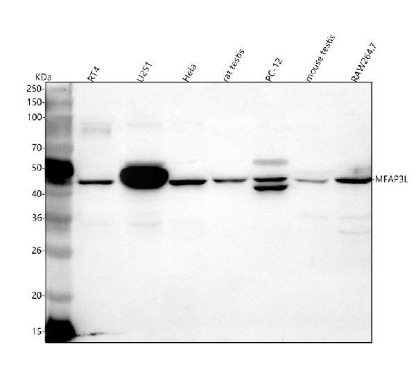
Western blot analysis of MFAP3L using anti-MFAP3L antibody (A12581).
Electrophoresis was performed on a 5-20% SDS-PAGE gel at 70V (Stacking gel) / 90V (Resolving gel) for 2-3 hours. The sample well of each lane was loaded with 30 ug of sample under reducing conditions.
Lane 1: human RT4 whole cell lysates,
Lane 2: human U251 whole cell lysates,
Lane 3: human Hela whole cell lysates,
Lane 4: rat testis tissue lysates,
Lane 5: rat PC-12 whole cell lysates,
Lane 6: mouse testis tissue lysates,
Lane 7: mouse RAW264.7 whole cell lysates.
After electrophoresis, proteins were transferred to a nitrocellulose membrane at 150 mA for 50-90 minutes. Blocked the membrane with 5% non-fat milk/TBS for 1.5 hour at RT. The membrane was incubated with rabbit anti-MFAP3L antigen affinity purified polyclonal antibody (Catalog # A12581) at 0.5 μg/mL overnight at 4°C, then washed with TBS-0.1%Tween 3 times with 5 minutes each and probed with a goat anti-rabbit IgG-HRP secondary antibody at a dilution of 1:5000 for 1.5 hour at RT. The signal is developed using an Enhanced Chemiluminescent detection (ECL) kit (Catalog # EK1002) with Tanon 5200 system. A specific band was detected for MFAP3L at approximately 45 kDa. The expected band size for MFAP3L is at 45 kDa.

Click image to see more details
IF analysis of MFAP3L using anti-MFAP3L antibody (A12581).
MFAP3L was detected in an immunocytochemical section of HELA cells. Enzyme antigen retrieval was performed using IHC enzyme antigen retrieval reagent (AR0022) for 15 mins. The cells were blocked with 10% goat serum. And then incubated with 5 μg/mL rabbit anti-MFAP3L Antibody (A12581) overnight at 4°C. DyLight®488 Conjugated Goat Anti-Rabbit IgG (BA1127) was used as secondary antibody at 1:500 dilution and incubated for 30 minutes at 37°C. The section was counterstained with DAPI. Visualize using a fluorescence microscope and filter sets appropriate for the label used.
Specific Publications For Anti-MFAP3L Antibody Picoband® (A12581)
Loading publications
Recommended Resources
Here are featured tools and databases that you might find useful.
- Boster's Pathways Library
- Protein Databases
- Bioscience Research Protocol Resources
- Data Processing & Analysis Software
- Photo Editing Software
- Scientific Literature Resources
- Research Paper Management Tools
- Molecular Biology Software
- Primer Design Tools
- Bioinformatics Tools
- Phylogenetic Tree Analysis
Customer Reviews
Have you used Anti-MFAP3L Antibody Picoband®?
Share your experimental results or join a short interview to earn up to $1,000 in product credits or other rewards.
0 Reviews For Anti-MFAP3L Antibody Picoband®
Customer Q&As
Have a question?
Find answers in Q&As, reviews.
Can't find your answer?
Submit your question
