Click image to see more details
Product Info Summary
| SKU: | A06832-1 |
|---|---|
| Size: | 100 µg/vial |
| Reactive Species: | Human |
| Host: | Rabbit |
| Application: | ELISA, Flow Cytometry, IF, ICC, WB |
Customers Who Bought This Also Bought

Product info
Product Name
Anti-MIS12 Antibody Picoband®
SKU/Catalog Number
A06832-1
Size
100 µg/vial
Form
Lyophilized
Description
Boster Bio Anti-MIS12 Antibody Picoband® catalog # A06832-1. Tested in ELISA, IF, ICC, WB, Flow Cytometry applications. This antibody reacts with Human. The brand Picoband indicates this is a premium antibody that guarantees superior quality, high affinity, and strong signals with minimal background in Western blot applications. Only our best-performing antibodies are designated as Picoband, ensuring unmatched performance.
Storage & Handling
At -20°C for one year from date of receipt. After reconstitution, at 4°C for one month. It can also be aliquotted and stored frozen at -20°C for six months. Avoid repeated freezing and thawing.
Cite This Product
Anti-MIS12 Antibody Picoband® (Boster Biological Technology, Pleasanton CA, USA, Catalog # A06832-1)
Host
Rabbit
Contents
Each vial contains 4 mg Trehalose, 0.9 mg NaCl, 0.2 mg Na2HPO4.
Clonality
Polyclonal
Isotype
Rabbit IgG
Immunogen
E.coli-derived human MIS12 recombinant protein (Position: M1-K187).
Cross-reactivity
No cross-reactivity with other proteins.
Reactive Species
A06832-1 is reactive to MIS12 in Human
Observed Molecular Weight
25 kDa
Calculated molecular weight
24.1 kDa
Background of MIS12
Protein MIS12 homolog is a protein that in humans is encoded by the MIS12 gene. Mis12 complex is composed of four subunits, Protein MIS12 homolog, Polyamine-modulated factor 1, Kinetochore-associated protein DSN1 homolog, and Kinetochore-associated protein NSL1 homolog (UniProt: Q9H081, Q6P1K2, Q9H410, Q96IY1, respectively) that are encoded by genes known as MIS12 (Gene ID: 79003), PMF1 (Gene ID: 100527963), DSN1 (Gene ID: 79980), and NSL1 (also known as C1orf48, DC31, DC8, MIS14) (Gene ID: 25936) in human. The MIS12 complex is a protein interaction hub for outer kinetochore assembly. This complex acts as the primary microtubule-binding interface at kinetochores and provides a platform to recruit regulatory proteins. In human Mis12 complex subunits are shown to localize with centromere protein A (CENP-A) at inner kinetochores and internally to Ndc80 at outer kinetochores. Mis12 complex plays an essential role in chromosome segregation in vertebrates and contributes to mitotic kinetochore assembly. Reduced levels of Mis12 complex proteins are shown to result in chromosome alignment defects in both human and chicken cells, but spindle bipolarity is not disturbed.
Antibody Validation
Boster validates all antibodies on WB, IHC, ICC, Immunofluorescence, and ELISA with known positive control and negative samples to ensure specificity and high affinity, including thorough antibody incubations.
Application & Images
Applications
A06832-1 is guaranteed for ELISA, Flow Cytometry, IF, ICC, WB Boster Guarantee
Assay Dilutions Recommendation
The recommendations below provide a starting point for assay optimization. The actual working concentration varies and should be decided by the user.
Western blot, 0.1-0.25 μg/ml, Human
Immunocytochemistry/Immunofluorescence, 5 μg/ml, Human
Flow Cytometry (Fixed), 1-3 μg/1x106 cells, Human
ELISA, 0.1-0.5 μg/ml, -
Positive Control
WB: human Hela whole cell, human 293T whole cell, human HepG2 whole cell, human Jurkat whole cell, human Hacat whole cell, human U251 whole cell, human SH-SY5Y whole cell, human K562 whole cell
ICC/IF: U2OS cell
FCM: JK cell
Validation Images & Assay Conditions
Click image to see more details
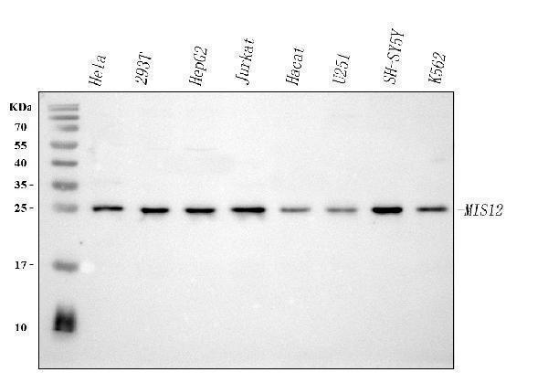
Western blot analysis of MIS12 using anti-MIS12 antibody (A06832-1).
Electrophoresis was performed on a 5-20% SDS-PAGE gel at 70V (Stacking gel) / 90V (Resolving gel) for 2-3 hours. The sample well of each lane was loaded with 30 ug of sample under reducing conditions.
Lane 1: human Hela whole cell lysates,
Lane 2: human 293T whole cell lysates,
Lane 3: human HepG2 whole cell lysates,
Lane 4: human Jurkat whole cell lysates,
Lane 5: human Hacat whole cell lysates,
Lane 6: human U251 whole cell lysates,
Lane 7: human SH-SY5Y whole cell lysates,
Lane 8: human K562 whole cell lysates.
After electrophoresis, proteins were transferred to a nitrocellulose membrane at 150 mA for 50-90 minutes. Blocked the membrane with 5% non-fat milk/TBS for 1.5 hour at RT. The membrane was incubated with rabbit anti-MIS12 antigen affinity purified polyclonal antibody (Catalog # A06832-1) at 0.25 μg/mL overnight at 4°C, then washed with TBS-0.1%Tween 3 times with 5 minutes each and probed with a goat anti-rabbit IgG-HRP secondary antibody at a dilution of 1:5000 for 1.5 hour at RT. The signal is developed using an Enhanced Chemiluminescent detection (ECL) kit (Catalog # EK1002) with Tanon 5200 system. A specific band was detected for MIS12 at approximately 25 kDa. The expected band size for MIS12 is at 24 kDa.

Click image to see more details
IF analysis of MIS12 using anti-MIS12 antibody (A06832-1).
MIS12 was detected in an immunocytochemical section of U2OS cells. Enzyme antigen retrieval was performed using IHC enzyme antigen retrieval reagent (AR0022) for 15 mins. The cells were blocked with 10% goat serum. And then incubated with 5 μg/mL rabbit anti-MIS12 Antibody (A06832-1) overnight at 4°C. Cy3 Conjugated Goat Anti-Rabbit IgG (BA1032) was used as secondary antibody at 1:500 dilution and incubated for 30 minutes at 37°C. The section was counterstained with DAPI. Visualize using a fluorescence microscope and filter sets appropriate for the label used.

Click image to see more details
Flow Cytometry analysis of JK cells using anti-MIS12 antibody (A06832-1).
Overlay histogram showing JK cells stained with A06832-1 (Blue line). To facilitate intracellular staining, cells were fixed with 4% paraformaldehyde and permeabilized with permeabilization buffer. The cells were blocked with 10% normal goat serum. And then incubated with rabbit anti-MIS12 Antibody (A06832-1, 1 μg/1x106 cells) for 30 min at 20°C. DyLight®488 conjugated goat anti-rabbit IgG (BA1127, 5-10 μg/1x106 cells) was used as secondary antibody for 30 minutes at 20°C. Isotype control antibody (Green line) was rabbit IgG (1 μg/1x106) used under the same conditions. Unlabelled sample (Red line) was also used as a control.
Specific Publications For Anti-MIS12 Antibody Picoband® (A06832-1)
Loading publications
Recommended Resources
Here are featured tools and databases that you might find useful.
- Boster's Pathways Library
- Protein Databases
- Bioscience Research Protocol Resources
- Data Processing & Analysis Software
- Photo Editing Software
- Scientific Literature Resources
- Research Paper Management Tools
- Molecular Biology Software
- Primer Design Tools
- Bioinformatics Tools
- Phylogenetic Tree Analysis
Customer Reviews
Have you used Anti-MIS12 Antibody Picoband®?
Share your experimental results or join a short interview to earn up to $1,000 in product credits or other rewards.
0 Reviews For Anti-MIS12 Antibody Picoband®
Customer Q&As
Have a question?
Find answers in Q&As, reviews.
Can't find your answer?
Submit your question
