Click image to see more details
-

-
-
-
-
+3
Product Info Summary
| SKU: | A05937-1 |
|---|---|
| Size: | 100 μg/vial |
| Reactive Species: | Human |
| Host: | Rabbit |
| Application: | ELISA, Flow Cytometry, IF, IHC, ICC, WB |
Customers Who Bought This Also Bought

Product info
Product Name
Anti-MTMR3 Antibody Picoband®
SKU/Catalog Number
A05937-1
Size
100 μg/vial
Form
Lyophilized
Description
Boster Bio Anti-MTMR3 Antibody Picoband® catalog # A05937-1. Tested in WB, IHC, IF, ICC, Flow Cytometry, ELISA applications. This antibody reacts with Human. The brand Picoband indicates this is a premium antibody that guarantees superior quality, high affinity, and strong signals with minimal background in Western blot applications. Only our best-performing antibodies are designated as Picoband, ensuring unmatched performance.
Storage & Handling
At -20°C for one year from date of receipt. After reconstitution, at 4°C for one month. It can also be aliquotted and stored frozen at -20°C for six months. Avoid repeated freezing and thawing.
Cite This Product
Anti-MTMR3 Antibody Picoband® (Boster Biological Technology, Pleasanton CA, USA, Catalog # A05937-1)
Host
Rabbit
Contents
Each vial contains 4 mg Trehalose, 0.9 mg NaCl, 0.2 mg Na2HPO4.
Clonality
Polyclonal
Immunogen
E.coli-derived human MTMR3 recombinant protein (Position: M1-E1074).
Reactive Species
A05937-1 is reactive to MTMR3 in Human
Observed Molecular Weight
150 kDa
Calculated molecular weight
133.6 kDa
Background of MTMR3
This gene encodes a member of the myotubularin dual specificity protein phosphatase gene family. The encoded protein is structurally similar to myotubularin but in addition contains a FYVE domain and an N-terminal PH-GRAM domain. The protein can self-associate and also form heteromers with another myotubularin related protein. The protein binds to phosphoinositide lipids through the PH-GRAM domain, and can hydrolyze phosphatidylinositol(3)-phosphate and phosphatidylinositol(3,5)-biphosphate in vitro. The encoded protein has been observed to have a perinuclear, possibly membrane-bound, distribution in cells, but it has also been found free in the cytoplasm. Multiple transcript variants encoding different isoforms have been found for this gene.
Antibody Validation
Boster validates all antibodies on WB, IHC, ICC, Immunofluorescence, and ELISA with known positive control and negative samples to ensure specificity and high affinity, including thorough antibody incubations.
Application & Images
Applications
A05937-1 is guaranteed for ELISA, Flow Cytometry, IF, IHC, ICC, WB Boster Guarantee
Assay Dilutions Recommendation
The recommendations below provide a starting point for assay optimization. The actual working concentration varies and should be decided by the user.
Western blot, 0.25-0.5 μg/ml, Human
Immunohistochemistry(Paraffin-embedded Section), 2-5 μg/ml, Human
Immunofluorescence, 5 μg/ml, Human
Immunocytochemistry/Immunofluorescence, 5 μg/ml, Human
Flow Cytometry (Fixed), 1-3 μg/1x106 cells, Human
ELISA, 0.1-0.5 μg/ml
Validation Images & Assay Conditions
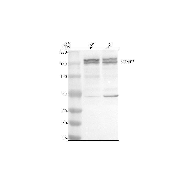
Click image to see more details
Western blot analysis of MTMR3 using anti-MTMR3 antibody (A05937-1).
Electrophoresis was performed on a 8% SDS-PAGE gel at 80V (Stacking gel) / 120V (Resolving gel) for 2 hours. The sample well of each lane was loaded with 30 ug of sample under reducing conditions.
Lane 1: human RT4 whole cell lysates,
Lane 2: human HEL whole cell lysates.
After electrophoresis, proteins were transferred to a nitrocellulose membrane at 150 mA for 50-90 minutes. Blocked the membrane with 5% non-fat milk/TBS for 1.5 hour at RT. The membrane was incubated with rabbit anti-MTMR3 antigen affinity purified polyclonal antibody (A05937-1) at 0.5 μg/mL overnight at 4°C, then washed with TBS-0.1%Tween 3 times with 5 minutes each and probed with a goat anti-rabbit IgG-HRP secondary antibody at a dilution of 1:5000 for 1.5 hour at RT. The signal is developed using an ECL Plus Western Blotting Substrate (Catalog # AR1196-200) with Tanon 5200 system. A specific band was detected for MTMR3 at approximately 150 kDa. The expected band size for MTMR3 is at 134 kDa.

Click image to see more details
IHC analysis of MTMR3 using anti-MTMR3 antibody (A05937-1).
MTMR3 was detected in a paraffin-embedded section of human breast cancer tissue. Heat mediated antigen retrieval was performed in EDTA buffer (pH 8.0, epitope retrieval solution). The tissue section was blocked with 10% goat serum. The tissue section was then incubated with 2 μg/ml rabbit anti-MTMR3 Antibody (A05937-1) overnight at 4°C. Peroxidase Conjugated Goat Anti-rabbit IgG was used as secondary antibody and incubated for 30 minutes at 37°C. The tissue section was developed using HRP Conjugated Rabbit IgG Super Vision Assay Kit (Catalog # SV0002) with DAB as the chromogen.

Click image to see more details
IHC analysis of MTMR3 using anti-MTMR3 antibody (A05937-1).
MTMR3 was detected in a paraffin-embedded section of human prostate cancer tissue. Heat mediated antigen retrieval was performed in EDTA buffer (pH 8.0, epitope retrieval solution). The tissue section was blocked with 10% goat serum. The tissue section was then incubated with 2 μg/ml rabbit anti-MTMR3 Antibody (A05937-1) overnight at 4°C. Peroxidase Conjugated Goat Anti-rabbit IgG was used as secondary antibody and incubated for 30 minutes at 37°C. The tissue section was developed using HRP Conjugated Rabbit IgG Super Vision Assay Kit (Catalog # SV0002) with DAB as the chromogen.

Click image to see more details
IF analysis of MTMR3 using anti-MTMR3 antibody (A05937-1).
MTMR3 was detected in a paraffin-embedded section of human breast cancer tissue. Heat mediated antigen retrieval was performed in EDTA buffer (pH 8.0, epitope retrieval solution). The tissue section was blocked with 10% goat serum. The tissue section was then incubated with 5 μg/mL rabbit anti-MTMR3 Antibody (A05937-1) overnight at 4°C. Cy3 Conjugated Goat Anti-Rabbit IgG (BA1032) was used as secondary antibody at 1:500 dilution and incubated for 30 minutes at 37°C. The section was counterstained with DAPI. Visualize using a fluorescence microscope and filter sets appropriate for the label used.

Click image to see more details
IF analysis of MTMR3 using anti-MTMR3 antibody (A05937-1) and anti-Beta Tubulin antibody (M01857-3).
MTMR3 was detected in an immunocytochemical section of SIHA cells. Enzyme antigen retrieval was performed using IHC enzyme antigen retrieval reagent (AR0022) for 15 mins. The cells were blocked with 10% goat serum. And then incubated with 5 μg/mL rabbit anti-MTMR3 Antibody (A05937-1) and mouse anti-Beta Tubulin antibody (M01857-3) overnight at 4°C. DyLight®488 Conjugated Goat Anti-Rabbit IgG (BA1127) and Cy3 Conjugated Goat Anti-Mouse IgG (BA1031) were used as secondary antibody at 1:500 dilution and incubated for 30 minutes at 37°C. Visualize using a fluorescence microscope and filter sets appropriate for the label used.

Click image to see more details
Flow Cytometry analysis of RT4 cells using anti-MTMR3 antibody (A05937-1).
Overlay histogram showing RT4 cells stained with A05937-1 (Blue line). The cells were fixed with 4% paraformaldehyde and blocked with 10% normal goat serum. And then incubated with rabbit anti-MTMR3 Antibody (A05937-1, 1 μg/1x106 cells) for 30 min at 20°C. DyLight®488 conjugated goat anti-rabbit IgG (BA1127, 5-10 μg/1x106 cells) was used as secondary antibody for 30 minutes at 20°C. Isotype control antibody (Green line) was rabbit IgG (1 μg/1x106) used under the same conditions. Unlabelled sample without incubation with primary antibody and secondary antibody (Red line) was used as a blank control.
Specific Publications For Anti-MTMR3 Antibody Picoband® (A05937-1)
Loading publications
Recommended Resources
Here are featured tools and databases that you might find useful.
- Boster's Pathways Library
- Protein Databases
- Bioscience Research Protocol Resources
- Data Processing & Analysis Software
- Photo Editing Software
- Scientific Literature Resources
- Research Paper Management Tools
- Molecular Biology Software
- Primer Design Tools
- Bioinformatics Tools
- Phylogenetic Tree Analysis
Customer Reviews
Have you used Anti-MTMR3 Antibody Picoband®?
Share your experimental results or join a short interview to earn up to $1,000 in product credits or other rewards.
0 Reviews For Anti-MTMR3 Antibody Picoband®
Customer Q&As
Have a question?
Find answers in Q&As, reviews.
Can't find your answer?
Submit your question
