Click image to see more details
Product Info Summary
| SKU: | A02674-2 |
|---|---|
| Size: | 100 μg/vial |
| Reactive Species: | Human, Mouse, Rat |
| Host: | Rabbit |
| Application: | ELISA, IF, IHC, WB |
Customers Who Bought This Also Bought

Product info
Product Name
Anti-MUC13 Antibody Picoband®
SKU/Catalog Number
A02674-2
Size
100 μg/vial
Form
Lyophilized
Description
Boster Bio Anti-MUC13 Antibody Picoband® catalog # A02674-2. Tested in ELISA, IF, IHC, WB applications. This antibody reacts with Human, Mouse, Rat. The brand Picoband indicates this is a premium antibody that guarantees superior quality, high affinity, and strong signals with minimal background in Western blot applications. Only our best-performing antibodies are designated as Picoband, ensuring unmatched performance.
Storage & Handling
Store at -20˚C for one year from date of receipt. After reconstitution, at 4˚C for one month. It can also be aliquotted and stored frozen at -20˚C for six months. Avoid repeated freeze-thaw cycles.
Cite This Product
Anti-MUC13 Antibody Picoband® (Boster Biological Technology, Pleasanton CA, USA, Catalog # A02674-2)
Host
Rabbit
Contents
Each vial contains 4 mg Trehalose, 0.9 mg NaCl and 0.2 mg Na2HPO4.
Clonality
Polyclonal
Isotype
Rabbit IgG
Immunogen
E.coli-derived human MUC13 recombinant protein (Position: E226-S312).
Cross-reactivity
No cross-reactivity with other proteins.
Reactive Species
A02674-2 is reactive to MUC13 in Human, Mouse, Rat
Observed Molecular Weight
75, 110-120 kDa
Calculated molecular weight
54.6 kDa
Background of MUC13
Epithelial mucins, such as MUC13, are a family of secreted and cell surface glycoproteins expressed by ductal and glandular epithelial tissues. It is mapped to 3q21.2. Mucins are a family of high molecular weight, heavily glycosylated proteins (glycoconjugates) produced by epithelial tissues in most animals. Mucins' key characteristic is their ability to form gels; therefore they are a key component in most gel-like secretions, serving functions from lubrication to cell signalling to forming chemical barriers. They often take an inhibitory role. Some mucins are associated with controlling mineralization, including nacre formation in mollusks,calcification in echinoderms and bone formation in vertebrates. They bind to pathogens as part of the immune system. Overexpression of the mucin proteins, especially MUC1, is associated with many types of cancer. Although some mucins are membrane-bound due to the presence of a hydrophobic membrane-spanning domain that favors retention in the plasma membrane, most mucins are secreted as principal components of mucus by mucous membranes or are secreted to become a component of saliva.
Antibody Validation
Boster validates all antibodies on WB, IHC, ICC, Immunofluorescence, and ELISA with known positive control and negative samples to ensure specificity and high affinity, including thorough antibody incubations.
Application & Images
Applications
A02674-2 is guaranteed for ELISA, IF, IHC, WB Boster Guarantee
Recommend Dilution
| Application | Dilution | Species |
|---|---|---|
| Western blot | 0.1-0.5μg/ml | Human, Mouse, Rat |
| Immunohistochemistry (Paraffin-embedded Section) | 2-5μg/ml | Human, Mouse |
| Immunofluorescence | 5μg/ml | Human |
| ELISA | 0.1-0.5μg/ml | - |
Tested application
Suggested blocking solution with 5% non-fat milk or BSA; (*)Recommended protein loading: 20-40 µg per lane
Use TE buffer pH 9.0 for antigen retrieval; (*) citrate buffer pH 6.0 is an alternative.
Validation Images & Assay Conditions
Click image to see more details
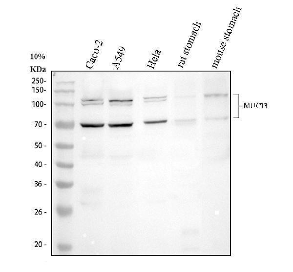
Western blot analysis of MUC13 using anti-MUC13 antibody (A02674-2).
Electrophoresis was performed on a 10% SDS-PAGE gel at 80V (Stacking gel) / 120V (Resolving gel) for 2 hours. The sample well of each lane was loaded with 30 ug of sample under reducing conditions.
Lane 1: human Caco-2 whole cell lysates,
Lane 2: human A549 whole cell lysates,
Lane 3: human Hela whole cell lysates,
Lane 4: rat stomach tissue lysates,
Lane 5: mouse stomach tissue lysates.
After electrophoresis, proteins were transferred to a nitrocellulose membrane at 150 mA for 50-90 minutes. Blocked the membrane with 5% non-fat milk/TBS for 1.5 hour at RT. The membrane was incubated with rabbit anti-MUC13 antigen affinity purified polyclonal antibody (A02674-2) at 0.5 μg/mL overnight at 4°C, then washed with TBS-0.1%Tween 3 times with 5 minutes each and probed with a goat anti-rabbit IgG-HRP secondary antibody (Catalog # BA1054) at a dilution of 1:5000 for 1.5 hour at RT. The signal is developed using an ECL Plus Western Blotting Substrate (Catalog # AR1196-200) with Tanon 5200 system. A specific band was detected for MUC13 at approximately 75, 110-120 kDa. The expected band size for MUC13 is at 55 kDa.

Click image to see more details
IHC analysis of MUC13 using anti-MUC13 antibody (A02674-2).
MUC13 was detected in a paraffin-embedded section of human colon cancer tissue. Heat mediated antigen retrieval was performed in EDTA buffer (pH 8.0, epitope retrieval solution). The tissue section was blocked with 10% goat serum. The tissue section was then incubated with 2 μg/ml rabbit anti-MUC13 Antibody (A02674-2) overnight at 4°C. Peroxidase Conjugated Goat Anti-rabbit IgG was used as secondary antibody and incubated for 30 minutes at 37°C. The tissue section was developed using HRP Conjugated Rabbit IgG Super Vision Assay Kit (Catalog # SV0002) with DAB as the chromogen.

Click image to see more details
IHC analysis of MUC13 using anti-MUC13 antibody (A02674-2).
MUC13 was detected in a paraffin-embedded section of mouse colon tissue. Heat mediated antigen retrieval was performed in EDTA buffer (pH 8.0, epitope retrieval solution). The tissue section was blocked with 10% goat serum. The tissue section was then incubated with 2 μg/ml rabbit anti-MUC13 Antibody (A02674-2) overnight at 4°C. Peroxidase Conjugated Goat Anti-rabbit IgG was used as secondary antibody and incubated for 30 minutes at 37°C. The tissue section was developed using HRP Conjugated Rabbit IgG Super Vision Assay Kit (Catalog # SV0002) with DAB as the chromogen.

Click image to see more details
IF analysis of AMUC13 using anti-MUC13 antibody (A02674-2).
MUC13 was detected in a paraffin-embedded section of human colon cancer tissue. Heat mediated antigen retrieval was performed in EDTA buffer (pH 8.0, epitope retrieval solution). The tissue section was blocked with 10% goat serum. The tissue section was then incubated with 5 μg/mL rabbit anti-MUC13 Antibody (A02674-2) overnight at 4°C. Cy3 Conjugated Goat Anti-Rabbit IgG (BA1032) was used as secondary antibody at 1:500 dilution and incubated for 30 minutes at 37°C. The section was counterstained with DAPI. Visualize using a fluorescence microscope and filter sets appropriate for the label used.
Specific Publications For Anti-MUC13 Antibody Picoband® (A02674-2)
Loading publications
Recommended Resources
Here are featured tools and databases that you might find useful.
- Boster's Pathways Library
- Protein Databases
- Bioscience Research Protocol Resources
- Data Processing & Analysis Software
- Photo Editing Software
- Scientific Literature Resources
- Research Paper Management Tools
- Molecular Biology Software
- Primer Design Tools
- Bioinformatics Tools
- Phylogenetic Tree Analysis
Customer Reviews
Have you used Anti-MUC13 Antibody Picoband®?
Share your experimental results or join a short interview to earn up to $1,000 in product credits or other rewards.
0 Reviews For Anti-MUC13 Antibody Picoband®
Customer Q&As
Have a question?
Find answers in Q&As, reviews.
Can't find your answer?
Submit your question
6 Customer Q&As for Anti-MUC13 Antibody Picoband®
Question
We are currently using anti-MUC13 antibody A02674-2 for rat tissue, and we are content with the ELISA results. The species of reactivity given in the datasheet says human, mouse, rat. Is it true that the antibody can work on goat tissues as well?
Verified Customer
Verified customer
Asked: 2020-03-27
Answer
The anti-MUC13 antibody (A02674-2) has not been validated for cross reactivity specifically with goat tissues, though there is a good chance of cross reactivity. We have an innovator award program that if you test this antibody and show it works in goat you can get your next antibody for free. Please contact me if I can help you with anything.
Boster Scientific Support
Answered: 2020-03-27
Question
I am looking for to test anti-MUC13 antibody A02674-2 on rat colon for research purposes, then I may be interested in using anti-MUC13 antibody A02674-2 for diagnostic purposes as well. Is the antibody suitable for diagnostic purposes?
L. Collins
Verified customer
Asked: 2019-11-18
Answer
The products we sell, including anti-MUC13 antibody A02674-2, are only intended for research use. They would not be suitable for use in diagnostic work. If you have the means to develop a product into diagnostic use, and are interested in collaborating with us and develop our product into an IVD product, please contact us for more discussions.
Boster Scientific Support
Answered: 2019-11-18
Question
Can you help my question with product A02674-2, anti-MUC13 antibody. I was wondering if it would be possible to conjugate this antibody with biotin. I would need it to be without BSA or sodium azide. I am planning on using a buffer exchange of sodium azide with PBS only. Would there be problems for me to conjugate the antibody and store it in -20 degrees in small aliquots?
Verified Customer
Verified customer
Asked: 2019-11-06
Answer
It is not recommended storing this antibody with PBS buffer only in -20 degrees. If you want to store it in -20 degrees it is best to add some cryoprotectant like glycerol. If you want carrier free A02674-2 anti-MUC13 antibody, we can provide it to you in a special formula with trehalose and/or glycerol. These molecules will not interfere with conjugation chemistry and provide a good level of protection for the antibody from degradation. Please be sure to specify this in your purchase order.
Boster Scientific Support
Answered: 2019-11-06
Question
Will A02674-2 anti-MUC13 antibody work on parafin embedded sections? If so, which fixation method do you recommend we use (PFA, paraformaldehyde, other)?
Verified Customer
Verified customer
Asked: 2019-09-24
Answer
As indicated on the product datasheet, A02674-2 anti-MUC13 antibody as been validated on IHC-P. It is best to use PFA for fixation because it has better tissue penetration ability. PFA needs to be prepared fresh before use. Long term stored PFA turns into formalin, as the PFA molecules congregate and become formalin.
Boster Scientific Support
Answered: 2019-09-24
Question
Is this A02674-2 anti-MUC13 antibody reactive to the isotypes of MUC13?
Verified Customer
Verified customer
Asked: 2019-04-04
Answer
The immunogen of A02674-2 anti-MUC13 antibody is E.coli-derived human MUC13 recombinant protein (Position: E226-S312). Could you tell me which isotype you are interested in so I can help see if the immunogen is part of this isotype?
Boster Scientific Support
Answered: 2019-04-04
Question
Will anti-MUC13 antibody A02674-2 work for IHC-P with colon?
R. Anderson
Verified customer
Asked: 2016-07-22
Answer
According to the expression profile of colon, MUC13 is highly expressed in colon. So, it is likely that anti-MUC13 antibody A02674-2 will work for IHC-P with colon.
Boster Scientific Support
Answered: 2016-07-22
