Click image to see more details
-

-
-
-
-
+3
Product Info Summary
| SKU: | A01246-4 |
|---|---|
| Size: | 100 μg/vial |
| Reactive Species: | Human, Mouse, Rat |
| Host: | Rabbit |
| Application: | ELISA, IP, IHC, WB |
Customers Who Bought This Also Bought

Product info
Product Name
Anti-Occludin/OCLN Antibody Picoband®
SKU/Catalog Number
A01246-4
Size
100 μg/vial
Form
Lyophilized
Description
Boster Bio Anti-Occludin/OCLN Antibody Picoband® catalog # A01246-4. Tested in WB, IHC, IP, ELISA applications. This antibody reacts with Human, Mouse, Rat. The brand Picoband indicates this is a premium antibody that guarantees superior quality, high affinity, and strong signals with minimal background in Western blot applications. Only our best-performing antibodies are designated as Picoband, ensuring unmatched performance.
Storage & Handling
At -20°C for one year from date of receipt. After reconstitution, at 4°C for one month. It can also be aliquotted and stored frozen at -20°C for six months. Avoid repeated freezing and thawing.
Cite This Product
Anti-Occludin/OCLN Antibody Picoband® (Boster Biological Technology, Pleasanton CA, USA, Catalog # A01246-4)
Host
Rabbit
Contents
Each vial contains 4 mg Trehalose, 0.9 mg NaCl, 0.2 mg Na2HPO4.
Clonality
Polyclonal
Immunogen
E.coli-derived human Occludin/OCLN recombinant protein (Position: S358-Q520). Human Occludin/OCLN shares 88.8% and 88.2% amino acid (aa) sequence identity with mouse and rat Occludin/OCLN, respectively.
Reactive Species
A01246-4 is reactive to OCLN in Human, Mouse, Rat
Observed Molecular Weight
59 kDa
Calculated molecular weight
59.1 kDa
Background of OCLN
Occludin is an integral membrane protein that is located at tight junctions. It is a member of a family of proteins containing the highly conserved Marvel domain, which contains 4 transmembrane-helix regions. This gene is mapped to 5q13.2. Occludin regulates TGF-beta receptor type I localization for efficient TGF-beta-dependent dissolution of tight junctions during epithelial-mesenchymal transitions. Human Occludin is an essential hepatitis C virus (HCV) cell entry factor that is able to render murine cells infectable with HCV glycoproteins. It has been found that occludin is involved in cell migration and functions to recruit active PI3 kinase to the leading edge, resulting in RAC1 activation and formation of lamellipodia.
Antibody Validation
Boster validates all antibodies on WB, IHC, ICC, Immunofluorescence, and ELISA with known positive control and negative samples to ensure specificity and high affinity, including thorough antibody incubations.
Application & Images
Applications
A01246-4 is guaranteed for ELISA, IP, IHC, WB Boster Guarantee
Recommend Dilution
| Application | Dilution | Species |
|---|---|---|
| Western blot | 0.25-0.5 μg/ml | Human |
| Immunohistochemistry(Paraffin-embedded Section) | 2-5 μg/ml | Human |
| Immunoprecipitation | 0.5-2 μg/ml | Human |
| ELISA | 0.1-0.5 μg/ml | Human |
Tested application
Suggested blocking solution with 5% non-fat milk or BSA; (*)Recommended protein loading: 20-40 µg per lane
Use TE buffer pH 9.0 for antigen retrieval; (*) citrate buffer pH 6.0 is an alternative.
Validation Images & Assay Conditions
Click image to see more details
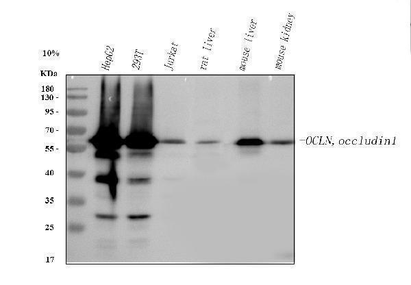
Western blot analysis of Occludin/OCLN using anti-Occludin/OCLN antibody (A01246-4).
Electrophoresis was performed on a 5-20% SDS-PAGE gel at 70V (Stacking gel) / 90V (Resolving gel) for 2-3 hours. The sample well of each lane was loaded with 30 ug of sample under reducing conditions.
Lane 1: human HepG2 whole cell lysates,
Lane 2: human 293T whole cell lysates,
Lane 3: human Jurkat whole cell lysates,
Lane 4: rat liver tissue lysates,
Lane 5: mouse liver tissue lysates,
Lane 6: mouse kidney tissue lysates.
After electrophoresis, proteins were transferred to a nitrocellulose membrane at 150 mA for 50-90 minutes. Blocked the membrane with 5% non-fat milk/TBS for 1.5 hour at RT. The membrane was incubated with rabbit anti-Occludin/OCLN antigen affinity purified polyclonal antibody (A01246-4) at 0.5 μg/mL overnight at 4°C, then washed with TBS-0.1%Tween 3 times with 5 minutes each and probed with a goat anti-rabbit IgG-HRP secondary antibody at a dilution of 1:5000 for 1.5 hour at RT. The signal is developed using an Enhanced Chemiluminescent detection (ECL) kit (Catalog # EK1002) with Tanon 5200 system. A specific band was detected for Occludin/OCLN at approximately 59 kDa. The expected band size for Occludin/OCLN is at 59 kDa.

Click image to see more details
IHC analysis of Occludin/OCLN using anti-Occludin/OCLN antibody (A01246-4).
Occludin/OCLN was detected in a paraffin-embedded section of human stomach tissue. Heat mediated antigen retrieval was performed in EDTA buffer (pH 8.0, epitope retrieval solution). The tissue section was blocked with 10% goat serum. The tissue section was then incubated with 2 μg/ml rabbit anti-Occludin/OCLN Antibody (A01246-4) overnight at 4°C. Peroxidase Conjugated Goat Anti-rabbit IgG was used as secondary antibody and incubated for 30 minutes at 37°C. The tissue section was developed using HRP Conjugated Rabbit IgG Super Vision Assay Kit (Catalog # SV0002) with DAB as the chromogen.

Click image to see more details
Effect of Huanglian-Jiedu decoction on intestinal barrier in APP/PS1 mice. A Representative image of HE staining in the mouse intestine, and representative images of immunohistochemistry of intestinal Occludin and ZO-1 (Scale bar = 50 μm). B Statistical analysis of the relative expression level of Occludin. C Statistical analysis of the relative expression level of ZO-1. D The expressions of ZO-1 and Occludin proteins in the intestine were detected by Western blotting. E Semi-quantitative analysis of the gray value of Occludin. F Semi-quantitative analysis of the gray value of ZO-1. Data are presented as mean ± SD (n = 3). ## P < 0.01 (vs control group), * P < 0.05 and ** P < 0.01 (vs model group)
Index in PubMed under a CC BY license. PMID: 40770806

Click image to see more details
Effects of MT water extract on regulating macrophage polarization, maintaining intestinal barrier integrity, and alleviating oxidative stress in the colons of DSS-induced UC mice. (A) IHC staining of iNOS, Arg1, occludin, and ZO-1 expressions in mouse colon tissues. Quantitative analysis of the integrated optical density (IOD) of (B) iNOS, (C) Arg1, (D) occludin, and (E) ZO-1 in each group. The levels of (F) IL-6, (G) IL-10, (H) MDA, and (I) GSH were measured and analyzed. Data were presented as means ± SD for three independent trials. One-way ANOVA followed by Tukey's multiple comparison test was performed to compare the differences between groups. *p < 0.05 versus control and #p < 0.05 versus DSS alone group. Arg1, arginase 1; DSS, dextran sulfate sodium; IL, interleukin; iNOS, inducible nitric oxide synthase; MDA, malondialdehyde; MT, Medulla Tetrapanacis.
Index in Food Frontiers under a CC BY license. DOI: 10.1002/fft2.70164

Click image to see more details
Immunoprecipitating Occludin/OCLN in HepG2 whole cell lysate.
Western blot analysis of Occludin/OCLN using anti-Occludin/OCLN antibody (A01246-4).
Lane 1: HepG2 whole cell lysates (30ug),
Lane 2: Rabbit control IgG instead of anti-Occludin/OCLN antibody in HepG2 whole cell lysate,
Lane 3: anti-Occludin/OCLN antibody (2μg) + HepG2 whole cell lysate (500μg).
After electrophoresis, proteins were transferred to a membrane. Then the membrane was incubated with rabbit anti-Occludin/OCLN antigen affinity purified polyclonal antibody (A01246-4) at a dilution of 0.5 μg/mL and probed with a mouse anti-rabbit IgG-HRP secondary antibody (Light Chain Specific). The signal is developed using ECL Plus Western Blotting Substrate (Catalog # AR1197). A specific band was detected for Occludin/OCLN at approximately 59 kDa. The expected band size for Occludin/OCLN is at 59 kDa.

Click image to see more details
IHC analysis of Occludin/OCLN using anti-Occludin/OCLN antibody (A01246-4).
Occludin/OCLN was detected in a paraffin-embedded section of human stomach tissue. Heat mediated antigen retrieval was performed in EDTA buffer (pH 8.0, epitope retrieval solution). The tissue section was blocked with 10% goat serum. The tissue section was then incubated with 2 μg/ml rabbit anti-Occludin/OCLN Antibody (A01246-4) overnight at 4°C. Peroxidase Conjugated Goat Anti-rabbit IgG was used as secondary antibody and incubated for 30 minutes at 37°C. The tissue section was developed using HRP Conjugated Rabbit IgG Super Vision Assay Kit (Catalog # SV0002) with DAB as the chromogen.
Specific Publications For Anti-Occludin/OCLN Antibody Picoband® (A01246-4)
Loading publications
Recommended Resources
Here are featured tools and databases that you might find useful.
- Boster's Pathways Library
- Protein Databases
- Bioscience Research Protocol Resources
- Data Processing & Analysis Software
- Photo Editing Software
- Scientific Literature Resources
- Research Paper Management Tools
- Molecular Biology Software
- Primer Design Tools
- Bioinformatics Tools
- Phylogenetic Tree Analysis
Customer Reviews
Have you used Anti-Occludin/OCLN Antibody Picoband®?
Share your experimental results or join a short interview to earn up to $1,000 in product credits or other rewards.
0 Reviews For Anti-Occludin/OCLN Antibody Picoband®
Customer Q&As
Have a question?
Find answers in Q&As, reviews.
Can't find your answer?
Submit your question
