Click image to see more details
Product Info Summary
| SKU: | A02664 |
|---|---|
| Size: | 100ug |
| Reactive Species: | Human |
| Host: | Goat |
| Application: | ELISA, WB |
Customers Who Bought This Also Bought


Product info
Product Name
Anti-p29 ING4 Antibody
SKU/Catalog Number
A02664
Size
100ug
Form
Liquid (sterile filtered)
Description
Boster Bio Anti-p29 ING4 Antibody (Catalog # A02664). Tested in ELISA, WB applications. This antibody reacts with Human.
Storage & Handling
Store vial at -20°C prior to opening. Aliquot contents and freeze at -20°C or below for extended storage. Avoid cycles of freezing and thawing. Centrifuge product if not completely clear after standing at room temperature. This product is stable for several weeks at 4°C as an undiluted liquid. Dilute only prior to immediate use. Expiration date is one (1) year from date of opening. (Ship on dry ice.)
Cite This Product
Anti-p29 ING4 Antibody (Boster Biological Technology, Pleasanton CA, USA, Catalog # A02664)
Host
Goat
Contents
0.02 M Potassium Phosphate, 0.15 M Sodium Chloride, pH 7.2, 0.01% (w/v) Sodium Azide
Clonality
Polyclonal
Isotype
IgG
Immunogen
This affinity purified antibody was prepared from whole goat serum produced by repeated immunizations with a synthetic peptide corresponding to an internal region near aa 150-175 of Human p29 ING4 protein (Inhibitor of growth family, member 4). This sequence only shows homology to isoform 1 for ING4.
Reactive Species
A02664 is reactive to ING4 in Human
Observed Molecular Weight
42 kDa
Calculated molecular weight
28.5 kDa
Background of ING4
p29 ING4 is a tumor suppressor protein similar to ING1 that may inhibit tumor progression by modulating the transcriptional output of signaling pathways which regulate cell proliferation. p29 ING4 has been shown to suppress brain tumor angio-genesis through transcriptional repression of RelA/ NFKB3 target genes when complexed with RelA. p29 ING4 may also specifically suppress loss of contact inhibition elicited by activated oncogenes such as MYC. p29 ING4 shows a nuclear localization and interacts with EP300, TP53, RelA, inhibits cell growth, and induces apoptosis. This protein contains a PHD-finger, which is a common motif in proteins involved in chromatin remodeling. Multiple alternatively spliced transcript variants have been observed. The accession number listed below is for variant (1) that encodes the longest isoform.
Antibody Validation
Boster validates all antibodies on WB, IHC, ICC, Immunofluorescence, and ELISA with known positive control and negative samples to ensure specificity and high affinity, including thorough antibody incubations.
Application & Images
Applications
A02664 is guaranteed for ELISA, WB Boster Guarantee
Recommend Dilution
| Application | Dilution | Species |
|---|---|---|
| ELISA: 1:3 | 000 - 1:12 | 000 |
| WB: 1:500 - 1:3 | 000 |
Validation Images & Assay Conditions
Click image to see more details
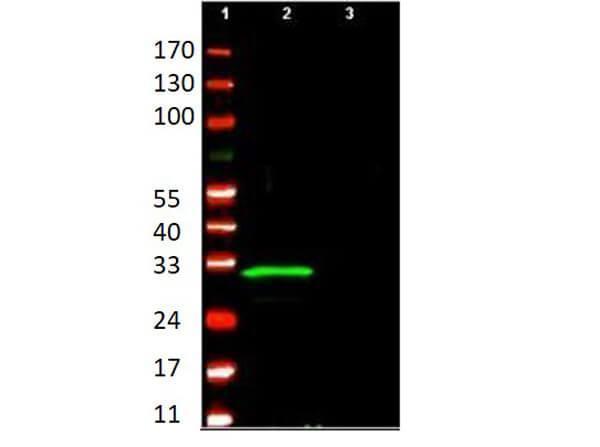
Anti-p29 ING4 polyclonal antibody detects ING4 protein by western blot. This antibody was used at 1.0 µg/ml to detect ING4 (lane 2) present in a U2OS whole cell lysate over expressing the protein. A control lysate (lane 3) shows no background staining. Comparison to MW markers (lane 1) indicates detection of a single band at ~29 kDa corresponding to ING4. A 4-20% TRIS-glycine gradient gel was used to separate the protein by SDS-PAGE under reducing conditions. The protein was transferred to nitrocellulose using standard methods. After blocking using 5% non-fat dry milk in PBS, the membrane was probed with the primary antibody overnight at 4° C followed by washes and reaction with a 1:20,000 dilution of IRDye™800 conjugated Rb-a-Goat IgG [H&L] (code 605-432-013) for 45 min at room temperature. LICOR's Odyssey® Infrared Imaging System was used to scan and process the image. Other detection systems will yield similar results.

Click image to see more details
Anti-p29 ING4 polyclonal antibody detects ING4 protein by western blot in over expressed cell lysates. This antibody was used at 1.0 µg/ml to detect ING4 expression in control (-) and transformed U2OS and HeLa cell lysates. A predominant band corresponding to p29 ING4 is only seen in lysates from transformed cells. Personnel Communication, Motoko Unoki, NCI, NIH.
Specific Publications For Anti-p29 ING4 Antibody (A02664)
Loading publications
Recommended Resources
Here are featured tools and databases that you might find useful.
- Boster's Pathways Library
- Protein Databases
- Bioscience Research Protocol Resources
- Data Processing & Analysis Software
- Photo Editing Software
- Scientific Literature Resources
- Research Paper Management Tools
- Molecular Biology Software
- Primer Design Tools
- Bioinformatics Tools
- Phylogenetic Tree Analysis
Customer Reviews
Have you used Anti-p29 ING4 Antibody?
Share your experimental results or join a short interview to earn up to $1,000 in product credits or other rewards.
0 Reviews For Anti-p29 ING4 Antibody
Customer Q&As
Have a question?
Find answers in Q&As, reviews.
Can't find your answer?
Submit your question
