Click image to see more details
-

-
-
-
-
+6
Product Info Summary
| SKU: | A01208-3 |
|---|---|
| Size: | 100 μg/vial |
| Reactive Species: | Human, Mouse, Rat |
| Host: | Rabbit |
| Application: | ELISA, Flow Cytometry, IHC, WB |
Customers Who Bought This Also Bought

Product info
Product Name
Anti-P2X7/P2RX7 Antibody Picoband®
SKU/Catalog Number
A01208-3
Size
100 μg/vial
Form
Lyophilized
Description
Boster Bio Anti-P2X7/P2RX7 Antibody Picoband® catalog # A01208-3. Tested in ELISA, Flow Cytometry, IHC, WB applications. This antibody reacts with Human, Mouse, Rat. The brand Picoband indicates this is a premium antibody that guarantees superior quality, high affinity, and strong signals with minimal background in Western blot applications. Only our best-performing antibodies are designated as Picoband, ensuring unmatched performance.
Storage & Handling
At -20°C for one year from date of receipt. After reconstitution, at 4°C for one month. It can also be aliquotted and stored frozen at -20°C for six months. Avoid repeated freezing and thawing.
Cite This Product
Anti-P2X7/P2RX7 Antibody Picoband® (Boster Biological Technology, Pleasanton CA, USA, Catalog # A01208-3)
Host
Rabbit
Contents
Each vial contains 4 mg Trehalose, 0.9 mg NaCl, 0.2 mg Na2HPO4.
Clonality
Polyclonal
Isotype
Rabbit IgG
Immunogen
E.coli-derived human P2X7/P2RX7 recombinant protein (Position: S47-K311).
Cross-reactivity
No cross-reactivity with other proteins.
Reactive Species
A01208-3 is reactive to P2RX7 in Human, Mouse, Rat
Observed Molecular Weight
70 kDa
Calculated molecular weight
68.6 kDa
Background of P2RX7
P2X purinoceptor 7 is a protein that in humans is encoded by the P2RX7 gene. The product of this gene belongs to the family of purinoceptors for ATP. This receptor functions as a ligand-gated ion channel and is responsible for ATP-dependent lysis of macrophages through the formation of membrane pores permeable to large molecules. Activation of this nuclear receptor by ATP in the cytoplasm may be a mechanism by which cellular activity can be coupled to changes in gene expression. Multiple alternatively spliced variants have been identified, most of which fit nonsense-mediated decay (NMD) criteria.
Antibody Validation
Boster validates all antibodies on WB, IHC, ICC, Immunofluorescence, and ELISA with known positive control and negative samples to ensure specificity and high affinity, including thorough antibody incubations.
Application & Images
Applications
A01208-3 is guaranteed for ELISA, Flow Cytometry, IHC, WB Boster Guarantee
Recommend Dilution
| Application | Dilution | Species |
|---|---|---|
| Western blot | 0.25-0.5 μg/ml | Mouse, Rat |
| Immunohistochemistry(Paraffin-embedded Section) | 2-5 μg/ml | Human, Rat |
| Flow Cytometry (Fixed) | 1-3 μg/1x106 cells | Human |
| ELISA | 0.1-0.5 μg/ml | - |
Tested application
Suggested blocking solution with 5% non-fat milk or BSA; (*)Recommended protein loading: 20-40 µg per lane
Use TE buffer pH 9.0 for antigen retrieval; (*) citrate buffer pH 6.0 is an alternative.
Validation Images & Assay Conditions
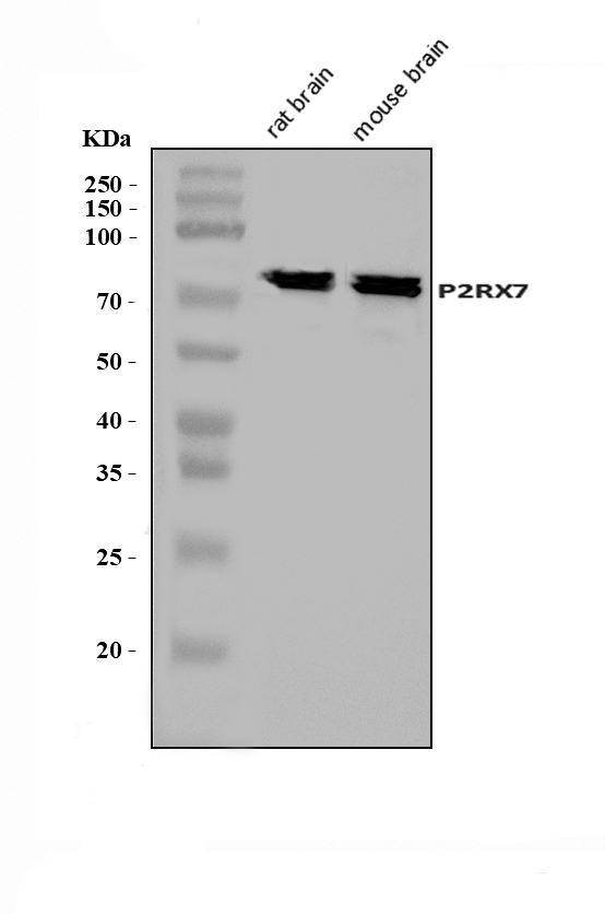
Click image to see more details
Western blot analysis of P2X7/P2RX7 using anti-P2X7/P2RX7 antibody (A01208-3).
Electrophoresis was performed on a 5-20% SDS-PAGE gel at 70V (Stacking gel) / 90V (Resolving gel) for 2-3 hours. The sample well of each lane was loaded with 30 ug of sample under reducing conditions.
Lane 1: rat brain tissue lysates,
Lane 2: mouse brain tissue lysates.
After electrophoresis, proteins were transferred to a nitrocellulose membrane at 150 mA for 50-90 minutes. Blocked the membrane with 5% non-fat milk/TBS for 1.5 hour at RT. The membrane was incubated with rabbit anti-P2X7/P2RX7 antigen affinity purified polyclonal antibody (Catalog # A01208-3) at 0.5 μg/mL overnight at 4°C, then washed with TBS-0.1%Tween 3 times with 5 minutes each and probed with a goat anti-rabbit IgG-HRP secondary antibody at a dilution of 1:5000 for 1.5 hour at RT. The signal is developed using an Enhanced Chemiluminescent detection (ECL) kit (Catalog # EK1002) with Tanon 5200 system. A specific band was detected for P2X7/P2RX7 at approximately 70 kDa. The expected band size for P2X7/P2RX7 is at 70 kDa.

Click image to see more details
IHC analysis of P2X7/P2RX7 using anti-P2X7/P2RX7 antibody (A01208-3).
P2X7/P2RX7 was detected in a paraffin-embedded section of human liver cancer tissue. Heat mediated antigen retrieval was performed in EDTA buffer (pH 8.0, epitope retrieval solution). The tissue section was blocked with 10% goat serum. The tissue section was then incubated with 2 μg/ml rabbit anti-P2X7/P2RX7 Antibody (A01208-3) overnight at 4°C. Biotinylated goat anti-rabbit IgG was used as secondary antibody and incubated for 30 minutes at 37°C. The tissue section was developed using Strepavidin-Biotin-Complex (SABC) (Catalog # SA1022) with DAB as the chromogen.

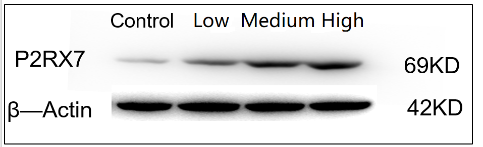
Click image to see more details
Western blot analysis of P2X7/P2RX7 using anti-P2X7/P2RX7 antibody (A01208-3).
Electrophoresis was performed on a 5-20% SDS-PAGE gel at 70V (Stacking gel) / 90V (Resolving gel) for 2-3 hours. The sample well of each lane was loaded with 30 ug of sample under reducing conditions.
Lane 1: Control group-mouse HT22 whole cell lysate,
Lane 2: Low-dose drug treatment-mouse HT22 whole cell lysate,
Lane 3: Medium-dose drug treatment-mouse HT22 whole cell lysate,
Lane 4: High-dose drug treatment-mouse HT22 whole cell lysate.
After electrophoresis, proteins were transferred to a nitrocellulose membrane at 150 mA for 50-90 minutes. Blocked the membrane with 5% non-fat milk/TBS for 1.5 hour at RT. The membrane was incubated with rabbit anti-P2X7/P2RX7 antigen affinity purified polyclonal antibody (Catalog # A01208-3) at 1:1000 overnight at 4°C, then washed with TBS-0.1%Tween 3 times with 5 minutes each and probed with a goat anti-rabbit IgG-HRP secondary antibody at a dilution of 1:2000 for 1 hour at RT. The signal is developed using an Enhanced Chemiluminescent detection (ECL) kit (Catalog # EK1002) with ChemiDoc MP system. A specific band was detected for P2X7/P2RX7 at approximately 69 kDa. The expected band size for P2X7/P2RX7 is at 70 kDa.

Click image to see more details
IHC analysis of P2X7/P2RX7 using anti-P2X7/P2RX7 antibody (A01208-3).
P2X7/P2RX7 was detected in a paraffin-embedded section of human gall bladder adenosquamous carcinomar tissue. Heat mediated antigen retrieval was performed in EDTA buffer (pH 8.0, epitope retrieval solution). The tissue section was blocked with 10% goat serum. The tissue section was then incubated with 2 μg/ml rabbit anti-P2X7/P2RX7 Antibody (A01208-3) overnight at 4°C. Biotinylated goat anti-rabbit IgG was used as secondary antibody and incubated for 30 minutes at 37°C. The tissue section was developed using Strepavidin-Biotin-Complex (SABC) (Catalog # SA1022) with DAB as the chromogen.

Click image to see more details
BBG treatment suppressed the P2X7R/NLRP3 pathway in CD4 + T cells in EAN. Proteins were obtained from sciatic nerves at the peak of EAN and subjected to western blot analysis and quantification. NLRP3 expression in immune cells was analyzed through flow cytometry and immunofluorescence. (A) Representative western blots of P2X7R, NLRP3, IL-1β and Caspase-1 protein expression in sciatic nerve. (B-E) Protein levels were quantified via densitometric analysis of immunoreactive bands in western blots using ImageJ software and were normalized to that of GAPDH. (F-G) Flow cytometry analysis and NLRP3 quantification on CD4 + T cells in splenic MNCs of control (unimmunized group), vehicle and BBG-treated rats. (H) Immunofluorescence photomicrographs of NLRP3 expression in CD4 + T cells in the sciatic nerves of control, vehicle, and BBG-treated EAN rats. (I) Number of NLRP3-positive cells in sciatic nerve sections obtained from control rats, vehicle and BBG-treated EAN rats. (J) NLRP3 quantification on CD4 + T cells. Scale bars, 20 μm. Experiments were performed in triplicate ( n = 6 per replicate) with similar results. Control group, unimmunized group; BBG-P, preventative BBG group; BBG-T, therapeutic BBG group. * p < 0.05, ** p < 0.01
Index in PubMed under a CC BY license. PMID: 38528529

Click image to see more details
IHC analysis of P2X7/P2RX7 using anti-P2X7/P2RX7 antibody (A01208-3).
P2X7/P2RX7 was detected in a paraffin-embedded section of rat brain tissue. Heat mediated antigen retrieval was performed in EDTA buffer (pH 8.0, epitope retrieval solution). The tissue section was blocked with 10% goat serum. The tissue section was then incubated with 2 μg/ml rabbit anti-P2X7/P2RX7 Antibody (A01208-3) overnight at 4°C. Biotinylated goat anti-rabbit IgG was used as secondary antibody and incubated for 30 minutes at 37°C. The tissue section was developed using Strepavidin-Biotin-Complex (SABC) (Catalog # SA1022) with DAB as the chromogen.

Click image to see more details
P2X7R expression was significantly elevated in CD4 + T cells in EAN rats. P2X7R expression in T cells and macrophages in control (unimmunized group) and EAN rats at day 18. (A-B) Fluorescence photomicrographs showing P2X7R expression and quantification in CD4 + T cells, CD8 + T cells and macrophages (CD68 + ) in the sciatic nerves of EAN and control rats. Scale bars, 20 μm. (C) Graph showing the number of P2X7R-positive cells in sciatic nerve sections obtained from control and EAN rats. (D-E) Flow cytometry analysis of P2X7R expression and quantification on CD4 + T cells, CD8 + T cells and macrophages in the spleen of EAN and control rats. Experiments were performed in triplicate ( n = 6 per replicate) with similar results. * p < 0.05, ** p < 0.01
Index in PubMed under a CC BY license. PMID: 38528529

Click image to see more details
P2X7R expression in blood of patients with GBS and healthy controls. Human peripheral blood mononuclear cells (PBMCs) were isolated from blood samples, and P2X7R expression was examined using flow cytometry and qRT-PCR. (A) P2X7R mRNA levels were elevated in the PBMCs collected from patients with GBS compared to those in PBMCs collected from the healthy controls ( n = 8 per group). (B-D) P2X7R expression increased on CD4 + T cells in the PBMCs collected from patients with GBS, but no significant increase was noted on CD8 + T cells or monocytes (CD14 + ) compared to healthy controls ( n = 7 per group). HC, healthy control; GBS, Guillain–Barre Syndrome. * p < 0.05, ** p < 0.01
Index in PubMed under a CC BY license. PMID: 38528529

Click image to see more details
IHC analysis of P2RX7 using anti-P2RX7 antibody (A01208-3).
P2RX7 was detected in a paraffin-embedded section of human cerebellum tissue. Heat mediated antigen retrieval was performed in EDTA buffer (pH 8.0, epitope retrieval solution). The tissue section was blocked with 10% goat serum. The tissue section was then incubated with 2 μg/ml rabbit anti-P2RX7 Antibody (A01208-3) overnight at 4°C. Peroxidase Conjugated Goat Anti-rabbit IgG was used as secondary antibody and incubated for 30 minutes at 37°C. The tissue section was developed using HRP Conjugated Rabbit IgG Super Vision Assay Kit (Catalog # SV0002) with DAB as the chromogen.

Click image to see more details
Flow Cytometry analysis of THP-1 cells using anti-P2X7/P2RX7 antibody (A01208-3).
Overlay histogram showing THP-1 cells stained with A01208-3 (Blue line). The cells were fixed with 4% paraformaldehyde and blocked with 10% normal goat serum. And then incubated with rabbit anti-P2X7/P2RX7 Antibody (A01208-3, 1 μg/1x106 cells) for 30 min at 20°C. DyLight®488 conjugated goat anti-rabbit IgG (BA1127, 5-10 μg/1x106 cells) was used as secondary antibody for 30 minutes at 20°C. Isotype control antibody (Green line) was rabbit IgG (1 μg/1x106) used under the same conditions. Unlabelled sample without incubation with primary antibody and secondary antibody (Red line) was used as a blank control.
Specific Publications For Anti-P2X7/P2RX7 Antibody Picoband® (A01208-3)
Loading publications
Recommended Resources
Here are featured tools and databases that you might find useful.
- Boster's Pathways Library
- Protein Databases
- Bioscience Research Protocol Resources
- Data Processing & Analysis Software
- Photo Editing Software
- Scientific Literature Resources
- Research Paper Management Tools
- Molecular Biology Software
- Primer Design Tools
- Bioinformatics Tools
- Phylogenetic Tree Analysis
Customer Reviews
Have you used Anti-P2X7/P2RX7 Antibody Picoband®?
Share your experimental results or join a short interview to earn up to $1,000 in product credits or other rewards.
1 Reviews For Anti-P2X7/P2RX7 Antibody Picoband®
This antibody is highly specific and efficient, suitable for Western blot detection of P2RX7 protein in HT22 cells, producing clear and single bands.
Excellent
| SKU | A01208-3 |
|---|---|
| Application | Western Blot |
| Sample | Mouse HT-22 cells |
| Sample Processing Description | After digestion, cells were collected by centrifugation and lysed in 1 mL of RIPA buffer containing protease inhibitors on ice for 1 h. The lysates were centrifuged, and the supernatants were collected. After protein quantification, 5× loading buffer was added, and the samples were denatured at 100 °C for 10 min before loading onto SDS-PAGE. |
| Other Reagents | Blocking buffer |
| Primary Antibody | P2X7/P2RX7 Antibody Picoband® |
| Primary Incubation | 1:1000, overnight at 4 ℃ |
| Secondary Antibody | HRP Conjugated AffiniPure Goat Anti-Rabbit IgG (H+L) |
| Secondary Incubation | 1:2000, 1 hour in room temperature |
| Detection | Substrate: ECL, Imaging system:ChemiDoc MP |
| Results Summary | The control represents HT22 cells without drug treatment, while Low, Medium, and High correspond to experimental groups treated with three different drug concentrations for 24 h. The Western blot results obtained with this antibody are clear and well-defined, allowing comparison of expression differences between groups. The expression of the target protein slightly increases with increasing drug concentration. |
Yu Tian, Institute of Materia Medica, Beijing
Verified customer
Submitted 2025-12-26
Customer Q&As
Have a question?
Find answers in Q&As, reviews.
Can't find your answer?
Submit your question
