Click image to see more details
Product Info Summary
| SKU: | A01903-1 |
|---|---|
| Size: | 100 μl/vial |
| Reactive Species: | Human, Mouse, Rat |
| Host: | Rabbit |
| Application: | ELISA, Flow Cytometry, IP, IF, IHC, ICC, WB |
Customers Who Bought This Also Bought

Product info
Product Name
Anti-PCM1 Antibody
SKU/Catalog Number
A01903-1
Size
100 μl/vial
Form
Liquid
Description
Boster Bio Anti-PCM1 Antibody catalog # A01903-1. Tested in WB, IHC, ICC, IF, IP, Flow Cytometry, ELISA applications. This antibody reacts with Human, Mouse, Rat.
Storage & Handling
12 months from date of receipt,-20℃ as supplied. 6 months 2 to 8℃ after reconstitution. Avoid repeated freezing and thawing.
Cite This Product
Anti-PCM1 Antibody (Boster Biological Technology, Pleasanton CA, USA, Catalog # A01903-1)
Host
Rabbit
Contents
500 μg/ml antibody with PBS, 0.02% NaN3, 1 mg stabilizing protein and 50% glycerol
*This antibody is supplied in a stabilized formulation.
Compatibility with conjugation reactions depends on the chemistry of the conjugation method used.
For conjugation methods that are not compatible with the stabilizing components present in this formulation, a carrier-free antibody format is required.
Clonality
Polyclonal
Immunogen
E.coli-derived human PCM1 recombinant protein (Position: M1-K259).
Reactive Species
A01903-1 is reactive to PCM1 in Human, Mouse, Rat
Calculated molecular weight
228.5 kDa
Background of PCM1
The protein encoded by this gene is a component of centriolar satellites, which are electron dense granules scattered around centrosomes. Inhibition studies show that this protein is essential for the correct localization of several centrosomal proteins, and for anchoring microtubules to the centrosome. Chromosomal aberrations involving this gene are associated with papillary thyroid carcinomas and a variety of hematological malignancies, including atypical chronic myeloid leukemia and T-cell lymphoma. Multiple transcript variants encoding different isoforms have been found for this gene.
Antibody Validation
Boster validates all antibodies on WB, IHC, ICC, Immunofluorescence, and ELISA with known positive control and negative samples to ensure specificity and high affinity, including thorough antibody incubations.
Application & Images
Applications
A01903-1 is guaranteed for ELISA, Flow Cytometry, IP, IF, IHC, ICC, WB Boster Guarantee
Recommend Dilution
| Application | Dilution | Species |
|---|---|---|
| Western blot | 1:500-2000 | |
| Immunohistochemistry | 1:50-400 | |
| Immunocytochemistry/Immunofluorescence | 1:50-400 | |
| Immunoprecipitation | 1:50 | |
| Flow Cytometry (Fixed) | 1-3μg/1x106 cells | |
| ELISA | 1:100-1000 |
Validation Images & Assay Conditions
Click image to see more details
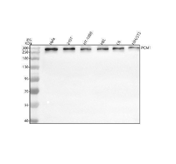
Western blot analysis of PCM1 using anti-PCM1 antibody (A01903-1).
Electrophoresis was performed on a 10% SDS-PAGE gel at 80V (Stacking gel) / 120V (Resolving gel) for 2 hours. The sample well of each lane was loaded with 30 ug of sample under reducing conditions.
Lane 1: human Hela whole cell lysates,
Lane 2: human 293T whole cell lysates,
Lane 3: human HT-1080 whole cell lysates,
Lane 4: human HEL whole cell lysates,
Lane 5: rat C6 whole cell lysates,
Lane 6: mouse NIH/3T3 whole cell lysates.
After electrophoresis, proteins were transferred to a nitrocellulose membrane at 150 mA for 50-90 minutes. Blocked the membrane with 5% non-fat milk/TBS for 1.5 hour at RT. The membrane was incubated with rabbit anti-PCM1 antigen affinity purified polyclonal antibody (A01903-1) at 1:1000 overnight at 4°C, then washed with TBS-0.1%Tween 3 times with 5 minutes each and probed with a goat anti-rabbit IgG-HRP secondary antibody at a dilution of 1:5000 for 1.5 hour at RT. The signal is developed using an ECL Plus Western Blotting Substrate (Catalog # AR1196-200) with Tanon 5200 system. A specific band was detected for PCM1 at approximately 280 kDa. The expected band size for PCM1 is at 229 kDa.

Click image to see more details
IHC analysis of PCM1 using anti-PCM1 antibody (A01903-1).
PCM1 was detected in a paraffin-embedded section of human colon cancer tissue. Heat mediated antigen retrieval was performed in EDTA buffer (pH 8.0, epitope retrieval solution). The tissue section was blocked with 10% goat serum. The tissue section was then incubated with 1:100 rabbit anti-PCM1 Antibody (A01903-1) overnight at 4°C. Peroxidase Conjugated Goat Anti-rabbit IgG was used as secondary antibody and incubated for 30 minutes at 37°C. The tissue section was developed using HRP Conjugated Rabbit IgG Super Vision Assay Kit (Catalog # SV0002) with DAB as the chromogen.

Click image to see more details
IHC analysis of PCM1 using anti-PCM1 antibody (A01903-1).
PCM1 was detected in a paraffin-embedded section of mouse brain tissue. Heat mediated antigen retrieval was performed in EDTA buffer (pH 8.0, epitope retrieval solution). The tissue section was blocked with 10% goat serum. The tissue section was then incubated with 1:100 rabbit anti-PCM1 Antibody (A01903-1) overnight at 4°C. Peroxidase Conjugated Goat Anti-rabbit IgG was used as secondary antibody and incubated for 30 minutes at 37°C. The tissue section was developed using HRP Conjugated Rabbit IgG Super Vision Assay Kit (Catalog # SV0002) with DAB as the chromogen.

Click image to see more details
IHC analysis of PCM1 using anti-PCM1 antibody (A01903-1).
PCM1 was detected in a paraffin-embedded section of rat brain tissue. Heat mediated antigen retrieval was performed in EDTA buffer (pH 8.0, epitope retrieval solution). The tissue section was blocked with 10% goat serum. The tissue section was then incubated with 1:100 rabbit anti-PCM1 Antibody (A01903-1) overnight at 4°C. Peroxidase Conjugated Goat Anti-rabbit IgG was used as secondary antibody and incubated for 30 minutes at 37°C. The tissue section was developed using HRP Conjugated Rabbit IgG Super Vision Assay Kit (Catalog # SV0002) with DAB as the chromogen.
Specific Publications For Anti-PCM1 Antibody (A01903-1)
Loading publications
Recommended Resources
Here are featured tools and databases that you might find useful.
- Boster's Pathways Library
- Protein Databases
- Bioscience Research Protocol Resources
- Data Processing & Analysis Software
- Photo Editing Software
- Scientific Literature Resources
- Research Paper Management Tools
- Molecular Biology Software
- Primer Design Tools
- Bioinformatics Tools
- Phylogenetic Tree Analysis
Customer Reviews
Have you used Anti-PCM1 Antibody?
Share your experimental results or join a short interview to earn up to $1,000 in product credits or other rewards.
0 Reviews For Anti-PCM1 Antibody
Customer Q&As
Have a question?
Find answers in Q&As, reviews.
Can't find your answer?
Submit your question
