Click image to see more details
Product Info Summary
| SKU: | RP1039 |
|---|---|
| Size: | 100 μg/vial |
| Reactive Species: | Human |
| Host: | Rabbit |
| Application: | IHC |
Customers Who Bought This Also Bought

Product info
Product Name
Anti-PD1/PDCD1 Antibody Picoband®
SKU/Catalog Number
RP1039
PB0165 is an alternative SKU for this antibody, used in previous lots.
Size
100 μg/vial
Form
Lyophilized
Description
Boster Bio Anti-PD1/PDCD1 Antibody catalog # RP1039. Tested in IHC applications. This antibody reacts with Human.
Storage & Handling
Store at -20˚C for one year from date of receipt. After reconstitution, at 4˚C for one month. It can also be aliquotted and stored frozen at -20˚C for six months. Avoid repeated freeze-thaw cycles.
Cite This Product
Anti-PD1/PDCD1 Antibody Picoband® (Boster Biological Technology, Pleasanton CA, USA, Catalog # RP1039)
Host
Rabbit
Contents
Each vial contains 4 mg Trehalose, 0.9 mg NaCl and 0.2 mg Na2HPO4.
Clonality
Polyclonal
Isotype
Rabbit IgG
Immunogen
E.coli-derived human PD1 recombinant protein (Position: P101-L288). Human PD1 shares 59% amino acid (aa) sequence identity with mouse PD1.
Cross-reactivity
No cross-reactivity with other proteins
Reactive Species
RP1039 is reactive to PDCD1 in Human
Calculated molecular weight
31.6 kDa
Background of PDCD1
PDCD1 (Programmed cell death 1), also called PD1, encodes a cell surface receptor that is a member of the B7 superfamily involved in immunomodulation. This gene is mapped to 2q37.3. PDCD1 acts as an inhibitory molecule on T cells after interacting with its ligands PDL1 and PDL2. The PDCD1 gene contains 5 exons. This protein is expressed in pro-B-cells and is thought to play a role in their differentiation. Using flow cytometric analysis, It has been found that expression of PDCD1 was upregulated on CD16-positive and CD16-negative monocytes, but not on dendritic cells, in viremic HIV-positive patients, but not in highly active antiretroviral therapy (HAART)-treated HIV-positive patients. PDCD1 upregulation in monocytes was induced by microbial Toll-like receptor ligands and inflammatory cytokines.
Antibody Validation
Boster validates all antibodies on WB, IHC, ICC, Immunofluorescence, and ELISA with known positive control and negative samples to ensure specificity and high affinity, including thorough antibody incubations.
Application & Images
Applications
RP1039 is guaranteed for IHC Boster Guarantee
Recommend Dilution
| Application | Dilution | Species |
|---|---|---|
| Immunohistochemistry (Paraffin-embedded Section) | 2-5μg/ml | Human |
Tested application
Use TE buffer pH 9.0 for antigen retrieval; (*) citrate buffer pH 6.0 is an alternative.
Validation Images & Assay Conditions
Click image to see more details
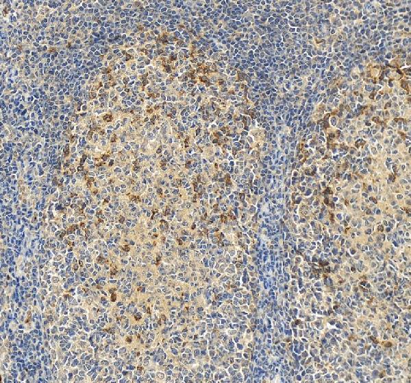
IHC analysis of PD-1/CD279/PDCD1 using anti-PD-1/CD279/PDCD1 antibody (RP1039).
PD-1/CD279/PDCD1 was detected in a paraffin-embedded section of human tonsil tissue. Heat mediated antigen retrieval was performed in EDTA buffer (pH 8.0, epitope retrieval solution). The tissue section was blocked with 10% goat serum. The tissue section was then incubated with 2 μg/ml rabbit anti-PD-1/CD279/PDCD1 Antibody (RP1039) overnight at 4°C. Peroxidase Conjugated Goat Anti-rabbit IgG was used as secondary antibody and incubated for 30 minutes at 37°C. The tissue section was developed using HRP Conjugated Rabbit IgG Super Vision Assay Kit (Catalog # SV0002) with DAB as the chromogen.

Click image to see more details
IHC analysis of PD1 using anti-PD1 antibody (RP1039).
PD1 was detected in a paraffin-embedded section of human tonsil tissue. Heat mediated antigen retrieval was performed in EDTA buffer (pH 8.0, epitope retrieval solution). The tissue section was blocked with 10% goat serum. The tissue section was then incubated with 2 μg/ml rabbit anti-PD1 Antibody (RP1039) overnight at 4°C. Peroxidase Conjugated Goat Anti-rabbit IgG was used as secondary antibody and incubated for 30 minutes at 37°C. The tissue section was developed using HRP Conjugated Rabbit IgG Super Vision Assay Kit (Catalog # SV0002) with DAB as the chromogen.
Specific Publications For Anti-PD1/PDCD1 Antibody Picoband® (RP1039)
Loading publications
Recommended Resources
Here are featured tools and databases that you might find useful.
- Boster's Pathways Library
- Protein Databases
- Bioscience Research Protocol Resources
- Data Processing & Analysis Software
- Photo Editing Software
- Scientific Literature Resources
- Research Paper Management Tools
- Molecular Biology Software
- Primer Design Tools
- Bioinformatics Tools
- Phylogenetic Tree Analysis
Customer Reviews
Have you used Anti-PD1/PDCD1 Antibody Picoband®?
Share your experimental results or join a short interview to earn up to $1,000 in product credits or other rewards.
0 Reviews For Anti-PD1/PDCD1 Antibody Picoband®
Customer Q&As
Have a question?
Find answers in Q&As, reviews.
Can't find your answer?
Submit your question
1 Customer Q&As for Anti-PD1/PDCD1 Antibody Picoband®
Question
We are currently using anti-PD1/PDCD1 antibody RP1039 for human tissue, and we are content with the WB results. The species of reactivity given in the datasheet says human, mouse. Is it true that the antibody can work on horse tissues as well?
Verified Customer
Verified customer
Asked: 2018-02-07
Answer
The anti-PD1/PDCD1 antibody (RP1039) has not been tested for cross reactivity specifically with horse tissues, though there is a good chance of cross reactivity. We have an innovator award program that if you test this antibody and show it works in horse you can get your next antibody for free. Please contact me if I can help you with anything.
Boster Scientific Support
Answered: 2018-02-07
