Click image to see more details
Product Info Summary
| SKU: | MP01105 |
|---|---|
| Size: | 100ug |
| Reactive Species: | Human, Mouse |
| Host: | Mouse |
| Application: | ELISA, IHC, WB |
Customers Who Bought This Also Bought

Product info
Product Name
Anti-Pdcd4 phospho S457 Monoclonal Antibody
SKU/Catalog Number
MP01105
Size
100ug
Form
Liquid (sterile filtered)
Description
Boster Bio Anti-Pdcd4 phospho S457 Monoclonal Antibody (Catalog # MP01105). Tested in ELISA, IHC, WB applications. This antibody reacts with Human, Mouse.
Storage & Handling
Store vial at -20°C prior to opening. Aliquot contents and freeze at -20°C or below for extended storage. Avoid cycles of freezing and thawing. Centrifuge product if not completely clear after standing at room temperature. This product is stable for several weeks at 4°C as an undiluted liquid. Dilute only prior to immediate use. Expiration date is one (1) year from date of opening. (Ship on dry ice.)
Cite This Product
Anti-Pdcd4 phospho S457 Monoclonal Antibody (Boster Biological Technology, Pleasanton CA, USA, Catalog # MP01105)
Host
Mouse
Contents
0.02 M Potassium Phosphate, 0.15 M Sodium Chloride, pH 7.2, 0.01% (w/v) Sodium Azide
Clonality
Monoclonal
Clone Number
Clone: 9G6
Isotype
IgG1 kappa
Immunogen
This monoclonal antibody was produced by repeated immunizations with a synthetic peptide corresponding to residues surrounding Ser457 of the human Pdcd4 protein.
Cross-reactivity
No cross reactivity with other proteins.
Reactive Species
MP01105 is reactive to PDCD4 in Human, Mouse
Observed Molecular Weight
42 kDa
Calculated molecular weight
51.7 kDa
Background of PDCD4
Programmed cell death 4 (Pdcd4) is a novel tumor suppressor. Pdcd4 directly inhibits the helicase activity of eukaryotic translation initiation factor 4A (eIF4A), a component of the translation initiation complex. Pdcd4 also suppresses the transactivation of activator protein-1 (AP-1)-responsive promoters by c-Jun. Pdcd4 contains two Akt phosphorylation sites, one at Ser67 and the other at Ser457. The phosphorylation of Pdcd4 by Akt causes nuclear translocation of Pdcd4 and a significant decrease in the ability of Pdcd4 to interfere with the transactivation of AP-1-responsive promoters by c-Jun.
Antibody Validation
Boster validates all antibodies on WB, IHC, ICC, Immunofluorescence, and ELISA with known positive control and negative samples to ensure specificity and high affinity, including thorough antibody incubations.
Application & Images
Applications
MP01105 is guaranteed for ELISA, IHC, WB Boster Guarantee
Assay Dilutions Recommendation
The recommendations below provide a starting point for assay optimization. The actual working concentration varies and should be decided by the user.
ELISA: 1:20,000 - 1:100,000
IHC: 1:1,000 - 1:5,000
WB: 1:2,000 - 1:10,000
Pdcd4 phospho S457 Antibody is tested by ELISA, immunohistochemistry, and western blotting. Specific conditions for reactivity should be optimized by the end user. Expect a band approximately 62 kDa in size corresponding to phosphorylated Pdcd4 protein by western blotting in the appropriate cell lysate or extract. This phospho-specific monoclonal antibody reacts with human Pdcd4 pS457 and shows minimal reactivity by ELISA against the non-phosphorylated form of the immunizing peptide.
Validation Images & Assay Conditions
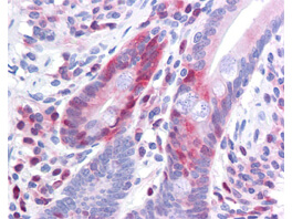
Click image to see more details
Boster Antibody 200-301-964 has been tested in immunohistochemistry, analyzed by an anatomic pathologist and validated for use in IHC applications against formalin-fixed, paraffin-embedded human tissues. The antibody was serially diluted and tested at a range of concentrations on at least 22 different human formalin-fixed, paraffin archival tissues, and positive and negative tissues were scored and compared to the published literature on the expression and function of the gene. A representative image from positively stained small intestine shows the localization of the anti Pdcd4 antibody as the precipitated red signal, with a hematoxylin purple nuclear counterstain. Image provided courtesy of LifeSpan Biosciences, Seattle, WA

Click image to see more details
Western blot using Boster's Protein A purified Mouse Monoclonal anti-Pdcd4 pS457 antibody shows detection of phosphorylated Pdcd4 (indicated by arrowhead at ~62 kDa) in NIH-3T3 cells after 5 min treatment with 30 ng/mL PDGF [lane 2]. No reactivity is seen for (non-phosphorylated) in unstimulated NIH 3T3 cells [lane 1]. The membrane was probed with the primary antibody at a 1:2,000 dilution, overnight at 4° C. For detection HRP conjugated Rb-a-Mouse IgG was used at a 1:20,000 dilution in blocking buffer for 1 h at 4° C followed by visualization using a Biospectrum® imaging system (UVP).

Click image to see more details
Western blot using Boster Immunochemicals Protein A purified Mouse Monoclonal anti-Pdcd4 pS457 antibody against recombinant PDCD4 protein. Membrane was blocked in 1% BSA-TBS-T for 30 min RT and probed with 1° Ab Ms-A-Pdcd4pS457 1:1000 (o/n 4°C in 1% BSA-TBS-T) followed by 2° Ab Peroxidase Conjugated Rabbit anti-Ms at 1:40,000 in 30 min RT. Bands at ~62 kD and ~32 kD were detected.
Specific Publications For Anti-Pdcd4 phospho S457 Monoclonal Antibody (MP01105)
Loading publications
Recommended Resources
Here are featured tools and databases that you might find useful.
- Boster's Pathways Library
- Protein Databases
- Bioscience Research Protocol Resources
- Data Processing & Analysis Software
- Photo Editing Software
- Scientific Literature Resources
- Research Paper Management Tools
- Molecular Biology Software
- Primer Design Tools
- Bioinformatics Tools
- Phylogenetic Tree Analysis
Customer Reviews
Have you used Anti-Pdcd4 phospho S457 Monoclonal Antibody?
Share your experimental results or join a short interview to earn up to $1,000 in product credits or other rewards.
0 Reviews For Anti-Pdcd4 phospho S457 Monoclonal Antibody
Customer Q&As
Have a question?
Find answers in Q&As, reviews.
Can't find your answer?
Submit your question
