Click image to see more details
Product Info Summary
| SKU: | A03763-1 |
|---|---|
| Size: | 100 μg/vial |
| Reactive Species: | Human, Mouse, Rat |
| Host: | Rabbit |
| Application: | IF, IHC, WB |
Customers Who Bought This Also Bought

Product info
Product Name
Anti-PDLIM5 Antibody Picoband®
SKU/Catalog Number
A03763-1
Size
100 μg/vial
Form
Lyophilized
Description
Boster Bio Anti-PDLIM5 Antibody Picoband® catalog # A03763-1. Tested in WB, IHC, IF applications. This antibody reacts with Human, Mouse, Rat. The brand Picoband indicates this is a premium antibody that guarantees superior quality, high affinity, and strong signals with minimal background in Western blot applications. Only our best-performing antibodies are designated as Picoband, ensuring unmatched performance.
Storage & Handling
At -20°C for one year from date of receipt. After reconstitution, at 4°C for one month. It can also be aliquotted and stored frozen at -20°C for six months. Avoid repeated freezing and thawing.
Cite This Product
Anti-PDLIM5 Antibody Picoband® (Boster Biological Technology, Pleasanton CA, USA, Catalog # A03763-1)
Host
Rabbit
Contents
Each vial contains 4 mg Trehalose, 0.9 mg NaCl, 0.2 mg Na2HPO4.
Clonality
Polyclonal
Immunogen
A synthetic peptide corresponding to a sequence in the middle region of human PDLIM5. Human PDLIM5 shares 95.5% amino acid (aa) sequence identity with both mouse and rat PDLIM5.
Reactive Species
A03763-1 is reactive to PDLIM5 in Human, Mouse, Rat
Observed Molecular Weight
64 kDa
Calculated molecular weight
63.9 kDa
Background of PDLIM5
This gene encodes a member of a family of proteins that possess a 100-amino acid PDZ domain at the N terminus and one to three LIM domains at the C-terminus. This family member functions as a scaffold protein that tethers protein kinases to the Z-disk in striated muscles. It is thought to function in cardiomyocyte expansion and in restraining postsynaptic growth of excitatory synapses. Alternative splicing of this gene results in multiple transcript variants.
Antibody Validation
Boster validates all antibodies on WB, IHC, ICC, Immunofluorescence, and ELISA with known positive control and negative samples to ensure specificity and high affinity, including thorough antibody incubations.
Application & Images
Applications
A03763-1 is guaranteed for IF, IHC, WB Boster Guarantee
Recommend Dilution
| Application | Dilution | Species |
|---|---|---|
| Western blot | 0.25-0.5 μg/ml | Human, Mouse, Rat |
| Immunohistochemistry(Paraffin-embedded Section) | 2-5 μg/ml | Human |
| Immunofluorescence | 5 μg/ml | Human |
Validation Images & Assay Conditions
Click image to see more details
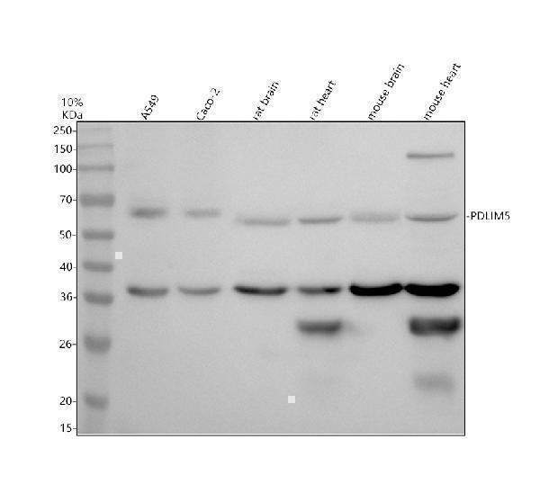
Western blot analysis of PDLIM5 using anti-PDLIM5 antibody (A03763-1).
Electrophoresis was performed on a 10% SDS-PAGE gel at 80V (Stacking gel) / 120V (Resolving gel) for 2 hours. The sample well of each lane was loaded with 30 ug of sample under reducing conditions.
Lane 1: human A549 whole cell lysates,
Lane 2: human Caco-2 whole cell lysates,
Lane 3: rat brain tissue lysates,
Lane 4: rat heart tissue lysates,
Lane 5: mouse brain tissue lysates,
Lane 6: mouse heart tissue lysates.
After electrophoresis, proteins were transferred to a nitrocellulose membrane at 150 mA for 50-90 minutes. Blocked the membrane with 5% non-fat milk/TBS for 1.5 hour at RT. The membrane was incubated with rabbit anti-PDLIM5 antigen affinity purified polyclonal antibody (A03763-1) at 0.5 μg/mL overnight at 4°C, then washed with TBS-0.1%Tween 3 times with 5 minutes each and probed with a goat anti-rabbit IgG-HRP secondary antibody at a dilution of 1:5000 for 1.5 hour at RT. The signal is developed using an ECL Plus Western Blotting Substrate (Catalog # AR1196-200) with Tanon 5200 system. A specific band was detected for PDLIM5 at approximately 64 kDa. The expected band size for PDLIM5 is at 64 kDa.

Click image to see more details
IHC analysis of PDLIM5 using anti-PDLIM5 antibody (A03763-1).
PDLIM5 was detected in a paraffin-embedded section of human prostate cancer tissue. Heat mediated antigen retrieval was performed in EDTA buffer (pH 8.0, epitope retrieval solution). The tissue section was blocked with 10% goat serum. The tissue section was then incubated with 2 μg/ml rabbit anti-PDLIM5 Antibody (A03763-1) overnight at 4°C. Peroxidase Conjugated Goat Anti-rabbit IgG was used as secondary antibody and incubated for 30 minutes at 37°C. The tissue section was developed using HRP Conjugated Rabbit IgG Super Vision Assay Kit (Catalog # SV0002) with DAB as the chromogen.

Click image to see more details
IF analysis of PDLIM5 using anti-PDLIM5 antibody (A03763-1).
PDLIM5 was detected in a paraffin-embedded section of human prostate cancer tissue. Heat mediated antigen retrieval was performed in EDTA buffer (pH 8.0, epitope retrieval solution). The tissue section was blocked with 10% goat serum. The tissue section was then incubated with 5 μg/mL rabbit anti-PDLIM5 Antibody (A03763-1) overnight at 4°C. Cy3 Conjugated Goat Anti-Rabbit IgG (BA1032) was used as secondary antibody at 1:500 dilution and incubated for 30 minutes at 37°C. The section was counterstained with DAPI. Visualize using a fluorescence microscope and filter sets appropriate for the label used.
Specific Publications For Anti-PDLIM5 Antibody Picoband® (A03763-1)
Loading publications
Recommended Resources
Here are featured tools and databases that you might find useful.
- Boster's Pathways Library
- Protein Databases
- Bioscience Research Protocol Resources
- Data Processing & Analysis Software
- Photo Editing Software
- Scientific Literature Resources
- Research Paper Management Tools
- Molecular Biology Software
- Primer Design Tools
- Bioinformatics Tools
- Phylogenetic Tree Analysis
Customer Reviews
Have you used Anti-PDLIM5 Antibody Picoband®?
Share your experimental results or join a short interview to earn up to $1,000 in product credits or other rewards.
0 Reviews For Anti-PDLIM5 Antibody Picoband®
Customer Q&As
Have a question?
Find answers in Q&As, reviews.
Can't find your answer?
Submit your question
