Click image to see more details
-

-
-
-
-
+7
Product Info Summary
| SKU: | P00007-2 |
|---|---|
| Size: | 100 μl |
| Reactive Species: | Human, Mouse, Rat |
| Host: | Rabbit |
| Application: | IP, IF, IHC, ICC, WB |
Customers Who Bought This Also Bought

Product info
Product Name
Anti-Phospho-STAT3 (Y705) Rabbit Monoclonal Antibody
SKU/Catalog Number
P00007-2
BM4835 is an alternative SKU for this antibody, used in previous lots.
Size
100 μl
Form
Liquid
Description
Boster Bio Anti-Phospho-STAT3 (Y705) Rabbit Monoclonal Antibody catalog # P00007-2. Tested in WB, IHC, ICC/IF, IP applications. This antibody reacts with Human, Mouse, Rat.
Storage & Handling
Store at -20°C for one year. For short term storage and frequent use, store at 4°C for up to one month. Avoid repeated freeze-thaw cycles.
Cite This Product
Anti-Phospho-STAT3 (Y705) Rabbit Monoclonal Antibody (Boster Biological Technology, Pleasanton CA, USA, Catalog # P00007-2)
Host
Rabbit
Contents
Rabbit IgG in stabilizing components, phosphate buffered saline, pH 7.4, 150mM NaCl, 0.02% sodium azide and 50% glycerol.
*This antibody is supplied in a stabilized formulation.
Compatibility with conjugation reactions depends on the chemistry of the conjugation method used.
For conjugation methods that are not compatible with the stabilizing components present in this formulation, a carrier-free antibody format is required.
Clonality
Monoclonal
Clone Number
IFA-19
Isotype
Rabbit IgG
Immunogen
A synthesized peptide derived from human STAT3
Reactive Species
P00007-2 is reactive to STAT3 in Human, Mouse, Rat
Observed Molecular Weight
88 kDa
Calculated molecular weight
88.1 kDa
Antibody Validation
Boster validates all antibodies on WB, IHC, ICC, Immunofluorescence, and ELISA with known positive control and negative samples to ensure specificity and high affinity, including thorough antibody incubations.
Application & Images
Applications
P00007-2 is guaranteed for IP, IF, IHC, ICC, WB Boster Guarantee
Recommend Dilution
WB 1:1000-5000
IHC 1:50-200
ICC/IF 1:50-200
IP 1:30
Tested application
Suggested blocking solution with 5% non-fat milk or BSA; (*)Recommended protein loading: 20-40 µg per lane
Use TE buffer pH 9.0 for antigen retrieval; (*) citrate buffer pH 6.0 is an alternative.
Validation Images & Assay Conditions
Click image to see more details
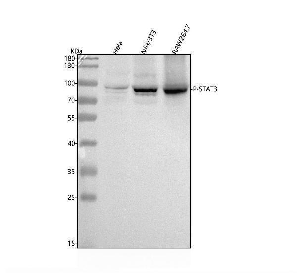
Western blot analysis of STAT3 using anti-STAT3 antibody (P00007-2).
Electrophoresis was performed on a 5-20% SDS-PAGE gel at 70V (Stacking gel) / 90V (Resolving gel) for 2-3 hours. The sample well of each lane was loaded with 30 ug of sample under reducing conditions.
Lane 1: human Hela whole cell lysates,
Lane 2: mouse NIH/3T3 whole cell lysates,
Lane 3: mouse RAW264.7 whole cell lysates.
After electrophoresis, proteins were transferred to a nitrocellulose membrane at 150 mA for 50-90 minutes. Blocked the membrane with 5% non-fat milk/TBS for 1.5 hour at RT. The membrane was incubated with rabbit anti-STAT3 antigen affinity purified monoclonal antibody (Catalog # P00007-2) at 1:5000 overnight at 4°C, then washed with TBS-0.1%Tween 3 times with 5 minutes each and probed with a goat anti-rabbit IgG-HRP secondary antibody at a dilution of 1:5000 for 1.5 hour at RT. The signal is developed using an Enhanced Chemiluminescent detection (ECL) kit (Catalog # EK1002) with Tanon 5200 system. A specific band was detected for STAT3 at approximately 88 kDa. The expected band size for STAT3 is at 88 kDa.

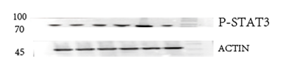
Click image to see more details
Western blot analysis of Phospho-STAT3 (Y705) using anti-Phospho-STAT3 (Y705) antibody (P00007-2).
Electrophoresis was performed on a 5-20% SDS-PAGE gel at 70V (Stacking gel) / 90V (Resolving gel) for 2-3 hours. The sample well of each lane was loaded with 30 ug of sample under reducing conditions.
Lane 1: Normal group-rat colon tissue lysates,
Lane 2: Model group-rat colon tissue lysates,
Lane 3: Triditional Chinese medicine treatment (low dose)-rat colon tissue lysates,
Lane 4: Triditional Chinese medicine treatment (medium dose)-rat colon tissue lysates,
Lane 5: Triditional Chinese medicine treatment(high dose)-rat colon tissue lysates,
Lane 6: Western medicine treatment-rat colon tissue lysates.
After electrophoresis, proteins were transferred to a nitrocellulose membrane at 150 mA for 50-90 minutes. Blocked the membrane with 5% non-fat milk/TBS for 1.5 hour at RT. The membrane was incubated with rabbit anti-Phospho-STAT3 (Y705) antigen affinity purified monoclonal antibody (Catalog # P00007-2) at 1:5000 overnight at 4°C, then washed with TBS-0.1%Tween 3 times with 5 minutes each and probed with a HRP Conjugated AffiniPure Goat Anti-rabbit IgG (H+L) secondary antibody at a dilution of 1:5000 for 1 hour at RT. The signal is developed using an Enhanced Chemiluminescent detection (ECL) kit (Catalog # EK1002) with ChemiDoc MP system. A specific band was detected for Phospho-STAT3 (Y705) at approximately 88 kDa. The expected band size for Phospho-STAT3 (Y705) is at 88 kDa.

Click image to see more details
IHC analysis of STAT3 using anti-STAT3 antibody (P00007-2).
STAT3 was detected in a paraffin-embedded section of human lung cancer tissue. Heat mediated antigen retrieval was performed in EDTA buffer (pH 8.0, epitope retrieval solution). The tissue section was blocked with 10% goat serum. The tissue section was then incubated with 1:50 rabbit anti-STAT3 Antibody (P00007-2) overnight at 4°C. Peroxidase Conjugated Goat Anti-rabbit IgG was used as secondary antibody and incubated for 30 minutes at 37°C. The tissue section was developed using HRP Conjugated Rabbit IgG Super Vision Assay Kit (Catalog # SV0002) with DAB as the chromogen.

Click image to see more details
Immunofluorescent analysis of HeLa cells treated with IFN-alpha, using Phospho-STAT3 (Y705) Antibody.

Click image to see more details
Ruxolitinib effectively reverse the PDGFRB Y562D -induced phenotypic modulation in HBVSMCs. Western blotting illustrates the alterations in p-JAK2 and p-STAT (p-STAT1 and p-STAT3) expression across different treatment groups ( A ). After normalization to the control group, the relative density of immunoblot bands in ( A ) is presented in scatter bar graph ( B ). Immunoblots ( C ) and relative density of bands ( D ) of markers associated with phenotypic modulation in HBVSMCs under different treatments are shown. A bar graph is used to present the rt-qPCR results for smooth muscle cell (SMC) markers and inflammatory markers in HBVSMCs under different treatment conditions ( E ). The results of RT-qPCR are analyzed using Tukey's multiple comparisons test. EDU assay demonstrate the proliferative capacity of HBVSMCs receiving different treatments ( F ) and statistical analysis of the proportion of Edu-positive cells in the different groups from 5 different fields of each group at × 200 magnification. Statistical differences are detected using Tukey's multiple comparisons test ( G ). Scratch assay shows the proliferative capacity of HBVSMCs underwent different treatment ( H ) and statistical analysis of the rate of wound healing (reduced area at 4H /area at 0H) in the different groups from 5 different fields of each group at × 200 magnification. Statistical differences are detected using Tukey's multiple comparisons test ( I ). ns, no significant; * p < 0.05; ** p < 0.01. The above experiments are all repeated three times
Index in PubMed under a CC BY license. PMID: 38741091

Click image to see more details
GO term, KEGG pathway, and GSEA enrichment analyses of HK2-related genes. A - B Heatmap of the top 50 positively and negatively related genes with HK2. C KEGG pathway enrichment analysis using the GSEA database. D - I Gene set enrichment plots of the NF-κB ( D ), complement and coagulation cascades ( E ), Toll-like receptor ( F ), NOD-like receptor ( G ), Th17 cell differentiation ( H ), and JAK-STAT ( I ) signalling pathways. J - M GO and KEGG enrichment analyses of HK2 positively or negatively related genes (Pearson’s rho ≥0.5, P < 0.05). N Expression levels of p-STAT3, STAT3, p-Akt, Akt in HK2-knockdown and control groups in T98 and GBM1 cell lines . Data shown as mean ± SEM. * P < 0.05, ** P < 0.01, *** P < 0.001, **** P < 0.0001
Index in PubMed under a CC BY license. PMID: 35982398

Click image to see more details
The JAK2-STAT pathway serves as the major downstream signaling pathway of the mutated PDGFB. mIF demonstrate the important downstream signaling pathway markers of PDGFRβ (p-JAK, p-Src, p-Erk1/2 and p-PLCγ) in FIA sections and NCA sections ( A ). Scale bar, 100 μm. Statistical analysis is performed to compare the mean fluorescence intensity of different signal markers between NCAs ( n = 4) and FIAs ( n = 5) ( C ). The expression of p-STAT1 and p-STAT3, which are downstream of p-JAK2, is detected by mIF in FIAs and NCAs. Scale bar, 50 μm ( B ). Statistical analysis of mean fluorescence intensity of these marks between NCAs ( n = 4) and FIAs ( n = 5) ( D ). Western blotting displays the expression of the important downstream signal pathway markers of PDGFRβ (p-JAK2, p-Src, p-Erk1/2 and p-PLCγ) in the HBVSMCs underwent different treatment in vitro ( E ). Relative density (normalized to Control) of immunoblot bands from ( E ) corresponding to pPLCγ, p-JAK2, p-Src, and p-ERK1/2 ( n = 3) ( F ). 'n' represented the number of samples. Three random fields were selected for statistical analysis in each sample, and the average value represented the detection value of this marker in this sample. Tukey's multiple comparisons test is conducted to evaluate the statistical differences. ns, no significant; ***padj < 0.001, ****padj < 0.0001
Index in PubMed under a CC BY license. PMID: 38741091

Click image to see more details
Curcumol combined with sorafenib inhibited the tumor growth of Huh7-R cell xenografts in vivo . (A) The gross appearance of the tumors in xenografted mice. (B) The tumor weight of xenografted mice after 14 days of intervention. (C) Tumor volumes in xenograft mice after 7 and 14 days of intervention. (D) Body weight of xenografted mice before and after 14 days of intervention. (E) HE staining of tumors. Scar bar = 100 μm. (F) Ki67 staining of tumors. Scar bar = 100 μm. (G) TUNEL staining of tumors. Scar bar = 50 μm. (H, I) Tumor tissues were stained for p-AKT (H) , and p-STAT3 (I) . Scar bar = 100 μm *p < 0.05, **p < 0.01, ***p < 0.001.
Index in PubMed under a CC BY license. PMID: 40242448

Click image to see more details
Experimental verification of the function of Curcumol in Sorafenib-resistant HCC cells. (A) Effect of Sorafenib (5, 10, 15, 20, 30, 40, and 50 μM) on Huh7-R and HepG2-R cells viability. (B) Effect of Curcumol (31.25, 62.5, 125, 250, 500, and 1000 μM) on Huh7-R and HepG2-R cells viability. (C) Effect of Curcumol combined with Sorafenib (5, 10, 15, 20, 30, 40, and 50 μM) on Huh7-R and HepG2-R cells viability. (D) Colony formation assay of Huh7-R and HepG2-R cells treated with Curcumol, Sorafenib, and Curcumol combined with Sorafenib, respectively. (E) Flow cytometry detection of apoptosis analysis of Huh7-R and HepG2-R cells treated with Curcumol, Sorafenib, and Curcumol combined with Sorafenib, respectively. (F) Transwell assay was performed to detect the effect of Curcumol on the migratory ability of Huh7-R cells. (G) Transwell assay to detect the effect of Curcumol on the invasive ability of Huh7-R cells. (H) Cell scratch assay of Huh7-R cells treated with Curcumol, Sorafenib, and Curcumol combined with Sorafenib, respectively. (I) The protein expression levels of PI3K\AKT pathway and JAK/STAT3 pathway following intervention with Curcumol combined with Sorafenib. *p < 0.05, **p < 0.01, ***p < 0.001.
Index in PubMed under a CC BY license. PMID: 40242448

Click image to see more details
Relationship between differentially expressed core targets and immune cell infiltration. (A) ALB (B) STAT3 (C) HSP90AA1 (D) HSP90AB1 (E) SRC.
Index in PubMed under a CC BY license. PMID: 40242448

Click image to see more details
Molecular docking pattern of Curcumol with target proteins. (A) ALB (B) STAT3 (C) HSP90AA1 (D) HSP90AB1 (E) SRC.
Index in PubMed under a CC BY license. PMID: 40242448
Specific Publications For Anti-Phospho-STAT3 (Y705) Rabbit Monoclonal Antibody (P00007-2)
Loading publications
Recommended Resources
Here are featured tools and databases that you might find useful.
- Boster's Pathways Library
- Protein Databases
- Bioscience Research Protocol Resources
- Data Processing & Analysis Software
- Photo Editing Software
- Scientific Literature Resources
- Research Paper Management Tools
- Molecular Biology Software
- Primer Design Tools
- Bioinformatics Tools
- Phylogenetic Tree Analysis
Customer Reviews
Have you used Anti-Phospho-STAT3 (Y705) Rabbit Monoclonal Antibody?
Share your experimental results or join a short interview to earn up to $1,000 in product credits or other rewards.
1 Reviews For Anti-Phospho-STAT3 (Y705) Rabbit Monoclonal Antibody
This antibody is highly efficient and specific, suitable for Western blot detection of STAT3 (Phospho-Y705) protein in rat colon tissue, with only minor nonspecific bands observed.
Excellent
| SKU | P00007-2 |
|---|---|
| Application | Western Blot |
| Sample | rat colon tissue |
| Sample Processing Description | RIPA lysis buffer with protease inhibitor PMSF (100:1) was used to lyse the sample for 10 minutes, followed by centrifugation at 12,000 rpm for 15 minutes. The supernatant was mixed with 5× loading buffer, denatured at 100°C for 10 minutes, and then loaded onto SDS-PAGE. |
| Other Reagents | Blocking buffer |
| Primary Antibody | Phospho-STAT3 (Y705) Rabbit Monoclonal Antibody |
| Primary Incubation | 1:1000, overnight at 4 ℃ |
| Secondary Antibody | HRP Conjugated AffiniPure Goat Anti-Rabbit IgG (H+L) |
| Secondary Incubation | 1:5000, 1 hour in room temperature |
| Detection | Substrate: ECL, Imaging system:ChemiDoc MP |
| Results Summary | The figure shows representative Western blot results of the target protein STAT3 (Phospho-Y705) and the internal control Actin in rat colon tissue from the normal control group, disease model group, low-, medium-, and high-dose traditional Chinese medicine–treated groups, and the western medicine–treated group. The target bands are clear and well defined, and the experimental results are satisfactory. |
Shiyu Zhang, LUTCM
Verified customer
Submitted 2026-01-07
Customer Q&As
Have a question?
Find answers in Q&As, reviews.
Can't find your answer?
Submit your question
5 Customer Q&As for Anti-Phospho-STAT3 (Y705) Rabbit Monoclonal Antibody
Question
you antibody using your anti-Phospho-STAT3 (Y705) Rabbit Monoclonal antibody for signaling by leptin studies. Has this antibody been tested with western blotting on hela cell lysate? We would like to see some validation images before ordering.
K. Carter
Verified customer
Asked: 2019-12-05
Answer
I appreciate your inquiry. This P00007-2 anti-Phospho-STAT3 (Y705) Rabbit Monoclonal antibody is tested on hela cell lysate. It is guaranteed to work for IP, IF, IHC, WB in human, mouse, rat. Our Boster guarantee will cover your intended experiment even if the sample type has not been be directly tested.
Boster Scientific Support
Answered: 2019-12-05
Question
Our lab were content with the WB result of your anti-Phospho-STAT3 (Y705) Rabbit Monoclonal antibody. However we have seen positive staining in placenta cytoplasm. nucleus using this antibody. Is that expected? Could you tell me where is STAT3 supposed to be expressed?
Verified Customer
Verified customer
Asked: 2019-08-05
Answer
Based on literature, placenta does express STAT3. Generally STAT3 expresses in cytoplasm. nucleus. Regarding which tissues have STAT3 expression, here are a few articles citing expression in various tissues:
Cervix carcinoma, Pubmed ID: 18669648, 18691976, 20068231
Erythroleukemia, Pubmed ID: 23186163
Kidney, and Pancreas, Pubmed ID: 15489334
Liver, Pubmed ID: 7701321, 24275569
Placenta, Pubmed ID: 7512451
Boster Scientific Support
Answered: 2019-08-05
Question
We have tried in the past anti-Phospho-STAT3 (Y705) Rabbit Monoclonal antibody for IHC on liver a few years ago. I am using mouse, and We intend to use the antibody for WB next. We are interested in examining liver as well as kidney pancreas in our next experiment. Do you have any suggestion on which antibody would work the best for WB?
Verified Customer
Verified customer
Asked: 2019-08-02
Answer
I have checked the website and datasheets of our anti-Phospho-STAT3 (Y705) Rabbit Monoclonal antibody and it appears that P00007-2 has been validated on mouse in both IHC and WB. Thus P00007-2 should work for your application. Our Boster satisfaction guarantee will cover this product for WB in mouse even if the specific tissue type has not been validated. We do have a comprehensive range of products for WB detection and you can check out our website bosterbio.com to find out more information about them.
Boster Scientific Support
Answered: 2019-08-02
Question
We have observed staining in mouse liver. What should we do? Is anti-Phospho-STAT3 (Y705) Rabbit Monoclonal antibody supposed to stain liver positively?
Verified Customer
Verified customer
Asked: 2018-02-21
Answer
From literature liver does express STAT3. From Uniprot.org, STAT3 is expressed in upper lobe of lung, placenta, kidney pancreas, liver, cervix carcinoma, erythroleukemia, among other tissues. Regarding which tissues have STAT3 expression, here are a few articles citing expression in various tissues:
Cervix carcinoma, Pubmed ID: 18669648, 18691976, 20068231
Erythroleukemia, Pubmed ID: 23186163
Kidney, and Pancreas, Pubmed ID: 15489334
Liver, Pubmed ID: 7701321, 24275569
Placenta, Pubmed ID: 7512451
Boster Scientific Support
Answered: 2018-02-21
Question
We are currently using anti-Phospho-STAT3 (Y705) Rabbit Monoclonal antibody P00007-2 for mouse tissue, and we are well pleased with the IP results. The species of reactivity given in the datasheet says human, mouse, rat. Is it likely that the antibody can work on primate tissues as well?
A. Bhatt
Verified customer
Asked: 2015-04-07
Answer
The anti-Phospho-STAT3 (Y705) Rabbit Monoclonal antibody (P00007-2) has not been tested for cross reactivity specifically with primate tissues, though there is a good chance of cross reactivity. We have an innovator award program that if you test this antibody and show it works in primate you can get your next antibody for free. Please contact me if I can help you with anything.
Boster Scientific Support
Answered: 2015-04-07
