Click image to see more details
-

-
-
-
-
+1
Product Info Summary
| SKU: | A01395-1 |
|---|---|
| Size: | 100 μg/vial |
| Reactive Species: | Human, Mouse, Rat |
| Host: | Rabbit |
| Application: | ELISA, IHC, WB |
Customers Who Bought This Also Bought

Product info
Product Name
Anti-Phospholamban/PLN Antibody Picoband®
SKU/Catalog Number
A01395-1
Size
100 μg/vial
Form
Lyophilized
Description
Boster Bio Anti-Phospholamban/PLN Antibody Picoband® catalog # A01395-1. Tested in ELISA, IHC, WB applications. This antibody reacts with Human, Mouse, Rat. The brand Picoband indicates this is a premium antibody that guarantees superior quality, high affinity, and strong signals with minimal background in Western blot applications. Only our best-performing antibodies are designated as Picoband, ensuring unmatched performance.
Storage & Handling
At -20°C for one year from date of receipt. After reconstitution, at 4°C for one month. It can also be aliquotted and stored frozen at -20°C for six months. Avoid repeated freezing and thawing.
Cite This Product
Anti-Phospholamban/PLN Antibody Picoband® (Boster Biological Technology, Pleasanton CA, USA, Catalog # A01395-1)
Host
Rabbit
Contents
Each vial contains 4 mg Trehalose, 0.9 mg NaCl, 0.2 mg Na2HPO4.
Clonality
Polyclonal
Isotype
Rabbit IgG
Immunogen
E.coli-derived human Phospholamban/PLN recombinant protein (Position: M1-C36).
Cross-reactivity
No cross-reactivity with other proteins
Reactive Species
A01395-1 is reactive to PLN in Human, Mouse, Rat
Observed Molecular Weight
23 kDa
Calculated molecular weight
6.1 kDa
Background of PLN
Phospholamban is a 52 amino acid integral membrane protein that regulates the Ca2+ pump in cardiac muscle and skeletal muscle cells. The subsequent activation of the Ca(2+) pump leads to enhanced muscle relaxation rates, thereby contributing to the inotropic response elicited in heart by beta-agonists. Phospholamban is also expressed in slow-twitch skeletal muscle and some smooth muscle cells. It is observed that human ventricle and quadriceps displayed high levels of phospholamban transcripts and proteins, with markedly lower expression observed in smooth muscles, while the right atrium also expressed low levels of phospholamban. The structure of the human phospholamban gene closely resembles that reported for chicken, rabbit, rat, and mouse. Comparison of the human to other mammalian phospholamban genes indicated a marked conservation of sequence for at least 217 bp upstream of the transcription start site.
Antibody Validation
Boster validates all antibodies on WB, IHC, ICC, Immunofluorescence, and ELISA with known positive control and negative samples to ensure specificity and high affinity, including thorough antibody incubations.
Application & Images
Applications
A01395-1 is guaranteed for ELISA, IHC, WB Boster Guarantee
Recommend Dilution
| Application | Dilution | Species |
|---|---|---|
| Western blot | 0.25-0.5 μg/ml | Mouse, Rat |
| Immunohistochemistry(Paraffin-embedded Section) | 2-5 μg/ml | Human, Mouse, Rat |
| ELISA | 0.1-0.5 μg/ml | - |
Tested application
Suggested blocking solution with 5% non-fat milk or BSA; (*)Recommended protein loading: 20-40 µg per lane
Use TE buffer pH 9.0 for antigen retrieval; (*) citrate buffer pH 6.0 is an alternative.
Validation Images & Assay Conditions
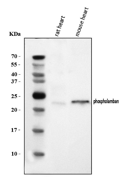
Click image to see more details
Western blot analysis of Phospholamban/PLN using anti-Phospholamban/PLN antibody (A01395-1).
Electrophoresis was performed on a 5-20% SDS-PAGE gel at 70V (Stacking gel) / 90V (Resolving gel) for 2-3 hours. The sample well of each lane was loaded with 30 ug of sample under reducing conditions.
Lane 1: rat heart tissue lysates,
Lane 2: mouse heart tissue lysates.
After electrophoresis, proteins were transferred to a nitrocellulose membrane at 150 mA for 50-90 minutes. Blocked the membrane with 5% non-fat milk/TBS for 1.5 hour at RT. The membrane was incubated with rabbit anti-Phospholamban/PLN antigen affinity purified polyclonal antibody (Catalog # A01395-1) at 0.5 μg/mL overnight at 4°C, then washed with TBS-0.1%Tween 3 times with 5 minutes each and probed with a goat anti-rabbit IgG-HRP secondary antibody at a dilution of 1:5000 for 1.5 hour at RT. The signal is developed using an Enhanced Chemiluminescent detection (ECL) kit (Catalog # EK1002) with Tanon 5200 system. A specific band was detected for Phospholamban/PLN at approximately 23 kDa. The expected band size for Phospholamban/PLN is at 6 kDa.

Click image to see more details
IHC analysis of Phospholamban/PLN using anti-Phospholamban/PLN antibody (A01395-1).
Phospholamban/PLN was detected in a paraffin-embedded section of human heart tissue. Heat mediated antigen retrieval was performed in EDTA buffer (pH 8.0, epitope retrieval solution). The tissue section was blocked with 10% goat serum. The tissue section was then incubated with 2 μg/ml rabbit anti-Phospholamban/PLN Antibody (A01395-1) overnight at 4°C. Biotinylated goat anti-rabbit IgG was used as secondary antibody and incubated for 30 minutes at 37°C. The tissue section was developed using Strepavidin-Biotin-Complex (SABC) (Catalog # SA1022) with DAB as the chromogen.

Click image to see more details
IHC analysis of PLB/PLN using anti-PLB/PLN antibody (A01395-1).
PLB/PLN was detected in a paraffin-embedded section of human heart tissue. Heat mediated antigen retrieval was performed in EDTA buffer (pH 8.0, epitope retrieval solution). The tissue section was blocked with 10% goat serum. The tissue section was then incubated with 2 μg/ml rabbit anti-PLB/PLN Antibody (A01395-1) overnight at 4°C. Peroxidase Conjugated Goat Anti-rabbit IgG was used as secondary antibody and incubated for 30 minutes at 37°C. The tissue section was developed using HRP Conjugated Rabbit IgG Super Vision Assay Kit (Catalog # SV0002) with DAB as the chromogen.

Click image to see more details
IHC analysis of Phospholamban/PLN using anti-Phospholamban/PLN antibody (A01395-1).
Phospholamban/PLN was detected in a paraffin-embedded section of mouse cardiac tissue. Heat mediated antigen retrieval was performed in EDTA buffer (pH 8.0, epitope retrieval solution). The tissue section was blocked with 10% goat serum. The tissue section was then incubated with 2 μg/ml rabbit anti-Phospholamban/PLN Antibody (A01395-1) overnight at 4°C. Biotinylated goat anti-rabbit IgG was used as secondary antibody and incubated for 30 minutes at 37°C. The tissue section was developed using Strepavidin-Biotin-Complex (SABC) (Catalog # SA1022) with DAB as the chromogen.

Click image to see more details
IHC analysis of Phospholamban/PLN using anti-Phospholamban/PLN antibody (A01395-1).
Phospholamban/PLN was detected in a paraffin-embedded section of rat cardiac tissue. Heat mediated antigen retrieval was performed in EDTA buffer (pH 8.0, epitope retrieval solution). The tissue section was blocked with 10% goat serum. The tissue section was then incubated with 2 μg/ml rabbit anti-Phospholamban/PLN Antibody (A01395-1) overnight at 4°C. Biotinylated goat anti-rabbit IgG was used as secondary antibody and incubated for 30 minutes at 37°C. The tissue section was developed using Strepavidin-Biotin-Complex (SABC) (Catalog # SA1022) with DAB as the chromogen.
Specific Publications For Anti-Phospholamban/PLN Antibody Picoband® (A01395-1)
Loading publications
Recommended Resources
Here are featured tools and databases that you might find useful.
- Boster's Pathways Library
- Protein Databases
- Bioscience Research Protocol Resources
- Data Processing & Analysis Software
- Photo Editing Software
- Scientific Literature Resources
- Research Paper Management Tools
- Molecular Biology Software
- Primer Design Tools
- Bioinformatics Tools
- Phylogenetic Tree Analysis
Customer Reviews
Have you used Anti-Phospholamban/PLN Antibody Picoband®?
Share your experimental results or join a short interview to earn up to $1,000 in product credits or other rewards.
0 Reviews For Anti-Phospholamban/PLN Antibody Picoband®
Customer Q&As
Have a question?
Find answers in Q&As, reviews.
Can't find your answer?
Submit your question
