Click image to see more details
Product Info Summary
| SKU: | A02188-3 |
|---|---|
| Size: | 100 μg/vial |
| Reactive Species: | Human, Rat |
| Host: | Rabbit |
| Application: | Flow Cytometry, IF, ICC, WB |
Customers Who Bought This Also Bought

Product info
Product Name
Anti-PSMB8 Antibody Picoband®
SKU/Catalog Number
A02188-3
Size
100 μg/vial
Form
Lyophilized
Description
Boster Bio Anti-PSMB8 Antibody Picoband® catalog # A02188-3. Tested in WB, ICC, IF, Flow Cytometry applications. This antibody reacts with Human, Rat. The brand Picoband indicates this is a premium antibody that guarantees superior quality, high affinity, and strong signals with minimal background in Western blot applications. Only our best-performing antibodies are designated as Picoband, ensuring unmatched performance.
Storage & Handling
At -20°C for one year from date of receipt. After reconstitution, at 4°C for one month. It can also be aliquotted and stored frozen at -20°C for six months. Avoid repeated freezing and thawing.
Cite This Product
Anti-PSMB8 Antibody Picoband® (Boster Biological Technology, Pleasanton CA, USA, Catalog # A02188-3)
Host
Rabbit
Contents
Each vial contains 4 mg Trehalose, 0.9 mg NaCl, 0.2 mg Na2HPO4.
Clonality
Polyclonal
Immunogen
A synthetic peptide corresponding to a sequence at the C-terminus of human PSMB8.
Reactive Species
A02188-3 is reactive to PSMB8 in Human, Rat
Observed Molecular Weight
23 kDa
Calculated molecular weight
30.4 kDa
Background of PSMB8
The proteasome is a multicatalytic proteinase complex with a highly ordered ring-shaped 20S core structure. The core structure is composed of 4 rings of 28 non-identical subunits; 2 rings are composed of 7 alpha subunits and 2 rings are composed of 7 beta subunits. Proteasomes are distributed throughout eukaryotic cells at a high concentration and cleave peptides in an ATP/ubiquitin-dependent process in a non-lysosomal pathway. An essential function of a modified proteasome, the immunoproteasome, is the processing of class I MHC peptides. This gene encodes a member of the proteasome B-type family, also known as the T1B family, that is a 20S core beta subunit. This gene is located in the class II region of the MHC (major histocompatibility complex). Expression of this gene is induced by gamma interferon and this gene product replaces catalytic subunit 3 (proteasome beta 5 subunit) in the immunoproteasome. Proteolytic processing is required to generate a mature subunit. Two alternative transcripts encoding two isoforms have been identified; both isoforms are processed to yield the same mature subunit.
Antibody Validation
Boster validates all antibodies on WB, IHC, ICC, Immunofluorescence, and ELISA with known positive control and negative samples to ensure specificity and high affinity, including thorough antibody incubations.
Application & Images
Applications
A02188-3 is guaranteed for Flow Cytometry, IF, ICC, WB Boster Guarantee
Recommend Dilution
| Application | Dilution | Species |
|---|---|---|
| Western blot | 0.25-0.5 μg/ml | Human, Rat |
| Immunocytochemistry/Immunofluorescence | 5 μg/ml | Human |
| Flow Cytometry (Fixed) | 1-3 μg/1x106 cells | Human |
Validation Images & Assay Conditions
Click image to see more details
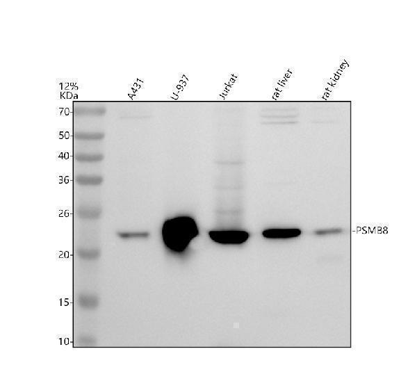
Western blot analysis of PSMB8 using anti-PSMB8 antibody (A02188-3).
Electrophoresis was performed on a 12% SDS-PAGE gel at 80V (Stacking gel) / 120V (Resolving gel) for 2 hours. The sample well of each lane was loaded with 30 ug of sample under reducing conditions.
Lane 1: human A431 whole cell lysates,
Lane 2: human U-937 whole cell lysates,
Lane 3: human Jurkat whole cell lysates,
Lane 4: rat liver tissue lysates,
Lane 5: rat kidney tissue lysates.
After electrophoresis, proteins were transferred to a nitrocellulose membrane at 150 mA for 50-90 minutes. Blocked the membrane with 5% non-fat milk/TBS for 1.5 hour at RT. The membrane was incubated with rabbit anti-PSMB8 antigen affinity purified polyclonal antibody (A02188-3) at 0.5 μg/mL overnight at 4°C, then washed with TBS-0.1%Tween 3 times with 5 minutes each and probed with a goat anti-rabbit IgG-HRP secondary antibody at a dilution of 1:5000 for 1.5 hour at RT. The signal is developed using an ECL Plus Western Blotting Substrate (Catalog # AR1196-200) with Tanon 5200 system. A specific band was detected for PSMB8 at approximately 23 kDa. The expected band size for PSMB8 is at 30 kDa.

Click image to see more details
IF analysis of PSMB8 using anti-PSMB8 antibody (A02188-3).
PSMB8 was detected in a paraffin-embedded section of U2OS tissue. Heat mediated antigen retrieval was performed in EDTA buffer (pH 8.0, epitope retrieval solution). The tissue section was blocked with 10% goat serum. The tissue section was then incubated with 5 μg/mL rabbit anti-PSMB8 Antibody (A02188-3) overnight at 4°C. Fluoro488 Conjugated Goat Anti-Rabbit IgG (BA1127) was used as secondary antibody at 1:500 dilution and incubated for 30 minutes at 37°C. The section was counterstained with DAPI. Visualize using a fluorescence microscope and filter sets appropriate for the label used.

Click image to see more details
Flow Cytometry analysis of Jurkat cells using anti-PSMB8 antibody (A02188-3).
Overlay histogram showing Jurkat cells stained with A02188-3 (Blue line). To facilitate intracellular staining, cells were fixed with 4% paraformaldehyde and permeabilized with permeabilization buffer. The cells were blocked with 10% normal goat serum. And then incubated with rabbit anti-PSMB8 Antibody (A02188-3, 1 μg/1x106 cells) for 30 min at 20°C. Fluoro488 conjugated goat anti-rabbit IgG (BA1127, 5-10 μg/1x106 cells) was used as secondary antibody for 30 minutes at 20°C. Isotype control antibody (Green line) was rabbit IgG (1 μg/1x106) used under the same conditions. Unlabelled sample without incubation with primary antibody and secondary antibody (Red line) was used as a blank control.
Specific Publications For Anti-PSMB8 Antibody Picoband® (A02188-3)
Loading publications
Recommended Resources
Here are featured tools and databases that you might find useful.
- Boster's Pathways Library
- Protein Databases
- Bioscience Research Protocol Resources
- Data Processing & Analysis Software
- Photo Editing Software
- Scientific Literature Resources
- Research Paper Management Tools
- Molecular Biology Software
- Primer Design Tools
- Bioinformatics Tools
- Phylogenetic Tree Analysis
Customer Reviews
Have you used Anti-PSMB8 Antibody Picoband®?
Share your experimental results or join a short interview to earn up to $1,000 in product credits or other rewards.
0 Reviews For Anti-PSMB8 Antibody Picoband®
Customer Q&As
Have a question?
Find answers in Q&As, reviews.
Can't find your answer?
Submit your question
