Click image to see more details
Product Info Summary
| SKU: | A04844-1 |
|---|---|
| Size: | 100 µg/vial |
| Reactive Species: | Human |
| Host: | Rabbit |
| Application: | ELISA, Flow Cytometry, WB |
Customers Who Bought This Also Bought

Product info
Product Name
Anti-PSTPIP1 Antibody Picoband®
SKU/Catalog Number
A04844-1
Size
100 µg/vial
Form
Lyophilized
Description
Boster Bio Anti-PSTPIP1 Antibody Picoband® catalog # A04844-1. Tested in WB, FCM, ELISA applications. This antibody reacts with Human. The brand Picoband indicates this is a premium antibody that guarantees superior quality, high affinity, and strong signals with minimal background in Western blot applications. Only our best-performing antibodies are designated as Picoband, ensuring unmatched performance.
Storage & Handling
At -20°C for one year from date of receipt. After reconstitution, at 4°C for one month. It can also be aliquotted and stored frozen at -20°C for six months. Avoid repeated freezing and thawing.
Cite This Product
Anti-PSTPIP1 Antibody Picoband® (Boster Biological Technology, Pleasanton CA, USA, Catalog # A04844-1)
Host
Rabbit
Contents
Each vial contains 4 mg Trehalose, 0.9 mg NaCl, 0.2 mg Na2HPO4.
Clonality
Polyclonal
Immunogen
E.coli-derived human PSTPIP1 recombinant protein (Position: M1-D394). Human PSTPIP1 shares 87.3% amino acid (aa) sequence identity with mouse PSTPIP1.
Reactive Species
A04844-1 is reactive to PSTPIP1 in Human
Observed Molecular Weight
45 kDa
Calculated molecular weight
47.6 kDa
Background of PSTPIP1
Proline-serine-threonine phosphatase-interacting protein 1 is an enzyme that in humans is encoded by the PSTPIP1 gene. This gene encodes a cytoskeletal protein that is highly expressed in hemopoietic tissues. This protein functions via its interaction with several different proteins involved in cytoskeletal organization and inflammatory processes. It binds to the cytoplasmic tail of CD2, an effector of T cell activation and adhesion, downregulating CD2-triggered adhesion. It binds PEST-type protein tyrosine phosphatases (PTP) and directs them to c-Abl kinase to mediate c-Abl dephosphorylation, thereby, regulating c-Abl activity. It also interacts with pyrin, which is found in association with the cytoskeleton in myeloid/monocytic cells and modulates immunoregulatory functions. Mutations in this gene are associated with PAPA (pyogenic sterile arthritis, pyoderma gangrenosum, and acne) syndrome. It is hypothesized that the disease-causing mutations compromise physiologic signaling necessary for the maintenance of a proper inflammatory response.
Antibody Validation
Boster validates all antibodies on WB, IHC, ICC, Immunofluorescence, and ELISA with known positive control and negative samples to ensure specificity and high affinity, including thorough antibody incubations.
Application & Images
Applications
A04844-1 is guaranteed for ELISA, Flow Cytometry, WB Boster Guarantee
Recommend Dilution
| Application | Dilution | Species |
|---|---|---|
| Western blot | 0.25-0.5 μg/ml | Human |
| Flow Cytometry (Fixed) | 1-3 μg/1x106 cells | Human |
| ELISA | 0.1-0.5 μg/ml | - |
Tested application
Suggested blocking solution with 5% non-fat milk or BSA; (*)Recommended protein loading: 20-40 µg per lane
Validation Images & Assay Conditions
Click image to see more details
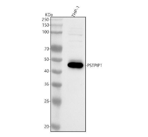
Western blot analysis of PSTPIP1 using anti-PSTPIP1 antibody (A04844-1).
Electrophoresis was performed on a 5-20% SDS-PAGE gel at 70V (Stacking gel) / 90V (Resolving gel) for 2-3 hours. The sample well of each lane was loaded with 30 ug of sample under reducing conditions.
Lane 1: human THP-1 whole cell lysates.
After electrophoresis, proteins were transferred to a nitrocellulose membrane at 150 mA for 50-90 minutes. Blocked the membrane with 5% non-fat milk/TBS for 1.5 hour at RT. The membrane was incubated with rabbit anti-PSTPIP1 antigen affinity purified polyclonal antibody (Catalog # A04844-1) at 0.5 μg/mL overnight at 4°C, then washed with TBS-0.1%Tween 3 times with 5 minutes each and probed with a goat anti-rabbit IgG-HRP secondary antibody at a dilution of 1:5000 for 1.5 hour at RT. The signal is developed using an Enhanced Chemiluminescent detection (ECL) kit (Catalog # EK1002) with Tanon 5200 system. A specific band was detected for PSTPIP1 at approximately 45 kDa. The expected band size for PSTPIP1 is at 48 kDa.

Click image to see more details
Flow Cytometry analysis of JK cells using anti-PSTPIP1 antibody (A04844-1).
Overlay histogram showing JK cells stained with A04844-1 (Blue line). To facilitate intracellular staining, cells were fixed with 4% paraformaldehyde and permeabilized with permeabilization buffer. The cells were blocked with 10% normal goat serum. And then incubated with rabbit anti-PSTPIP1 Antibody (A04844-1, 1 μg/1x106 cells) for 30 min at 20°C. DyLight®488 conjugated goat anti-rabbit IgG (BA1127, 5-10 μg/1x106 cells) was used as secondary antibody for 30 minutes at 20°C. Isotype control antibody (Green line) was rabbit IgG (1 μg/1x106) used under the same conditions. Unlabelled sample (Red line) was also used as a control.
Specific Publications For Anti-PSTPIP1 Antibody Picoband® (A04844-1)
Loading publications
Recommended Resources
Here are featured tools and databases that you might find useful.
- Boster's Pathways Library
- Protein Databases
- Bioscience Research Protocol Resources
- Data Processing & Analysis Software
- Photo Editing Software
- Scientific Literature Resources
- Research Paper Management Tools
- Molecular Biology Software
- Primer Design Tools
- Bioinformatics Tools
- Phylogenetic Tree Analysis
Customer Reviews
Have you used Anti-PSTPIP1 Antibody Picoband®?
Share your experimental results or join a short interview to earn up to $1,000 in product credits or other rewards.
0 Reviews For Anti-PSTPIP1 Antibody Picoband®
Customer Q&As
Have a question?
Find answers in Q&As, reviews.
Can't find your answer?
Submit your question
