Click image to see more details
-

-
-
-
-
+2
Product Info Summary
| SKU: | A14523-1 |
|---|---|
| Size: | 100 µg/vial |
| Reactive Species: | Human, Mouse, Rat |
| Host: | Rabbit |
| Application: | ELISA, IP, IF, IHC, WB |
Customers Who Bought This Also Bought

Product info
Product Name
Anti-PUS7L Antibody Picoband®
SKU/Catalog Number
A14523-1
Size
100 µg/vial
Form
Lyophilized
Description
Boster Bio Anti-PUS7L Antibody Picoband® catalog # A14523-1. Tested in ELISA, IP, IF, IHC, WB applications. This antibody reacts with Human, Mouse, Rat. The brand Picoband indicates this is a premium antibody that guarantees superior quality, high affinity, and strong signals with minimal background in Western blot applications. Only our best-performing antibodies are designated as Picoband, ensuring unmatched performance.
Storage & Handling
At -20°C for one year from date of receipt. After reconstitution, at 4°C for one month. It can also be aliquotted and stored frozen at -20°C for six months. Avoid repeated freezing and thawing.
Cite This Product
Anti-PUS7L Antibody Picoband® (Boster Biological Technology, Pleasanton CA, USA, Catalog # A14523-1)
Host
Rabbit
Contents
Each vial contains 4 mg Trehalose, 0.9 mg NaCl, 0.2 mg Na2HPO4.
Clonality
Polyclonal
Isotype
Rabbit IgG
Immunogen
E.coli-derived human PUS7L recombinant protein (Position: R165-H699).
Cross-reactivity
No cross-reactivity with other proteins
Reactive Species
A14523-1 is reactive to PUS7L in Human, Mouse, Rat
Observed Molecular Weight
81 kDa
Calculated molecular weight
80.7 kDa
Background of PUS7L
Pseudouridylate synthase 7 homolog-like protein is an enzyme that in humans is encoded by the PUS7L gene. PUS7L (pseudouridylate synthase 7 homolog (S. cerevisiae) -like) is a 701 amino acid protein that belongs to the pseudouridine synthase truD family and contains one TRUD domain. The PUS7L gene is conserved in chimpanzee, canine, bovine, mouse, chicken and zebrafish, and maps to human chromosome 12q12. Encoding over 1,100 genes within 132 million bases, chromosome 12 makes up about 4.5% of the human genome. A number of skeletal deformities are linked to chromosome 12 including hypochondrogenesis, achondrogenesis and Kniest dysplasia. Noonan syndrome, which includes heart and facial developmental defects among the primary symptoms, is caused by a mutant form of PTPN11 gene product, SH-PTP2. Chromosome 12 is also home to a homeobox gene cluster which encodes crucial transcription factors for morphogenesis, and the natural killer complex gene cluster encoding C-type lectin proteins which mediate the NK cell response to MHC I interaction.
Antibody Validation
Boster validates all antibodies on WB, IHC, ICC, Immunofluorescence, and ELISA with known positive control and negative samples to ensure specificity and high affinity, including thorough antibody incubations.
Application & Images
Applications
A14523-1 is guaranteed for ELISA, IP, IF, IHC, WB Boster Guarantee
Recommend Dilution
| Application | Dilution | Species |
|---|---|---|
| Western blot | 0.25-0.5 μg/ml | Human |
| Immunohistochemistry(Paraffin-embedded Section) | 2-5 μg/ml | Mouse, Rat |
| Immunofluorescence | 5 μg/ml | Mouse, Rat |
| Immunoprecipitation | 0.5-2 μg/ml | Human |
| ELISA | 0.1-0.5 μg/ml | - |
Tested application
Suggested blocking solution with 5% non-fat milk or BSA; (*)Recommended protein loading: 20-40 µg per lane
Use TE buffer pH 9.0 for antigen retrieval; (*) citrate buffer pH 6.0 is an alternative.
Validation Images & Assay Conditions
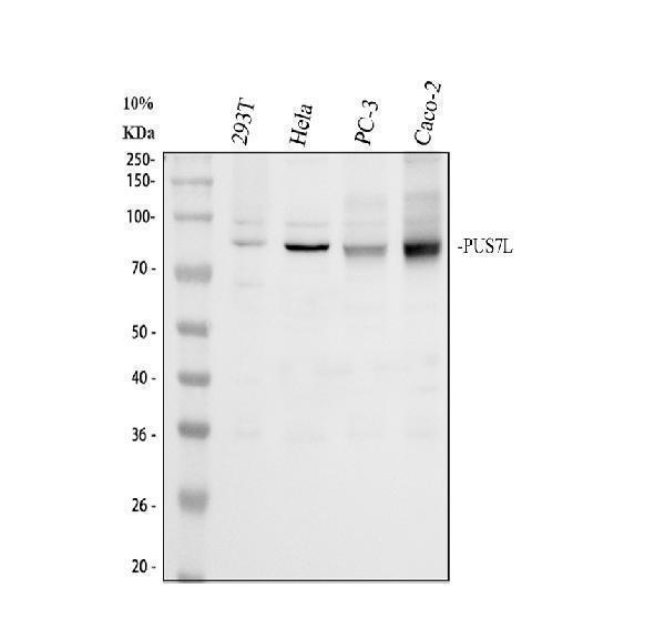
Click image to see more details
Western blot analysis of PUS7L using anti-PUS7L antibody (A14523-1).
Electrophoresis was performed on a 10% SDS-PAGE gel at 80V (Stacking gel) / 120V (Resolving gel) for 2 hours. The sample well of each lane was loaded with 30 ug of sample under reducing conditions.
Lane 1: human 293T whole cell lysates,
Lane 2: human Hela whole cell lysates,
Lane 3: human PC-3 whole cell lysates,
Lane 4: human CACO-2 whole cell lysates.
After electrophoresis, proteins were transferred to a nitrocellulose membrane at 150 mA for 50-90 minutes. Blocked the membrane with 5% non-fat milk/TBS for 1.5 hour at RT. The membrane was incubated with rabbit anti-PUS7L antigen affinity purified polyclonal antibody (Catalog # A14523-1) at 0.5 μg/mL overnight at 4°C, then washed with TBS-0.1%Tween 3 times with 5 minutes each and probed with a goat anti-rabbit IgG-HRP secondary antibody at a dilution of 1:5000 for 1.5 hour at RT. The signal is developed using an ECL Plus Western Blotting Substrate (Catalog # AR1196-200) with Tanon 5200 system. A specific band was detected for PUS7L at approximately 81 kDa. The expected band size for PUS7L is at 81 kDa.

Click image to see more details
IHC analysis of PUS7L using anti-PUS7L antibody (A14523-1).
PUS7L was detected in a paraffin-embedded section of mouse brain tissue. Heat mediated antigen retrieval was performed in EDTA buffer (pH 8.0, epitope retrieval solution). The tissue section was blocked with 10% goat serum. The tissue section was then incubated with 2 μg/ml rabbit anti-PUS7L Antibody (A14523-1) overnight at 4°C. Peroxidase Conjugated Goat Anti-rabbit IgG was used as secondary antibody and incubated for 30 minutes at 37°C. The tissue section was developed using HRP Conjugated Rabbit IgG Super Vision Assay Kit (Catalog # SV0002) with DAB as the chromogen.

Click image to see more details
IHC analysis of PUS7L using anti-PUS7L antibody (A14523-1).
PUS7L was detected in a paraffin-embedded section of rat brain tissue. Heat mediated antigen retrieval was performed in EDTA buffer (pH 8.0, epitope retrieval solution). The tissue section was blocked with 10% goat serum. The tissue section was then incubated with 2 μg/ml rabbit anti-PUS7L Antibody (A14523-1) overnight at 4°C. Peroxidase Conjugated Goat Anti-rabbit IgG was used as secondary antibody and incubated for 30 minutes at 37°C. The tissue section was developed using HRP Conjugated Rabbit IgG Super Vision Assay Kit (Catalog # SV0002) with DAB as the chromogen.

Click image to see more details
IF analysis of PUS7L using anti-PUS7L antibody (A14523-1).
PUS7L was detected in a paraffin-embedded section of mouse brain tissue. Heat mediated antigen retrieval was performed in EDTA buffer (pH 8.0, epitope retrieval solution). The tissue section was blocked with 10% goat serum. The tissue section was then incubated with 5 μg/mL rabbit anti-PUS7L Antibody (A14523-1) overnight at 4°C. Cy3 Conjugated Goat Anti-Rabbit IgG (BA1032) was used as secondary antibody at 1:500 dilution and incubated for 30 minutes at 37°C. The section was counterstained with DAPI. Visualize using a fluorescence microscope and filter sets appropriate for the label used.

Click image to see more details
IF analysis of PUS7L using anti-PUS7L antibody (A14523-1).
PUS7L was detected in a paraffin-embedded section of rat brain tissue. Heat mediated antigen retrieval was performed in EDTA buffer (pH 8.0, epitope retrieval solution). The tissue section was blocked with 10% goat serum. The tissue section was then incubated with 5 μg/mL rabbit anti-PUS7L Antibody (A14523-1) overnight at 4°C. Cy3 Conjugated Goat Anti-Rabbit IgG (BA1032) was used as secondary antibody at 1:500 dilution and incubated for 30 minutes at 37°C. The section was counterstained with DAPI. Visualize using a fluorescence microscope and filter sets appropriate for the label used.

Click image to see more details
Immunoprecipitating (IP) PUS7L in Hela whole cell lysate.
Western blot analysis of PUS7L using anti-PUS7L antibody (A14523-1);
Lane 1: Hela whole cell lysates (30ug);
Lane 2: Rabbit control IgG instead of anti-PUS7L antibody in Hela whole cell lysate;
Lane 3: anti-PUS7L antibody (2μg) + Hela whole cell lysate (500μg).
After electrophoresis, proteins were transferred to a membrane. Then the membrane was incubated with rabbit anti-PUS7L antigen affinity purified polyclonal antibody (A14523-1) at a dilution of 0.5 μg/mL and probed with a goat anti-rabbit IgG-HRP secondary antibody (Catalog # BA1054). The signal is developed using ECL Plus Western Blotting Substrate (Catalog # AR1196-200). A specific band was detected for PUS7L at approximately 81 kDa. The expected band size for PUS7L is at 81 kDa.
Specific Publications For Anti-PUS7L Antibody Picoband® (A14523-1)
Loading publications
Recommended Resources
Here are featured tools and databases that you might find useful.
- Boster's Pathways Library
- Protein Databases
- Bioscience Research Protocol Resources
- Data Processing & Analysis Software
- Photo Editing Software
- Scientific Literature Resources
- Research Paper Management Tools
- Molecular Biology Software
- Primer Design Tools
- Bioinformatics Tools
- Phylogenetic Tree Analysis
Customer Reviews
Have you used Anti-PUS7L Antibody Picoband®?
Share your experimental results or join a short interview to earn up to $1,000 in product credits or other rewards.
0 Reviews For Anti-PUS7L Antibody Picoband®
Customer Q&As
Have a question?
Find answers in Q&As, reviews.
Can't find your answer?
Submit your question
