Click image to see more details
-

-
-
-
-
+6
Product Info Summary
| SKU: | A01923-1 |
|---|---|
| Size: | 100 μg/vial |
| Reactive Species: | Human, Mouse, Rat |
| Host: | Rabbit |
| Application: | ELISA, Flow Cytometry, IF, IHC, ICC, WB |
Customers Who Bought This Also Bought

Product info
Product Name
Anti-RP2 Antibody Picoband®
SKU/Catalog Number
A01923-1
Size
100 μg/vial
Form
Lyophilized
Description
Boster Bio Anti-RP2 Antibody Picoband® catalog # A01923-1. Tested in ELISA, Flow Cytometry, IF, IHC, ICC, WB applications. This antibody reacts with Human, Mouse, Rat. The brand Picoband indicates this is a premium antibody that guarantees superior quality, high affinity, and strong signals with minimal background in Western blot applications. Only our best-performing antibodies are designated as Picoband, ensuring unmatched performance.
Storage & Handling
Store at -20˚C for one year from date of receipt. After reconstitution, at 4˚C for one month. It can also be aliquotted and stored frozen at -20˚C for six months. Avoid repeated freeze-thaw cycles.
Cite This Product
Anti-RP2 Antibody Picoband® (Boster Biological Technology, Pleasanton CA, USA, Catalog # A01923-1)
Host
Rabbit
Contents
Each vial contains 4mg Trehalose, 0.9mg NaCl, 0.2mg Na2HPO4, 0.05mg NaN3.
Clonality
Polyclonal
Isotype
Rabbit IgG
Immunogen
E. coli-derived human RP2 recombinant protein (Position: D244-M348).
Cross-reactivity
No cross-reactivity with other proteins.
Reactive Species
A01923-1 is reactive to RP2 in Human, Mouse, Rat
Observed Molecular Weight
40 kDa
Calculated molecular weight
39.6 kDa
Background of RP2
Protein XRP2 is a protein that in humans is encoded by the RP2 gene. It is mapped to Xp11.3. The RP2 locus has been implicated as one cause of X-linked retinitis pigmentosa. The predicted gene product shows homology with human cofactor C, a protein involved in the ultimate step of beta-tubulin folding. Progressive retinal degeneration may therefore be due to the accumulation of incorrectly folded photoreceptor or neuron-specific tubulin isoforms followed by progressive cell death. The RP2 protein is also involved in regulating the function and extension of outer segment of cone photoreceptors in mice.
Antibody Validation
Boster validates all antibodies on WB, IHC, ICC, Immunofluorescence, and ELISA with known positive control and negative samples to ensure specificity and high affinity, including thorough antibody incubations.
Application & Images
Applications
A01923-1 is guaranteed for ELISA, Flow Cytometry, IF, IHC, ICC, WB Boster Guarantee
Recommend Dilution
| Application | Dilution | Species |
|---|---|---|
| Western blot | 0.1-0.5μg/ml | Human, Mouse, Rat |
| Immunohistochemistry (Paraffin-embedded Section) | 0.5-1μg/ml | Human, Mouse, Rat |
| Immunocytochemistry/Immunofluorescence | 2μg/ml | Human |
| Flow Cytometry (Fixed) | 1-3μg/1x106 cells | Human |
| ELISA | 0.1-0.5μg/ml | - |
Tested application
Suggested blocking solution with 5% non-fat milk or BSA; (*)Recommended protein loading: 20-40 µg per lane
Use TE buffer pH 9.0 for antigen retrieval; (*) citrate buffer pH 6.0 is an alternative.
Validation Images & Assay Conditions
Click image to see more details
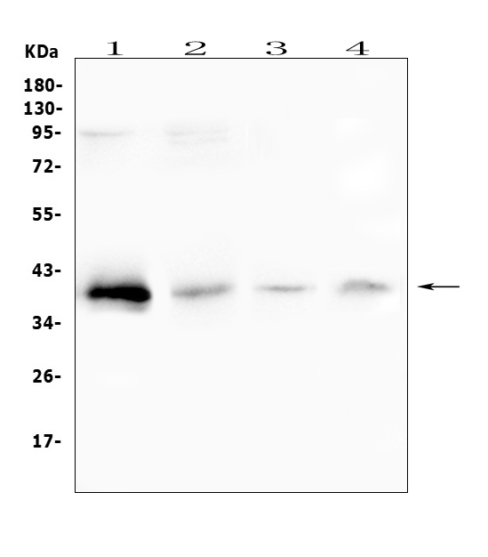
Western blot analysis of RP2 using anti-RP2 antibody (A01923-1).
Electrophoresis was performed on a 5-20% SDS-PAGE gel at 70V (Stacking gel) / 90V (Resolving gel) for 2-3 hours. The sample well of each lane was loaded with 50ug of sample under reducing conditions.
Lane 1: human placenta tissue lysates,
Lane 2: human SGC-7901 whole cell lysate,
Lane 3: rat lung tissue lysates,
Lane 4: mouse lung tissue lysates.
After Electrophoresis, proteins were transferred to a Nitrocellulose membrane at 150mA for 50-90 minutes. Blocked the membrane with 5% Non-fat Milk/ TBS for 1.5 hour at RT. The membrane was incubated with rabbit anti-RP2 antigen affinity purified polyclonal antibody (Catalog # A01923-1) at 0.5 μg/mL overnight at 4°C, then washed with TBS-0.1%Tween 3 times with 5 minutes each and probed with a goat anti-rabbit IgG-HRP secondary antibody at a dilution of 1:10000 for 1.5 hour at RT. The signal is developed using an Enhanced Chemiluminescent detection (ECL) kit (Catalog # EK1002) with Tanon 5200 system. A specific band was detected for RP2 at approximately 40KD. The expected band size for RP2 is at 40KD.

Click image to see more details
IF analysis of RP2 using anti-RP2 antibody (A01923-1).
RP2 was detected in immunocytochemical section of U20S cell. Enzyme antigen retrieval was performed using IHC enzyme antigen retrieval reagent (AR0022) for 15 mins. The cells were blocked with 10% goat serum. And then incubated with 2μg/mL rabbit anti-RP2 Antibody (A01923-1) overnight at 4°C. DyLight®488 Conjugated Goat Anti-Rabbit IgG (BA1127) was used as secondary antibody at 1:100 dilution and incubated for 30 minutes at 37°C. The section was counterstained with DAPI. Visualize using a fluorescence microscope and filter sets appropriate for the label used.

Click image to see more details
IHC analysis of RP2 using anti-RP2 antibody (A01923-1).
RP2 was detected in paraffin-embedded section of mouse spleen tissue. Heat mediated antigen retrieval was performed in citrate buffer (pH6, epitope retrieval solution) for 20 mins. The tissue section was blocked with 10% goat serum. The tissue section was then incubated with 1μg/ml rabbit anti-RP2 Antibody (A01923-1) overnight at 4°C. Biotinylated goat anti-rabbit IgG was used as secondary antibody and incubated for 30 minutes at 37°C. The tissue section was developed using Strepavidin-Biotin-Complex (SABC)(Catalog # SA1022) with DAB as the chromogen.

Click image to see more details
IHC analysis of RP2 using anti-RP2 antibody (A01923-1).
RP2 was detected in paraffin-embedded section of rat spleen tissue. Heat mediated antigen retrieval was performed in citrate buffer (pH6, epitope retrieval solution) for 20 mins. The tissue section was blocked with 10% goat serum. The tissue section was then incubated with 1μg/ml rabbit anti-RP2 Antibody (A01923-1) overnight at 4°C. Biotinylated goat anti-rabbit IgG was used as secondary antibody and incubated for 30 minutes at 37°C. The tissue section was developed using Strepavidin-Biotin-Complex (SABC)(Catalog # SA1022) with DAB as the chromogen.

Click image to see more details
IHC analysis of RP2 using anti-RP2 antibody (A01923-1).
RP2 was detected in paraffin-embedded section of human lung cancer tissue. Heat mediated antigen retrieval was performed in citrate buffer (pH6, epitope retrieval solution) for 20 mins. The tissue section was blocked with 10% goat serum. The tissue section was then incubated with 1μg/ml rabbit anti-RP2 Antibody (A01923-1) overnight at 4°C. Biotinylated goat anti-rabbit IgG was used as secondary antibody and incubated for 30 minutes at 37°C. The tissue section was developed using Strepavidin-Biotin-Complex (SABC)(Catalog # SA1022) with DAB as the chromogen.

Click image to see more details
IHC analysis of RP2 using anti-RP2 antibody (A01923-1).
RP2 was detected in paraffin-embedded section of human mammary cancer tissue. Heat mediated antigen retrieval was performed in citrate buffer (pH6, epitope retrieval solution) for 20 mins. The tissue section was blocked with 10% goat serum. The tissue section was then incubated with 1μg/ml rabbit anti-RP2 Antibody (A01923-1) overnight at 4°C. Biotinylated goat anti-rabbit IgG was used as secondary antibody and incubated for 30 minutes at 37°C. The tissue section was developed using Strepavidin-Biotin-Complex (SABC)(Catalog # SA1022) with DAB as the chromogen.

Click image to see more details
IHC analysis of RP2 using anti-RP2 antibody (A01923-1).
RP2 was detected in paraffin-embedded section of human placenta tissue. Heat mediated antigen retrieval was performed in citrate buffer (pH6, epitope retrieval solution) for 20 mins. The tissue section was blocked with 10% goat serum. The tissue section was then incubated with 1μg/ml rabbit anti-RP2 Antibody (A01923-1) overnight at 4°C. Biotinylated goat anti-rabbit IgG was used as secondary antibody and incubated for 30 minutes at 37°C. The tissue section was developed using Strepavidin-Biotin-Complex (SABC)(Catalog # SA1022) with DAB as the chromogen.

Click image to see more details
Flow Cytometry analysis of A431 cells using anti-RP2 antibody (A01923-1).
Overlay histogram showing A431 cells stained with A01923-1 (Blue line). To facilitate intracellular staining, cells were fixed with 4% paraformaldehyde and permeabilized with permeabilization buffer. The cells were blocked with 10% normal goat serum. And then incubated with rabbit anti-RP2 Antibody (A01923-1,1μg/1x106 cells) for 30 min at 20°C. DyLight®488 conjugated goat anti-rabbit IgG (BA1127, 5-10μg/1x106 cells) was used as secondary antibody for 30 minutes at 20°C. Isotype control antibody (Green line) was rabbit IgG (1μg/1x106) used under the same conditions. Unlabelled sample without incubation with primary antibody and secondary antibody (Red line) was used as a blank control.

Click image to see more details
Flow Cytometry analysis of THP-1 cells using anti-RP2 antibody (A01923-1).
Overlay histogram showing THP-1 cells stained with A01923-1 (Blue line). To facilitate intracellular staining, cells were fixed with 4% paraformaldehyde and permeabilized with permeabilization buffer. The cells were blocked with 10% normal goat serum. And then incubated with rabbit anti-RP2 Antibody (A01923-1,1μg/1x106 cells) for 30 min at 20°C. DyLight®488 conjugated goat anti-rabbit IgG (BA1127, 5-10μg/1x106 cells) was used as secondary antibody for 30 minutes at 20°C. Isotype control antibody (Green line) was rabbit IgG (1μg/1x106) used under the same conditions. Unlabelled sample without incubation with primary antibody and secondary antibody (Red line) was used as a blank control.
Specific Publications For Anti-RP2 Antibody Picoband® (A01923-1)
Loading publications
Recommended Resources
Here are featured tools and databases that you might find useful.
- Boster's Pathways Library
- Protein Databases
- Bioscience Research Protocol Resources
- Data Processing & Analysis Software
- Photo Editing Software
- Scientific Literature Resources
- Research Paper Management Tools
- Molecular Biology Software
- Primer Design Tools
- Bioinformatics Tools
- Phylogenetic Tree Analysis
Customer Reviews
Have you used Anti-RP2 Antibody Picoband®?
Share your experimental results or join a short interview to earn up to $1,000 in product credits or other rewards.
0 Reviews For Anti-RP2 Antibody Picoband®
Customer Q&As
Have a question?
Find answers in Q&As, reviews.
Can't find your answer?
Submit your question
5 Customer Q&As for Anti-RP2 Antibody Picoband®
Question
Will anti-RP2 antibody A01923-1 work for Flow Cytometry with bone marrow?
Verified Customer
Verified customer
Asked: 2020-03-13
Answer
According to the expression profile of bone marrow, RP2 is highly expressed in bone marrow. So, it is likely that anti-RP2 antibody A01923-1 will work for Flow Cytometry with bone marrow.
Boster Scientific Support
Answered: 2020-03-13
Question
Will anti-RP2 antibody A01923-1 work on primate Flow Cytometry with bone marrow?
Verified Customer
Verified customer
Asked: 2020-02-20
Answer
Our lab technicians have not validated anti-RP2 antibody A01923-1 on primate. You can run a BLAST between primate and the immunogen sequence of anti-RP2 antibody A01923-1 to see if they may cross-react. If the sequence homology is close, then you can perform a pilot test. Keep in mind that since we have not validated primate samples, this use of the antibody is not covered by our guarantee. However we have an innovator award program that if you test this antibody and show it works in primate bone marrow in Flow Cytometry, you can get your next antibody for free.
Boster Scientific Support
Answered: 2020-02-20
Question
Is a blocking peptide available for product anti-RP2 antibody (A01923-1)?
Verified Customer
Verified customer
Asked: 2019-08-23
Answer
We do provide the blocking peptide for product anti-RP2 antibody (A01923-1). If you would like to place an order for it please contact support@bosterbio.com and make a special request.
Boster Scientific Support
Answered: 2019-08-23
Question
Is this A01923-1 anti-RP2 antibody reactive to the isotypes of RP2?
Verified Customer
Verified customer
Asked: 2019-06-17
Answer
The immunogen of A01923-1 anti-RP2 antibody is E. coli-derived human RP2 recombinant protein (Position: D244-M348). Could you tell me which isotype you are interested in so I can help see if the immunogen is part of this isotype?
Boster Scientific Support
Answered: 2019-06-17
Question
Our lab want to know about to test anti-RP2 antibody A01923-1 on rat bone marrow for research purposes, then I may be interested in using anti-RP2 antibody A01923-1 for diagnostic purposes as well. Is the antibody suitable for diagnostic purposes?
B. Jones
Verified customer
Asked: 2018-10-15
Answer
The products we sell, including anti-RP2 antibody A01923-1, are only intended for research use. They would not be suitable for use in diagnostic work. If you have the means to develop a product into diagnostic use, and are interested in collaborating with us and develop our product into an IVD product, please contact us for more discussions.
Boster Scientific Support
Answered: 2018-10-15
