Click image to see more details
-

-
-
-
-
+5
Product Info Summary
| SKU: | PB9438 |
|---|---|
| Size: | 100 μg/vial |
| Reactive Species: | Human, Mouse, Rat |
| Host: | Rabbit |
| Application: | IF, IHC, WB |
Customers Who Bought This Also Bought

Product info
Product Name
Anti-Serotonin transporter/SLC6A4 Antibody Picoband®
SKU/Catalog Number
PB9438
PB0442 is an alternative SKU for this antibody, used in previous lots.
Size
100 μg/vial
Form
Lyophilized
Description
Boster Bio Anti-Serotonin transporter/SLC6A4 Antibody Picoband® catalog # PB9438. Tested in IF, IHC, WB applications. This antibody reacts with Human, Mouse, Rat. The brand Picoband indicates this is a premium antibody that guarantees superior quality, high affinity, and strong signals with minimal background in Western blot applications. Only our best-performing antibodies are designated as Picoband, ensuring unmatched performance.
Storage & Handling
Store at -20˚C for one year from date of receipt. After reconstitution, at 4˚C for one month. It can also be aliquotted and stored frozen at -20˚C for six months. Avoid repeated freeze-thaw cycles.
Cite This Product
Anti-Serotonin transporter/SLC6A4 Antibody Picoband® (Boster Biological Technology, Pleasanton CA, USA, Catalog # PB9438)
Host
Rabbit
Contents
Each vial contains 4mg Trehalose, 0.9mg NaCl, 0.2mg Na2HPO4, 0.01mg NaN3.
Clonality
Polyclonal
Isotype
Rabbit IgG
Immunogen
A synthetic peptide corresponding to a sequence at the N-terminus of human SLC6A4, different from the related mouse and rat sequences by six amino acids.
Cross-reactivity
No cross-reactivity with other proteins
Reactive Species
PB9438 is reactive to SLC6A4 in Human, Mouse, Rat
Observed Molecular Weight
70-90 kDa
Calculated molecular weight
70.3 kDa
Background of SLC6A4
SLC6A4 (Solute carrier family 6, member 4), also known as SERT (serotonin transporter), is a monoamine transporter protein. The SLC6A4 gene spans 31 kb and contains 14 exons. This protein integral membrane protein transports the neurotransmitter serotonin from synaptic spaces into presynaptic neurons. This transport of serotonin by the SERT protein terminates the action of serotonin and recycles it in a sodium-dependent manner. This protein is the target of many antidepressant medications, including those of the SSRI class. It is a member of the sodium: neurotransmitter symporter family. A repeat length polymorphism in the promoter of this gene has been shown to affect the rate of serotonin uptake and may play a role in sudden infant death syndrome, aggressive behavior in Alzheimer disease patients, post-traumatic stress disorder and depression-susceptibility in people experiencing emotional trauma.
Antibody Validation
Boster validates all antibodies on WB, IHC, ICC, Immunofluorescence, and ELISA with known positive control and negative samples to ensure specificity and high affinity, including thorough antibody incubations.
Application & Images
Applications
PB9438 is guaranteed for IF, IHC, WB Boster Guarantee
Recommend Dilution
| Application | Dilution | Species |
|---|---|---|
| Western blot | 0.1-0.5μg/ml | Human, Mouse, Rat |
| Immunohistochemistry (Paraffin-embedded Section) | 2-5μg/ml | Mouse, Rat |
| Immunofluorescence | 5 μg/ml | Mouse, Rat |
Tested application
Suggested blocking solution with 5% non-fat milk or BSA; (*)Recommended protein loading: 20-40 µg per lane
Use TE buffer pH 9.0 for antigen retrieval; (*) citrate buffer pH 6.0 is an alternative.
Validation Images & Assay Conditions
Click image to see more details
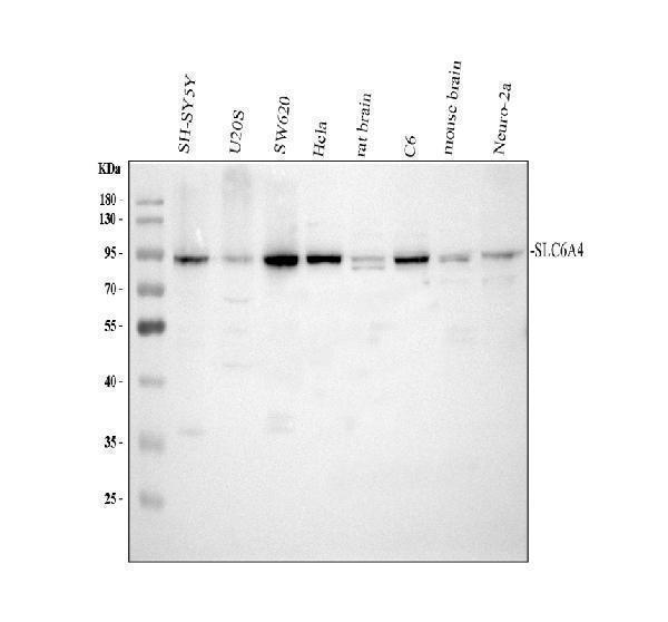
Western blot analysis of SLC6A4 using anti-SLC6A4 antibody (PB9438).
Electrophoresis was performed on a 10% SDS-PAGE gel at 80V (Stacking gel) / 120V (Resolving gel) for 2 hours. The sample well of each lane was loaded with 30 ug of sample under reducing conditions.
Lane 1: human SH-SY5Y whole cell lysates,
Lane 2: human U2OS whole cell lysates,
Lane 3: human SW620 whole cell lysates,
Lane 4: human Hela whole cell lysates,
Lane 5: rat brain tissue lysates,
Lane 6: rat C6 whole cell lysates,
Lane 7: mouse brain tissue lysates,
Lane 8: mouse Neuro-2a whole cell lysates.
After electrophoresis, proteins were transferred to a nitrocellulose membrane at 150 mA for 50-90 minutes. Blocked the membrane with 5% non-fat milk/TBS for 1.5 hour at RT. The membrane was incubated with rabbit anti-SLC6A4 antigen affinity purified polyclonal antibody (PB9438) at 0.5 μg/mL overnight at 4°C, then washed with TBS-0.1%Tween 3 times with 5 minutes each and probed with a goat anti-rabbit IgG-HRP secondary antibody (Catalog # BA1054) at a dilution of 1:5000 for 1.5 hour at RT. The signal is developed using an ECL Plus Western Blotting Substrate (Catalog # AR1196-200) with Tanon 5200 system. A specific band was detected for SLC6A4 at approximately 70-90 kDa. The expected band size for SLC6A4 is at 70 kDa.

Click image to see more details
Western blot analysis of SLC6A4 using anti-SLC6A4 antibody (PB9438).
Electrophoresis was performed on a 10% SDS-PAGE gel at 80V (Stacking gel) / 120V (Resolving gel) for 2 hours. The sample well of each lane was loaded with 30 ug of sample under reducing conditions.
Lane 1: human HepG2 whole cell lysates,
Lane 2: human CACO-2 whole cell lysates,
Lane 3: human A549 whole cell lysates,
Lane 4: human SH-SY5Y whole cell lysates.
After electrophoresis, proteins were transferred to a nitrocellulose membrane at 150 mA for 50-90 minutes. Blocked the membrane with 5% non-fat milk/TBS for 1.5 hour at RT. The membrane was incubated with rabbit anti-SLC6A4 antigen affinity purified polyclonal antibody (PB9438) at 0.5 μg/mL overnight at 4°C, then washed with TBS-0.1%Tween 3 times with 5 minutes each and probed with a goat anti-rabbit IgG-HRP secondary antibody (Catalog # BA1054) at a dilution of 1:5000 for 1.5 hour at RT. The signal is developed using an ECL Plus Western Blotting Substrate (Catalog # AR1196-200) with Tanon 5200 system. A specific band was detected for SLC6A4 at approximately 70-90 kDa. The expected band size for SLC6A4 is at 70 kDa.

Click image to see more details
Western blot analysis of SLC6A4 using anti-SLC6A4 antibody (PB9438).
Electrophoresis was performed on a 10% SDS-PAGE gel at 80V (Stacking gel) / 120V (Resolving gel) for 2 hours. The sample well of each lane was loaded with 30 ug of sample under reducing conditions.
Lane 1: rat testis tissue lysates,
Lane 2: mouse testis tissue lysates.
After electrophoresis, proteins were transferred to a nitrocellulose membrane at 150 mA for 50-90 minutes. Blocked the membrane with 5% non-fat milk/TBS for 1.5 hour at RT. The membrane was incubated with rabbit anti-SLC6A4 antigen affinity purified polyclonal antibody (PB9438) at 0.5 μg/mL overnight at 4°C, then washed with TBS-0.1%Tween 3 times with 5 minutes each and probed with a goat anti-rabbit IgG-HRP secondary antibody (Catalog # BA1054) at a dilution of 1:5000 for 1.5 hour at RT. The signal is developed using an ECL Plus Western Blotting Substrate (Catalog # AR1196-200) with Tanon 5200 system. A specific band was detected for SLC6A4 at approximately 70-90 kDa. The expected band size for SLC6A4 is at 70 kDa.

Click image to see more details
Western blot analysis of SLC6A4 using anti-SLC6A4 antibody (PB9438) .
Electrophoresis was performed on a 10% SDS-PAGE gel at 80V (Stacking gel) / 120V (Resolving gel) for 2 hours. The sample well of each lane was loaded with 30 ug of sample under reducing conditions.
Lane 2: human 293T whole cell lysatess.
Lane 3: human MCF-7 whole cell lysatess.
Lane 4: human A549 whole cell lysatess.
After electrophoresis, proteins were transferred to a nitrocellulose membrane at 150 mA for 50-90 minutes. Blocked the membrane with 5% non-fat milk/TBS for 1.5 hour at RT. The membrane was incubated with rabbit anti-SLC6A4 antigen affinity purified polyclonal antibody (PB9438) at 0.5 μg/mL overnight at 4°C, then washed with TBS-0.1%Tween 3 times with 10 minutes each and probed with a goat anti-rabbit IgG-HRP secondary antibody at a dilution of 1:5000 for 1.5 hour at RT. The signal is developed using an an ECL Plus Western Blotting Substrate (Catalog # AR1196-200) with Tanon 5200 system. A specific band was detected for SLC6A4 at approximately 95 kDa. The expected band size for SLC6A4 is at 95 kDa.

Click image to see more details
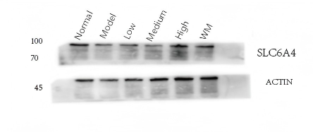
Western blot analysis of SLC6A4 using anti-SLC6A4 antibody (PB9438) .
Electrophoresis was performed on a 10% SDS-PAGE gel at 80V (Stacking gel) / 120V (Resolving gel) for 2 hours. The sample well of each lane was loaded with 30 ug of sample under reducing conditions.
Lane 1: Normal group-rat colon tissue lysates,
Lane 2: Model group-rat colon tissue lysates,
Lane 3: Triditional Chinese medicine treatment (low dose)-rat colon tissue lysates,
Lane 4: Triditional Chinese medicine treatment (medium dose)-rat colon tissue lysates,
Lane 5: Triditional Chinese medicine treatment(high dose)-rat colon tissue lysates,
Lane 6: Western medicine treatment-rat colon tissue lysates.
After electrophoresis, proteins were transferred to a nitrocellulose membrane at 150 mA for 50-90 minutes. Blocked the membrane with 5% non-fat milk/TBS for 1.5 hour at RT. The membrane was incubated with rabbit anti-SLC6A4 antigen affinity purified polyclonal antibody (PB9438) at 1:1000 overnight at 4°C, then washed with TBS-0.1%Tween 3 times with 10 minutes each and probed with a HRP Conjugated AffiniPure Goat Anti-rabbit IgG (H+L) secondary antibody at a dilution of 1:5000 for 1 hour at RT. The signal is developed using an an ECL Plus Western Blotting Substrate (Catalog # AR1196-200) with ChemiDoc MP system. A specific band was detected for SLC6A4 at approximately 95 kDa. The expected band size for SLC6A4 is at 95 kDa.

Click image to see more details
IHC analysis of SLC6A4 using anti-SLC6A4 antibody (PB9438).
SLC6A4 was detected in a paraffin-embedded section of mouse brain tissue. Heat mediated antigen retrieval was performed in EDTA buffer (pH 8.0, epitope retrieval solution). The tissue section was blocked with 10% goat serum. The tissue section was then incubated with 2 μg/ml rabbit anti-SLC6A4 Antibody (PB9438) overnight at 4°C. Peroxidase Conjugated Goat Anti-rabbit IgG was used as secondary antibody and incubated for 30 minutes at 37°C. The tissue section was developed using HRP Conjugated Rabbit IgG Super Vision Assay Kit (Catalog # SV0002) with DAB as the chromogen.

Click image to see more details
IHC analysis of SLC6A4 using anti-SLC6A4 antibody (PB9438).
SLC6A4 was detected in a paraffin-embedded section of rat brain tissue. Heat mediated antigen retrieval was performed in EDTA buffer (pH 8.0, epitope retrieval solution). The tissue section was blocked with 10% goat serum. The tissue section was then incubated with 2 μg/ml rabbit anti-SLC6A4 Antibody (PB9438) overnight at 4°C. Peroxidase Conjugated Goat Anti-rabbit IgG was used as secondary antibody and incubated for 30 minutes at 37°C. The tissue section was developed using HRP Conjugated Rabbit IgG Super Vision Assay Kit (Catalog # SV0002) with DAB as the chromogen.

Click image to see more details
IF analysis of SLC6A4 using anti-SLC6A4 antibody (PB9438).
SLC6A4 was detected in paraffin-embedded section of mouse brain tissue. Heat mediated antigen retrieval was performed in EDTA buffer (pH8.0, epitope retrieval solution). The tissue section was blocked with 10% goat serum. The tissue section was then incubated with 5μg/mL rabbit anti-SLC6A4 Antibody (PB9438) overnight at 4°C. Cy3 Conjugated Goat Anti-Rabbit IgG (BA1032) was used as secondary antibody at 1:500 dilution and incubated for 30 minutes at 37°C. The section was counterstained with DAPI. Visualize using a fluorescence microscope and filter sets appropriate for the label used.

Click image to see more details
IF analysis of SLC6A4 using anti-SLC6A4 antibody (PB9438).
SLC6A4 was detected in paraffin-embedded section of rat brain tissue. Heat mediated antigen retrieval was performed in EDTA buffer (pH8.0, epitope retrieval solution). The tissue section was blocked with 10% goat serum. The tissue section was then incubated with 5μg/mL rabbit anti-SLC6A4 Antibody (PB9438) overnight at 4°C. Cy3 Conjugated Goat Anti-Rabbit IgG (BA1032) was used as secondary antibody at 1:500 dilution and incubated for 30 minutes at 37°C. The section was counterstained with DAPI. Visualize using a fluorescence microscope and filter sets appropriate for the label used.
Specific Publications For Anti-Serotonin transporter/SLC6A4 Antibody Picoband® (PB9438)
Loading publications
Recommended Resources
Here are featured tools and databases that you might find useful.
- Boster's Pathways Library
- Protein Databases
- Bioscience Research Protocol Resources
- Data Processing & Analysis Software
- Photo Editing Software
- Scientific Literature Resources
- Research Paper Management Tools
- Molecular Biology Software
- Primer Design Tools
- Bioinformatics Tools
- Phylogenetic Tree Analysis
Customer Reviews
Have you used Anti-Serotonin transporter/SLC6A4 Antibody Picoband®?
Share your experimental results or join a short interview to earn up to $1,000 in product credits or other rewards.
1 Reviews For Anti-Serotonin transporter/SLC6A4 Antibody Picoband®
This antibody is highly efficient and specific, suitable for Western blot detection of SLC6A4 protein in rat colon tissue, with only slight nonspecific bands observed.
Excellent
| SKU | PB9438 |
|---|---|
| Application | Western Blot |
| Sample | rat colon tissue |
| Sample Processing Description | RIPA lysis buffer with protease inhibitor PMSF (100:1) was used to lyse the sample for 10 minutes, followed by centrifugation at 12,000 rpm for 15 minutes. The supernatant was mixed with 5× loading buffer, denatured at 100°C for 10 minutes, and then loaded onto SDS-PAGE. |
| Other Reagents | Blocking buffer |
| Primary Antibody | Serotonin transporter/SLC6A4 Antibody Picoband® |
| Primary Incubation | 1:1000, overnight at 4 ℃ |
| Secondary Antibody | HRP Conjugated AffiniPure Goat Anti-Rabbit IgG (H+L) |
| Secondary Incubation | 1:5000, 1 hour in room temperature |
| Detection | Substrate: ECL, Imaging system:ChemiDoc MP |
| Results Summary | The figure shows representative Western blot results of the target protein SLC6A4 and the internal control Actin in rat colon tissue from the normal control group, disease model group, low-, medium-, and high-dose traditional Chinese medicine–treated groups, and the western medicine–treated group. The target bands are clear and well defined, and the experimental results are satisfactory. |
Shiyu Zhang, LUTCM
Verified customer
Submitted 2026-01-07
Customer Q&As
Have a question?
Find answers in Q&As, reviews.
Can't find your answer?
Submit your question
2 Customer Q&As for Anti-Serotonin transporter/SLC6A4 Antibody Picoband®
Question
We are currently using anti-Serotonin transporter/SLC6A4 antibody PB9438 for human tissue, and we are content with the IHC results. The species of reactivity given in the datasheet says human, rat. Is it true that the antibody can work on zebrafish tissues as well?
Verified Customer
Verified customer
Asked: 2019-10-03
Answer
The anti-Serotonin transporter/SLC6A4 antibody (PB9438) has not been tested for cross reactivity specifically with zebrafish tissues, but there is a good chance of cross reactivity. We have an innovator award program that if you test this antibody and show it works in zebrafish you can get your next antibody for free. Please contact me if I can help you with anything.
Boster Scientific Support
Answered: 2019-10-03
Question
For the Western Blots, were the Hela Cells transfected? Keywords: Hela Cell Transfection, Anti-SLC6A4 Antibody
Verified Customer
Verified customer
Asked: 2014-11-17
Answer
We do not transfect our cell lines for testings. The samples are not enriched/spiked.
Boster Scientific Support
Answered: 2014-11-17
