Click image to see more details
Product Info Summary
| SKU: | PA1585 |
|---|---|
| Size: | 100 μg/vial |
| Reactive Species: | Human, Mouse, Rat |
| Host: | Rabbit |
| Application: | WB |
Customers Who Bought This Also Bought

Product info
Product Name
Anti-SPARC Antibody Picoband®
SKU/Catalog Number
PA1585
BA1403-2 is an alternative SKU for this antibody, used in previous lots.
Size
100 μg/vial
Form
Lyophilized
Description
Boster Bio Anti-SPARC Antibody catalog # PA1585. Tested in WB applications. This antibody reacts with Human, Mouse, Rat. The brand Picoband indicates this is a premium antibody that guarantees superior quality, high affinity, and strong signals with minimal background in Western blot applications. Only our best-performing antibodies are designated as Picoband, ensuring unmatched performance.
Storage & Handling
Store at -20˚C for one year from date of receipt. After reconstitution, at 4˚C for one month. It can also be aliquotted and stored frozen at -20˚C for six months. Avoid repeated freeze-thaw cycles.
Cite This Product
Anti-SPARC Antibody Picoband® (Boster Biological Technology, Pleasanton CA, USA, Catalog # PA1585)
Host
Rabbit
Contents
Each vial contains 4 mg Trehalose, 0.9 mg NaCl and 0.2 mg Na2HPO4.
Clonality
Polyclonal
Isotype
Rabbit IgG
Immunogen
A synthetic peptide corresponding to a sequence at the C-terminus of human SPARC.
Cross-reactivity
No cross-reactivity with other proteins
Reactive Species
PA1585 is reactive to SPARC in Human, Mouse, Rat
Observed Molecular Weight
38 kDa
Calculated molecular weight
34.6 kDa
Background of SPARC
SPARC, secreted protein acidic and rich in cysteine, also known as Osteonectin is a protein that in humans is encoded by the SPARC gene. The human SPARC gene is 26.5 kb long, and contains 10 exons and 9 introns and is located on chromosome 5q31-q33. SPARC is a glycoprotein of 40 kD. SPARC is an acidic, cysteine-rich glycoprotein consisting of a single polypeptide chain that can be broken into 4 domains: 1) an Ca++ binding domains near the glutamic acidic-rich region at the amino terminus (domain I), 2) a cysteine- rich (domain II), 3) a hydrophilic region (domain III) and 4) an EF hand motif at the carboxy terminus region (domain IV). Osteonectin is a glycoprotein in the bone that binds sodium. It is secreted by osteoblasts during bone formation, initiating mineralization and promoting mineral crystal formation. Osteonectin also shows affinity for collagen in addition to bone mineral calcium. A correlation between osteonectin over expression and ampullary cancers and chronic pancreatitis has been found.
Antibody Validation
Boster validates all antibodies on WB, IHC, ICC, Immunofluorescence, and ELISA with known positive control and negative samples to ensure specificity and high affinity, including thorough antibody incubations.
Application & Images
Applications
PA1585 is guaranteed for WB Boster Guarantee
Recommend Dilution
| Application | Dilution | Species |
|---|---|---|
| Western blot | 0.1-0.5μg/ml | Human, Mouse, Rat |
Tested application
Suggested blocking solution with 5% non-fat milk or BSA; (*)Recommended protein loading: 20-40 µg per lane
Validation Images & Assay Conditions
Click image to see more details
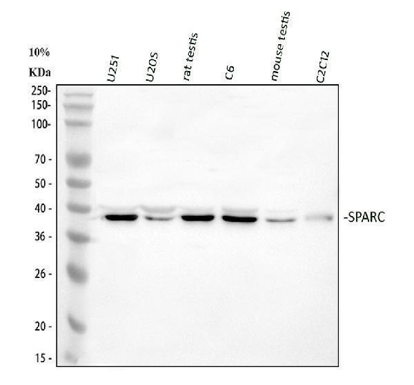
Western blot analysis of SPARC using anti-SPARC antibody (PA1585).
Electrophoresis was performed on a 108% SDS-PAGE gel at 80V (Stacking gel) / 120V (Resolving gel) for 2 hours. The sample well of each lane was loaded with 30 ug of sample under reducing conditions.
Lane 1: human U251 whole cell lysates,
Lane 2: human U2OS whole cell lysates,
Lane 3: rat testis tissue lysates,
Lane 4: rat C6 whole cell lysates,
Lane 5: mouse testis tissue lysates,
Lane 6: mouse C2C12 whole cell lysates.
After electrophoresis, proteins were transferred to a nitrocellulose membrane at 150 mA for 50-90 minutes. Blocked the membrane with 5% non-fat milk/TBS for 1.5 hour at RT. The membrane was incubated with rabbit anti-SPARC antigen affinity purified polyclonal antibody (PA1585) at 0.5 μg/mL overnight at 4°C, then washed with TBS-0.1%Tween 3 times with 5 minutes each and probed with a goat anti-rabbit IgG-HRP secondary antibody (Catalog # BA1054) at a dilution of 1:5000 for 1.5 hour at RT. The signal is developed using an ECL Plus Western Blotting Substrate (Catalog # AR1196-200) with Tanon 5200 system. A specific band was detected for SPARC at approximately 38 kDa. The expected band size for SPARC is at 35 kDa.
Specific Publications For Anti-SPARC Antibody Picoband® (PA1585)
Loading publications
Recommended Resources
Here are featured tools and databases that you might find useful.
- Boster's Pathways Library
- Protein Databases
- Bioscience Research Protocol Resources
- Data Processing & Analysis Software
- Photo Editing Software
- Scientific Literature Resources
- Research Paper Management Tools
- Molecular Biology Software
- Primer Design Tools
- Bioinformatics Tools
- Phylogenetic Tree Analysis
Customer Reviews
Have you used Anti-SPARC Antibody Picoband®?
Share your experimental results or join a short interview to earn up to $1,000 in product credits or other rewards.
0 Reviews For Anti-SPARC Antibody Picoband®
Customer Q&As
Have a question?
Find answers in Q&As, reviews.
Can't find your answer?
Submit your question
5 Customer Q&As for Anti-SPARC Antibody Picoband®
Question
We are currently using anti-SPARC antibody PA1585 for human tissue, and we are well pleased with the IHC results. The species of reactivity given in the datasheet says human. Is it likely that the antibody can work on horse tissues as well?
Verified Customer
Verified customer
Asked: 2020-03-30
Answer
The anti-SPARC antibody (PA1585) has not been tested for cross reactivity specifically with horse tissues, though there is a good chance of cross reactivity. We have an innovator award program that if you test this antibody and show it works in horse you can get your next antibody for free. Please contact me if I can help you with anything.
Boster Scientific Support
Answered: 2020-03-30
Question
We ordered your anti-SPARC antibody for WB on pns skin last year. I am using human, and We want to use the antibody for IHC next. We want examining pns skin as well as muscle in our next experiment. Could give a recommendation on which antibody would work the best for IHC?
Verified Customer
Verified customer
Asked: 2019-12-30
Answer
I looked at the website and datasheets of our anti-SPARC antibody and I see that PA1585 has been tested on human in both WB and IHC. Thus PA1585 should work for your application. Our Boster satisfaction guarantee will cover this product for IHC in human even if the specific tissue type has not been validated. We do have a comprehensive range of products for IHC detection and you can check out our website bosterbio.com to find out more information about them.
Boster Scientific Support
Answered: 2019-12-30
Question
My boss were happy with the WB result of your anti-SPARC antibody. However we have been able to see positive staining in muscle extracellular using this antibody. Is that expected? Could you tell me where is SPARC supposed to be expressed?
Verified Customer
Verified customer
Asked: 2019-12-24
Answer
From literature, muscle does express SPARC. Generally SPARC expresses in secreted, extracellular space, extracellular. Regarding which tissues have SPARC expression, here are a few articles citing expression in various tissues:
Muscle, PNS, and Skin, Pubmed ID: 15489334
Placenta, Pubmed ID: 3410046
Boster Scientific Support
Answered: 2019-12-24
Question
I am looking for using your anti-SPARC antibody for receptor-mediated endocytosis studies. Has this antibody been tested with western blotting on intestinal cancer tissue? We would like to see some validation images before ordering.
Verified Customer
Verified customer
Asked: 2018-12-13
Answer
Thanks for your inquiry. This PA1585 anti-SPARC antibody is validated on hela cell lysate, intestinal cancer tissue. It is guaranteed to work for IHC, WB in human. Our Boster guarantee will cover your intended experiment even if the sample type has not been be directly tested.
Boster Scientific Support
Answered: 2018-12-13
Question
We have been able to see staining in human placenta. Are there any suggestions? Is anti-SPARC antibody supposed to stain placenta positively?
M. Parker
Verified customer
Asked: 2014-10-03
Answer
According to literature placenta does express SPARC. According to Uniprot.org, SPARC is expressed in tibia, placenta, muscle, pns skin, among other tissues. Regarding which tissues have SPARC expression, here are a few articles citing expression in various tissues:
Muscle, PNS, and Skin, Pubmed ID: 15489334
Placenta, Pubmed ID: 3410046
Boster Scientific Support
Answered: 2014-10-03
