Click image to see more details
-

-
-
-
-
+3
Product Info Summary
| SKU: | A11249 |
|---|---|
| Size: | 100 μl/vial |
| Reactive Species: | Human, Mouse, Rat |
| Host: | Rabbit |
| Application: | ELISA, Flow Cytometry, IP, IF, IHC, ICC, WB |
Customers Who Bought This Also Bought

Product info
Product Name
Anti-SYNJ2BP antibody
SKU/Catalog Number
A11249
Size
100 μl/vial
Form
Liquid
Description
Boster Bio Anti-SYNJ2BP antibody catalog # A11249. Tested in WB, IHC, ICC, IF, IP, Flow Cytometry, ELISA applications. This antibody reacts with Human, Mouse, Rat.
Storage & Handling
12 months from date of receipt,-20℃ as supplied. 6 months 2 to 8℃ after reconstitution. Avoid repeated freezing and thawing.
Cite This Product
Anti-SYNJ2BP antibody (Boster Biological Technology, Pleasanton CA, USA, Catalog # A11249)
Host
Rabbit
Contents
500 μg/ml antibody with PBS, 0.02% NaN3, 1 mg stabilizing protein and 50% glycerol
This antibody is supplied in a stabilized formulation.
Compatibility with conjugation reactions depends on the chemistry of the conjugation method used.
For conjugation methods that are not compatible with the stabilizing components present in this formulation, a carrier-free antibody format is required.
Clonality
Polyclonal
Immunogen
E.coli-derived human SYNJ2BP recombinant protein (Position: M1-P115)
Reactive Species
A11249 is reactive to SYNJ2BP in Human, Mouse, Rat
Calculated molecular weight
15.9 kDa
Antibody Validation
Boster validates all antibodies on WB, IHC, ICC, Immunofluorescence, and ELISA with known positive control and negative samples to ensure specificity and high affinity, including thorough antibody incubations.
Application & Images
Applications
A11249 is guaranteed for ELISA, Flow Cytometry, IP, IF, IHC, ICC, WB Boster Guarantee
Recommend Dilution
| Application | Dilution | Species |
|---|---|---|
| Western blot | 1:500-2000 | |
| Immunohistochemistry | 1:50-400 | |
| Immunocytochemistry/Immunofluorescence | 1:50-400 | |
| Immunoprecipitation | 1:50 | |
| Flow Cytometry (Fixed) | 1-3μg/1x106 cells | |
| ELISA | 1:100-1000 |
Validation Images & Assay Conditions
Click image to see more details
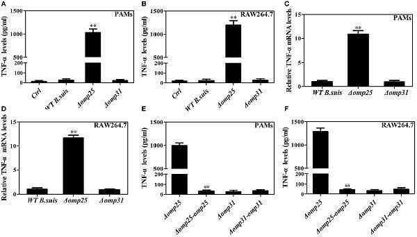
Deficiency of omp25 enhances B. suis -induced tumor necrosis factor (TNF)-α production in porcine alveolar macrophages (PAMs) and RAW264.7 cells. (A,B) PAMs and RAW264.7 cells were infected with wild-type (WT) B. suis , Omp25-deficient mutant (Δ omp25 B. suis ), Omp31-deficient mutant (Δ omp31 B. suis ) or were uninfected (ctrl), and TNF-α secretion was measured at 24 h post-infection in culture supernatants by enzyme-linked immunosorbent assay (ELISA). (C,D) PAMs and RAW264.7 cells were infected with WT B. suis , Δ omp25 , or Δ omp31 and cultured for 6 h, Q-PCR was used to measure TNF-α mRNAs levels. (E,F) PAMs and RAW264.7 cells were infected with Δ omp25 , the complemented Δ omp25 strain of B. suis (Δ omp25 - omp25 B. suis ), Δ omp31 , or the complemented Δ omp31 strain of B. suis (Δ omp31 - omp31 B. suis ), followed by ELISA detection of TNF-α in culture supernatants. The results are mean ± SEM of three independent experiments. ** P < 0.01 versus WT B. suis - infected cells. ## P < 0.01 versus Δ omp25 B. suis -infected cells.
Index in PubMed under a CC BY license. PMID: 29387067

Click image to see more details
Omp25, but not Omp31, inhibits LPS-induced tumor necrosis factor (TNF)-α production in porcine alveolar macrophages (PAMs) and RAW264.7 cells. (A,B) Evaluation of the expression of Omp25 and Omp31 in PAMs and RAW264.7 cells infected with LV-Blank, lentivirus expressing Omp25 (LV-Omp25), or LV-Omp31. PAMs and RAW264.7 cells were, respectively, infected with 100 multiplicities of infection (MOIs) of lentivirus for 24 h, and then treated with or without LPS for 24 h. The expression of protein was detected by western blotting. (C,D) Omp25 inhibits LPS-induced TNF-α production in PAMs and RAW264.7 cells. Cells were infected and expression of TNF-α was detected by enzyme-linked immunosorbent assay in culture supernatants. (E) Omp25 decreases the levels of TNF-α mRNA in LPS-treated PAMs and RAW264.7 cells. Cells were, respectively, infected with 100 MOIs of lentivirus for 24 h and stimulated with LPS for 6 h, Q-PCR was used to measure the levels of TNF-α mRNA. Values are mean ± SEM of three independent experiments. * P < 0.05, ** P < 0.01 versus LV-Blank-infected cells in the same processing.
Index in PubMed under a CC BY license. PMID: 29387067

Click image to see more details
Omp25 inhibits the transcriptional expression of tumor necrosis factor (TNF)-α by suppressing NF-κB pathway activation. (A–D) Porcine alveolar macrophages (PAMs) and RAW264.7 cells were, respectively, transfected with pCI-neo, pCI-neo-Omp31, or pCI-neo-Omp25 along with TNF-α or NF-κB luciferase reporter plasmids for 24 h; cells were stimulated with or without LPS for another 24 h, and TNF-α promoter activities (A,B) and the relative transcriptional activities of NF-κB (C,D) were examined. (E,F) PAMs and RAW264.7 cells were infected with 100 multiplicities of infection of LV-Blank, lentivirus expressing Omp25 (LV-Omp25), or LV-Omp31 for 24 h and the expression levels of cytoplasmic p-IκB, IκB, or p65 and nucleoprotein p65 at 0, 0.5, 1, and 3 h following LPS stimulation were determined by western blotting. The results are mean ± SEM of three independent experiments. * P < 0.05 versus LV-Blank-infected cells; # P < 0.05, ## P < 0.01 versus control (Ctrl) for same transfection.
Index in PubMed under a CC BY license. PMID: 29387067

Click image to see more details
Omp25 upregulates miR-130a-3p, -146a, -181a, -181b, or -301a-3p in porcine alveolar macrophages (PAMs) and miR-146a, -181a, -181b, -301a-3p, or -351-5p in RAW264.7 cells. (A,B) Expression profiling of microRNAs in Omp25-expressing PAMs and RAW264.7 cells. Quantitative polymerase chain reaction (Q-PCR) assay was used to measure the levels of 17 specific miRNAs normalized by RNU6B at 24 h following infection. (C–H) Q-PCR was used to measure the kinetics of miR-130a-3p, miR-146a, miR-181a, miR-181b, miR-301a-3p, and miR-155 expression in PAMs infected with LV-Blank or lentivirus expressing Omp25 (LV-Omp25). (I–N) Q-PCR was used to measure the kinetics of miR-146a, miR-181a, miR-181b, miR-301a-3p, miR-351-5p, and miR-155 expression in RAW264.7 cells infected with LV-Blank or LV-Omp25. Results are mean ± SEM of three independent experiments. * P < 0.05, ** P < 0.01 versus LV-Blank-infected cells for same miRNAs or same time point.
Index in PubMed under a CC BY license. PMID: 29387067

Click image to see more details
Omp25-induced miR-146a and miR-351-5p inhibit the transcriptional expression of tumor necrosis factor (TNF)-α by targeting to TRAF6 and IRAK1. (A,B) Porcine alveolar macrophages (PAMs) and RAW264.7 cells were infected with LV-Blank and lentivirus expressing Omp25 (LV-Omp25), and western blotting was used to determine the expressions of TRAF6, IRAK1, and IRAK2 at 0, 24, and 48 h. (C,D) PAMs and RAW264.7 cells were transfected with anti-miRNA control or indicated anti-miRNAs; then, cells were infected with LV-Blank or LV-Omp25 for 24 h, following LPS stimulation for another 1 h, and cells were lysed and examined for TRAF6, IRAK1, IRAK2, p-IκB, and IκB by western blotting. (E,F) Cells were transfected with anti-miRNA control or indicated anti-miRNAs and then infected with LV-Blank or LV-Omp25 for 24 h; following LPS treatment for 6 h, quantitative polymerase chain reaction was used to measure the level of TNF-α mRNA. The results are mean ± SEM of three independent experiments. * P < 0.05, ** P < 0.01 versus LV-Blank-infected cells; ## P < 0.01 versus LV-Omp25-infected cells with anti-control (Anti-Ctrl).
Index in PubMed under a CC BY license. PMID: 29387067

Click image to see more details
miR-146a, miR-181a, and miR-301a-3p participate in the regulation of tumor necrosis factor (TNF)-α in both porcine alveolar macrophages (PAMs) and RAW264.7 cells, whereas miR-130a-3p and miR-351-5p differentially regulate TNF-α expression in porcine and murine cells. (A,B) PAMs and RAW264.7 cells were transfected with anti-control, or indicated anti-miRNA, or anti-miRNAs mix (4 miRNA inhibitors); then, cells were infected with LV-Blank or lentivirus expressing Omp25 (LV-Omp25) for 24 h, and the levels of TNF-α were measured by enzyme-linked immunosorbent assay. The results are mean ± SEM of three independent experiments. * P < 0.05, ** P < 0.01 versus LV-Blank-infected cells; # P < 0.05, ## P < 0.01 versus LV-Omp25-infected cells with anti-control (Anti-Ctrl); && P < 0.01 versus LV-Omp25-infected cells with anti-mix (Anti-mix).
Index in PubMed under a CC BY license. PMID: 29387067

Click image to see more details
Deficiency of Omp25 decreases B. suis -induced miR-130a-3p, miR-146a, miR-181a, miR-301a-3p, or miR-351-5p whereas inhibition of these miRNAs upregulates tumor necrosis factor (TNF)-α and promotes the intracellular clearance of wild-type (WT). B. suis . (A–D) Porcine alveolar macrophages (PAMs) were infected with WT B. suis or Δ omp25 B. suis for 0, 6, 12, and 24 h, and quantitative polymerase chain reaction (Q-PCR) was used to analyze the levels of indicated miRNAs. (E) PAMs were transfected anti-control or anti-miRNAs mix; cells were infected with WT B. suis for 24 h, and TNF-α production was measured by enzyme-linked immunosorbent assay (ELISA). (F–I) RAW264.7 cells were infected with WT B. suis or Δ omp25 B. suis for 24 h, and cells were harvested to examine the expression of indicated miRNAs by Q-PCR at 0, 6, 12, and 24 h. (J) RAW264.7 cells were treated as in (E) and followed by ELISA measurement of TNF-α in culture supernatants. (K) Cells were transfected with anti-miRNA control or anti-miRNAs mix and infected with WT B. suis for 24 h; following stimulation with LPS for another 24 h, TNF-α production was measured by ELISA. (L) Cells were transfected anti-miRNA control or anti-miRNAs mix; cells were infected with WT B. suis for 48 h and the numbers of viable intracellular bacteria were determined as described in Section “Materials and Methods.” The results are mean ± SEM of three independent experiments. * P < 0.05, ** P < 0.01 versus WT B. suis -infected cells at same time point; ## P < 0.01 versus Anti-Ctrl.
Index in PubMed under a CC BY license. PMID: 29387067
Specific Publications For Anti-SYNJ2BP antibody (A11249)
Loading publications
Recommended Resources
Here are featured tools and databases that you might find useful.
- Boster's Pathways Library
- Protein Databases
- Bioscience Research Protocol Resources
- Data Processing & Analysis Software
- Photo Editing Software
- Scientific Literature Resources
- Research Paper Management Tools
- Molecular Biology Software
- Primer Design Tools
- Bioinformatics Tools
- Phylogenetic Tree Analysis
Customer Reviews
Have you used Anti-SYNJ2BP antibody?
Share your experimental results or join a short interview to earn up to $1,000 in product credits or other rewards.
0 Reviews For Anti-SYNJ2BP antibody
Customer Q&As
Have a question?
Find answers in Q&As, reviews.
Can't find your answer?
Submit your question
