Click image to see more details
Product Info Summary
| SKU: | PB9827 |
|---|---|
| Size: | 100 μg/vial |
| Reactive Species: | Human, Mouse, Rat |
| Host: | Rabbit |
| Application: | Flow Cytometry, IHC, WB |
Customers Who Bought This Also Bought

Product info
Product Name
Anti-Transferrin/TF Antibody Picoband®
SKU/Catalog Number
PB9827
Size
100 μg/vial
Form
Lyophilized
Description
Boster Bio Anti-Transferrin/TF Antibody Picoband® catalog # PB9827. Tested in Flow Cytometry, IHC, WB applications. This antibody reacts with Human, Mouse, Rat. The brand Picoband indicates this is a premium antibody that guarantees superior quality, high affinity, and strong signals with minimal background in Western blot applications. Only our best-performing antibodies are designated as Picoband, ensuring unmatched performance.
Storage & Handling
Store at -20˚C for one year from date of receipt. After reconstitution, at 4˚C for one month. It can also be aliquotted and stored frozen at -20˚C for six months. Avoid repeated freeze-thaw cycles.
Cite This Product
Anti-Transferrin/TF Antibody Picoband® (Boster Biological Technology, Pleasanton CA, USA, Catalog # PB9827)
Host
Rabbit
Contents
Each vial contains 4mg Trehalose, 0.9mg NaCl, 0.2mg Na2HPO4, 0.01mg NaN3.
Clonality
Polyclonal
Isotype
Rabbit IgG
Immunogen
A synthetic peptide corresponding to a sequence at the N-terminus of human Transferrin, different from the related mouse and rat sequences by five amino acids.
Cross-reactivity
No cross-reactivity with other proteins
Reactive Species
PB9827 is reactive to TF in Human, Mouse, Rat
Observed Molecular Weight
77 kDa
Calculated molecular weight
77.1 kDa
Background of TF
Transferrins are iron-binding blood plasma glycoproteins that control the level of free iron in biological fluids. In humans, it is encoded by the TF gene. Transferrin consists of a polypeptide chain containing 679 amino acids in humans. The protein is composed of alpha helices and beta sheets to form two domains. The N- and C- terminal sequences are represented by globular lobes and between the two lobes is an iron-binding site. Transferrin is a glycoprotein that binds iron very tightly but reversibly. Although iron bound to transferrin is less than 0.1% (4 mg) of the total body iron, it is the most important iron pool, with the highest rate of turnover (25 mg/24 h). And Transferrin has a molecular weight of around 80 kDa and contains 2 specific high-affinity Fe (III) binding sites. The affinity of transferrin for Fe (III) is extremely high (1023 M−1 at pH 7.4) but decreases progressively with decreasing pH below neutrality.
Antibody Validation
Boster validates all antibodies on WB, IHC, ICC, Immunofluorescence, and ELISA with known positive control and negative samples to ensure specificity and high affinity, including thorough antibody incubations.
Application & Images
Applications
PB9827 is guaranteed for Flow Cytometry, IHC, WB Boster Guarantee
Recommend Dilution
| Application | Dilution | Species |
|---|---|---|
| Western blot | 0.1-0.5μg/ml | Human, Mouse, Rat |
| Immunohistochemistry (Paraffin-embedded Section) | 2-5μg/ml | Human |
| Flow Cytometry(Fixed) | 1-3 μg/1x106 cells | Human |
Tested application
Suggested blocking solution with 5% non-fat milk or BSA; (*)Recommended protein loading: 20-40 µg per lane
Use TE buffer pH 9.0 for antigen retrieval; (*) citrate buffer pH 6.0 is an alternative.
Validation Images & Assay Conditions
Click image to see more details
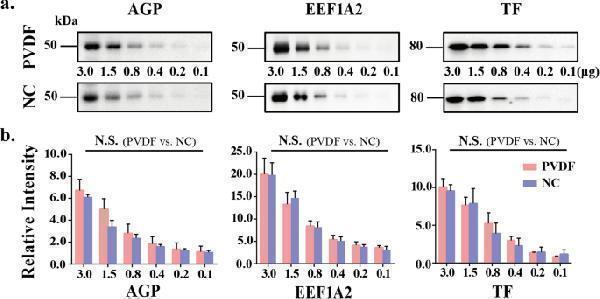
Comparison of the binding ability of PVDF membrane and NC membrane to medium molecular weight proteins. ( a ) The pooled sera proteins (0.1–3.0 μg) were subjected to 8% SDS-PAGE. The electroblotted membranes are PVDF membrane (up) and NC membrane (down), respectively. The membranes were incubated with anti-alpha-1-acid glycoprotein (AGP), anti-eukaryotic transformation extension factor 1 alpha 2 (EEF1A2) and anti-transferrin (TF) antibodies. ( b ) Staining intensities were statistically analyzed (n = 3 individual experiments). Pink bar, PVDF membrane; Blue bar, NC membrane. Band intensities were analyzed and compared using Image Lab software (Bio-Rad Laboratories) and GraphPad Prism version 6. N.S., not significant. All values are means ± S.E. (error bars).
Index in PubMed under a CC BY license. PMID: 34103620

Click image to see more details
Western blot analysis of Transferrin using anti-Transferrin antibody (PB9827).
Electrophoresis was performed on a 5-20% SDS-PAGE gel at 70V (Stacking gel) / 90V (Resolving gel) for 2-3 hours. The sample well of each lane was loaded with 30 ug of sample under reducing conditions.
Lane 1: human placenta tissue lysates,
Lane 2: human hepatocellular carcinoma tumor tissue (HCCT) lysates,
Lane 3: human hepatocellular carcinoma paracancerous tissue (HCCP) lysates,
Lane 4: human HepG2 whole cell lysates,
Lane 5: rat liver tissue lysates,
Lane 6: mouse liver tissue lysates,
Lane 7: mouse Hepa1/6 whole cell lysates.
After electrophoresis, proteins were transferred to a nitrocellulose membrane at 150 mA for 50-90 minutes. Blocked the membrane with 5% non-fat milk/TBS for 1.5 hour at RT. The membrane was incubated with rabbit anti-Transferrin antigen affinity purified polyclonal antibody (Catalog # PB9827) at 0.5 μg/mL overnight at 4°C, then washed with TBS-0.1%Tween 3 times with 5 minutes each and probed with a goat anti-rabbit IgG-HRP secondary antibody at a dilution of 1:5000 for 1.5 hour at RT. The signal is developed using an Enhanced Chemiluminescent detection (ECL) kit (Catalog # EK1002) with Tanon 5200 system. A specific band was detected for Transferrin at approximately 77 kDa. The expected band size for Transferrin is at 77 kDa.

Click image to see more details
IHC analysis of Transferrin using anti-Transferrin antibody (PB9827).
Transferrin was detected in a paraffin-embedded section of human esophageal cancer tissue. Heat mediated antigen retrieval was performed in EDTA buffer (pH 8.0, epitope retrieval solution). The tissue section was blocked with 10% goat serum. The tissue section was then incubated with 2 μg/ml rabbit anti-Transferrin Antibody (PB9827) overnight at 4°C. Peroxidase Conjugated Goat Anti-rabbit IgG was used as secondary antibody and incubated for 30 minutes at 37°C. The tissue section was developed using HRP Conjugated Rabbit IgG Super Vision Assay Kit (Catalog # SV0002) with DAB as the chromogen.

Click image to see more details
Flow Cytometry analysis of HepG2 cells using anti-Transferrin antibody (PB9827).
Overlay histogram showing HepG2 cells stained with PB9827 (Blue line). The cells were fixed with 4% paraformaldehyde and blocked with 10% normal goat serum. And then incubated with rabbit anti-Transferrin Antibody (PB9827, 1 μg/1x106 cells) for 30 min at 20°C. DyLight®488 conjugated goat anti-rabbit IgG (BA1127, 5-10 μg/1x106 cells) was used as secondary antibody for 30 minutes at 20°C. Isotype control antibody (Green line) was rabbit IgG (1 μg/1x106) used under the same conditions. Unlabelled sample without incubation with primary antibody and secondary antibody (Red line) was used as a blank control.
Specific Publications For Anti-Transferrin/TF Antibody Picoband® (PB9827)
Loading publications
Recommended Resources
Here are featured tools and databases that you might find useful.
- Boster's Pathways Library
- Protein Databases
- Bioscience Research Protocol Resources
- Data Processing & Analysis Software
- Photo Editing Software
- Scientific Literature Resources
- Research Paper Management Tools
- Molecular Biology Software
- Primer Design Tools
- Bioinformatics Tools
- Phylogenetic Tree Analysis
Customer Reviews
Have you used Anti-Transferrin/TF Antibody Picoband®?
Share your experimental results or join a short interview to earn up to $1,000 in product credits or other rewards.
0 Reviews For Anti-Transferrin/TF Antibody Picoband®
Customer Q&As
Have a question?
Find answers in Q&As, reviews.
Can't find your answer?
Submit your question
5 Customer Q&As for Anti-Transferrin/TF Antibody Picoband®
Question
We have been able to see staining in human brain. What should we do? Is anti-Transferrin/TF antibody supposed to stain brain positively?
Verified Customer
Verified customer
Asked: 2019-09-13
Answer
From what I have seen in literature brain does express TF. From what I have seen in Uniprot.org, TF is expressed in inferior vagus x ganglion, liver, brain, fetal liver, brain, cajal-retzius cell fetal brain cortex, heart, bile, plasma, saliva, cerebrospinal fluid, among other tissues. Regarding which tissues have TF expression, here are a few articles citing expression in various tissues:
Bile, Pubmed ID: 15084671
Brain, Pubmed ID: 9272172, 15489334
Cerebrospinal fluid, Pubmed ID: 19838169
Heart, Pubmed ID: 7498159
Liver, Pubmed ID: 1809186, 19159218, 24275569
Plasma, Pubmed ID: 14760718, 16335952
Saliva, Pubmed ID: 16740002
Boster Scientific Support
Answered: 2019-09-13
Question
We are currently using anti-Transferrin/TF antibody PB9827 for human tissue, and we are satisfied with the IHC results. The species of reactivity given in the datasheet says human, mouse, rat. Is it possible that the antibody can work on dog tissues as well?
O. Edwards
Verified customer
Asked: 2017-10-10
Answer
The anti-Transferrin/TF antibody (PB9827) has not been validated for cross reactivity specifically with dog tissues, though there is a good chance of cross reactivity. We have an innovator award program that if you test this antibody and show it works in dog you can get your next antibody for free. Please contact me if I can help you with anything.
Boster Scientific Support
Answered: 2017-10-10
Question
We ordered your anti-Transferrin/TF antibody for IHC on bile in a previous experiment. I am using mouse, and We intend to use the antibody for WB next. We want examining bile as well as cerebrospinal fluid in our next experiment. Do you have any suggestion on which antibody would work the best for WB?
J. Carter
Verified customer
Asked: 2017-09-25
Answer
I viewed the website and datasheets of our anti-Transferrin/TF antibody and I see that PB9827 has been tested on mouse in both IHC and WB. Thus PB9827 should work for your application. Our Boster satisfaction guarantee will cover this product for WB in mouse even if the specific tissue type has not been validated. We do have a comprehensive range of products for WB detection and you can check out our website bosterbio.com to find out more information about them.
Boster Scientific Support
Answered: 2017-09-25
Question
I am interested in using your anti-Transferrin/TF antibody for cellular response to iron ion studies. Has this antibody been tested with western blotting on human placenta tissue? We would like to see some validation images before ordering.
Verified Customer
Verified customer
Asked: 2017-06-09
Answer
Thank you for your inquiry. This PB9827 anti-Transferrin/TF antibody is tested on rat thymus tissue, tissue lysate, human placenta tissue, mouse liver. It is guaranteed to work for IHC, WB in human, mouse, rat. Our Boster guarantee will cover your intended experiment even if the sample type has not been be directly tested.
Boster Scientific Support
Answered: 2017-06-09
Question
We were well pleased with the WB result of your anti-Transferrin/TF antibody. However we have been able to see positive staining in heart secreted. using this antibody. Is that expected? Could you tell me where is TF supposed to be expressed?
B. Krishna
Verified customer
Asked: 2017-04-27
Answer
From literature, heart does express TF. Generally TF expresses in secreted. Regarding which tissues have TF expression, here are a few articles citing expression in various tissues:
Bile, Pubmed ID: 15084671
Brain, Pubmed ID: 9272172, 15489334
Cerebrospinal fluid, Pubmed ID: 19838169
Heart, Pubmed ID: 7498159
Liver, Pubmed ID: 1809186, 19159218, 24275569
Plasma, Pubmed ID: 14760718, 16335952
Saliva, Pubmed ID: 16740002
Boster Scientific Support
Answered: 2017-04-27
