Click image to see more details
Product Info Summary
| SKU: | M06313 |
|---|---|
| Size: | 100 μl |
| Reactive Species: | Human, Monkey, Mouse, Rat, Zebrafish |
| Host: | Rabbit |
| Application: | Flow Cytometry, IP, IF, IHC, ICC, WB |
Customers Who Bought This Also Bought

Product info
Product Name
Anti-Tubulin gamma Rabbit Monoclonal Antibody
SKU/Catalog Number
M06313
BM4273 is an alternative SKU for this antibody, used in previous lots.
Size
100 μl
Form
Liquid
Description
Boster Bio Anti-Tubulin gamma Rabbit Monoclonal Antibody catalog # M06313. Tested in WB, IHC, ICC/IF, IP, Flow Cytometry applications. This antibody reacts with Human, Monkey, Mouse, Rat, Zebrafish.
Storage & Handling
Store at -20°C for one year. For short term storage and frequent use, store at 4°C for up to one month. Avoid repeated freeze-thaw cycles.
Cite This Product
Anti-Tubulin gamma Rabbit Monoclonal Antibody (Boster Biological Technology, Pleasanton CA, USA, Catalog # M06313)
Host
Rabbit
Contents
Rabbit IgG in stabilizing components, phosphate buffered saline, pH 7.4, 150mM NaCl, 0.02% sodium azide and 50% glycerol.
*This antibody is supplied in a stabilized formulation.
Compatibility with conjugation reactions depends on the chemistry of the conjugation method used.
For conjugation methods that are not compatible with the stabilizing components present in this formulation, a carrier-free antibody format is required.
Clonality
Monoclonal
Clone Number
DAE-20
Isotype
Rabbit IgG
Immunogen
A synthesized peptide derived from human Tubulin gamma
Reactive Species
M06313 is reactive to TUBG1 in Human, Monkey, Mouse, Rat, Zebrafish
Observed Molecular Weight
47 kDa
Calculated molecular weight
51.2 kDa
Antibody Validation
Boster validates all antibodies on WB, IHC, ICC, Immunofluorescence, and ELISA with known positive control and negative samples to ensure specificity and high affinity, including thorough antibody incubations.
Application & Images
Applications
M06313 is guaranteed for Flow Cytometry, IP, IF, IHC, ICC, WB Boster Guarantee
Recommend Dilution
WB 1:500-2000
IHC 1:50-200
ICC/IF 1:50-200
IP 1:50
FC 1:50
Tested application
Suggested blocking solution with 5% non-fat milk or BSA; (*)Recommended protein loading: 20-40 µg per lane
Use TE buffer pH 9.0 for antigen retrieval; (*) citrate buffer pH 6.0 is an alternative.
Validation Images & Assay Conditions
Click image to see more details
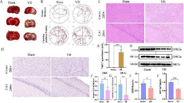
Construction of animal models and analysis of pathological damage. (A) TTC staining plots of the Sham and VD group. (B) Motion trajectory diagram. (C) Representative images stained with HE (scale bars = 50 μm). (D) Representative histopathological images obtained from TUNEL staining (scale bars = 50 μm), showing changes in the cortex hippocampus CA1. (E) Quantification of TUNEL+ cells in the hippocampal CA1 (n = 3). (F) Expression profile of CD31 between Sham and VD groups (qPCR). **P < 0.01. (G) Expression profile of HIF-1α between Sham and VD groups (qPCR; n = 6). ***P < 0.001. (H) WB strips. (I) Expression profile of CD31 between Sham and VD groups (WB). *P < 0.05. (J) Expression profile of HIF-1α between Sham and VD groups (WB; n = 3). ***P < 0.001.
Index in PubMed under a CC BY license. PMID: 40988927

Click image to see more details
Experimental validation of the key genes in vivo . (A) Representative images of immunofluorescence. (B–F) Expression profile of CCL2, VEGFA, SPP1, ANGPT2, and ANGPTL4 between Sham and VD groups (qPCR; n = 6). *P < 0.05; **P < 0.01; ***P < 0.001. (G) Immunofluorescence quantification of CD31. ***P < 0.001. (H) WB representative strips of the 5 key genes. (I–M) Protein expression level of CCL2, VEGFA, SPP1, ANGPT2, and ANGPTL4 between Sham and VD groups (WB; n = 3). *P < 0.05; **P < 0.01; ***P < 0.001.
Index in PubMed under a CC BY license. PMID: 40988927

Click image to see more details
Western blot analysis of TUBG1 using anti-TUBG1 antibody (M06313).
Electrophoresis was performed on a 10% SDS-PAGE gel at 80V (Stacking gel) / 120V (Resolving gel) for 2 hours. The sample well of each lane was loaded with 30 ug of sample under reducing conditions.
Lane 1: human Hela whole cell lysates,
Lane 2: human MCF-7 whole cell lysates,
Lane 3: monkey COS-7 whole cell lysates,
Lane 4: fish embryo tissue lysates,
Lane 5: rat brain tissue lysates,
Lane 6: rat RH35 whole cell lysates,
Lane 7: mouse brain tissue lysates,
Lane 8: mouse HEPA1-6 whole cell lysates.
After electrophoresis, proteins were transferred to a nitrocellulose membrane at 150 mA for 50-90 minutes. Blocked the membrane with 5% non-fat milk/TBS for 1.5 hour at RT. The membrane was incubated with rabbit anti-TUBG1 antigen affinity purified monoclonal antibody (M06313) at 1:500 overnight at 4°C, then washed with TBS-0.1%Tween 3 times with 5 minutes each and probed with a goat anti-rabbit IgG-HRP secondary antibody at a dilution of 1:5000 for 1.5 hour at RT. The signal is developed using an ECL Plus Western Blotting Substrate (Catalog # AR1196-200) with Tanon 5200 system. A specific band was detected for TUBG1 at approximately 47 kDa. The expected band size for TUBG1 is at 51 kDa.

Click image to see more details
Immunohistochemical analysis of paraffin-embedded human breast cancer, using Tubulin gamma Antibody.
Specific Publications For Anti-Tubulin gamma Rabbit Monoclonal Antibody (M06313)
Loading publications
Recommended Resources
Here are featured tools and databases that you might find useful.
- Boster's Pathways Library
- Protein Databases
- Bioscience Research Protocol Resources
- Data Processing & Analysis Software
- Photo Editing Software
- Scientific Literature Resources
- Research Paper Management Tools
- Molecular Biology Software
- Primer Design Tools
- Bioinformatics Tools
- Phylogenetic Tree Analysis
Customer Reviews
Have you used Anti-Tubulin gamma Rabbit Monoclonal Antibody?
Share your experimental results or join a short interview to earn up to $1,000 in product credits or other rewards.
0 Reviews For Anti-Tubulin gamma Rabbit Monoclonal Antibody
Customer Q&As
Have a question?
Find answers in Q&As, reviews.
Can't find your answer?
Submit your question
4 Customer Q&As for Anti-Tubulin gamma Rabbit Monoclonal Antibody
Question
Would anti-Tubulin gamma Rabbit Monoclonal antibody M06313 work on zebrafish IF with skin?
Verified Customer
Verified customer
Asked: 2020-03-04
Answer
Our lab technicians have not tested anti-Tubulin gamma Rabbit Monoclonal antibody M06313 on zebrafish. You can run a BLAST between zebrafish and the immunogen sequence of anti-Tubulin gamma Rabbit Monoclonal antibody M06313 to see if they may cross-react. If the sequence homology is close, then you can perform a pilot test. Keep in mind that since we have not validated zebrafish samples, this use of the antibody is not covered by our guarantee. However we have an innovator award program that if you test this antibody and show it works in zebrafish skin in IF, you can get your next antibody for free.
Boster Scientific Support
Answered: 2020-03-04
Question
We are currently using anti-Tubulin gamma Rabbit Monoclonal antibody M06313 for mouse tissue, and we are happy with the WB results. The species of reactivity given in the datasheet says human, mouse, rat. Is it likely that the antibody can work on bovine tissues as well?
Verified Customer
Verified customer
Asked: 2020-02-24
Answer
The anti-Tubulin gamma Rabbit Monoclonal antibody (M06313) has not been tested for cross reactivity specifically with bovine tissues, but there is a good chance of cross reactivity. We have an innovator award program that if you test this antibody and show it works in bovine you can get your next antibody for free. Please contact me if I can help you with anything.
Boster Scientific Support
Answered: 2020-02-24
Question
I see that the anti-Tubulin gamma Rabbit Monoclonal antibody M06313 works with IF, what is the protocol used to produce the result images on the product page?
Verified Customer
Verified customer
Asked: 2020-01-07
Answer
You can find protocols for IF on the "support/technical resources" section of our navigation menu. If you have any further questions, please send an email to support@bosterbio.com
Boster Scientific Support
Answered: 2020-01-07
Question
Will M06313 anti-Tubulin gamma Rabbit Monoclonal antibody work on parafin embedded sections? If so, which fixation method do you recommend we use (PFA, paraformaldehyde, other)?
Verified Customer
Verified customer
Asked: 2019-04-25
Answer
You can see on the product datasheet, M06313 anti-Tubulin gamma Rabbit Monoclonal antibody as been validated on IF. It is best to use PFA for fixation because it has better tissue penetration ability. PFA needs to be prepared fresh before use. Long term stored PFA turns into formalin, as the PFA molecules congregate and become formalin.
Boster Scientific Support
Answered: 2019-04-25
