Click image to see more details
Product Info Summary
| SKU: | A00270-1 |
|---|---|
| Size: | 100 μg/vial |
| Reactive Species: | Human |
| Host: | Rabbit |
| Application: | ELISA, IHC |
Customers Who Bought This Also Bought

Product info
Product Name
Anti-Von Willebrand Factor/VWF Antibody
SKU/Catalog Number
A00270-1
Size
100 μg/vial
Form
Lyophilized
Description
Boster Bio Anti-Von Willebrand Factor/VWF Antibody catalog # A00270-1. Tested in ELISA, IHC applications. This antibody reacts with Human.
Storage & Handling
At -20°C for one year from date of receipt. After reconstitution, at 4°C for one month. It can also be aliquotted and stored frozen at -20°C for six months. Avoid repeated freezing and thawing.
Cite This Product
Anti-Von Willebrand Factor/VWF Antibody (Boster Biological Technology, Pleasanton CA, USA, Catalog # A00270-1)
Host
Rabbit
Contents
Each vial contains 4 mg Trehalose, 0.9 mg NaCl, 0.2 mg Na2HPO4.
Clonality
Polyclonal
Isotype
Rabbit IgG
Immunogen
E.coli-derived human Von Willebrand Factor/VWF recombinant protein (Position: R1287-Q2770).
Cross-reactivity
No cross-reactivity with other proteins.
Reactive Species
A00270-1 is reactive to VWF in Human
Calculated molecular weight
309.3 kDa
Background of VWF
Von Willebrand factor (VWF) is a blood glycoprotein involved in hemostasis. It is mapped to 12p13.31. The VWF gene encodes von Willebrand factor (VWF), a large multimeric glycoprotein that plays a central role in the blood coagulation system, serving both as a major mediator of platelet-vessel wall interaction and platelet adhesion, and as a carrier for coagulation factor VIII. VWF released from endothelial cell Weibel-Palade bodies bound particularly avidly to the extracellular matrix. VWF deficiency or dysfunction (von Willebrand disease) leads to a bleeding tendency, which is most apparent in tissues having high blood flow shear in narrow vessels.
Antibody Validation
Boster validates all antibodies on WB, IHC, ICC, Immunofluorescence, and ELISA with known positive control and negative samples to ensure specificity and high affinity, including thorough antibody incubations.
Application & Images
Applications
A00270-1 is guaranteed for ELISA, IHC Boster Guarantee
Recommend Dilution
| Application | Dilution | Species |
|---|---|---|
| Immunohistochemistry(Paraffin-embedded Section) | 2-5 μg/ml | Human |
| ELISA | 0.1-0.5 μg/ml | - |
Tested application
Use TE buffer pH 9.0 for antigen retrieval; (*) citrate buffer pH 6.0 is an alternative.
Validation Images & Assay Conditions
Click image to see more details
IHC analysis of Von Willebrand Factor/VWF using anti-Von Willebrand Factor/VWF antibody (A00270-1).
Von Willebrand Factor/VWF was detected in a paraffin-embedded section of human renal cancer tissue. Heat mediated antigen retrieval was performed in EDTA buffer (pH 8.0, epitope retrieval solution). The tissue section was blocked with 10% goat serum. The tissue section was then incubated with 2 μg/ml rabbit anti-Von Willebrand Factor/VWF Antibody (A00270-1) overnight at 4°C. Peroxidase Conjugated Goat Anti-rabbit IgG was used as secondary antibody and incubated for 30 minutes at 37°C. The tissue section was developed using HRP Conjugated Rabbit IgG Super Vision Assay Kit (Catalog # SV0002) with DAB as the chromogen.

Click image to see more details
IHC analysis of Von Willebrand Factor/VWF using anti-Von Willebrand Factor/VWF antibody (A00270-1).
Von Willebrand Factor/VWF was detected in a paraffin-embedded section of human tonsil tissue. Heat mediated antigen retrieval was performed in EDTA buffer (pH 8.0, epitope retrieval solution). The tissue section was blocked with 10% goat serum. The tissue section was then incubated with 2 μg/ml rabbit anti-Von Willebrand Factor/VWF Antibody (A00270-1) overnight at 4°C. Peroxidase Conjugated Goat Anti-rabbit IgG was used as secondary antibody and incubated for 30 minutes at 37°C. The tissue section was developed using HRP Conjugated Rabbit IgG Super Vision Assay Kit (Catalog # SV0002) with DAB as the chromogen.

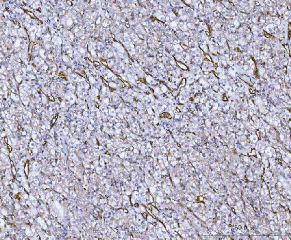
Click image to see more details
IHC analysis of Von Willebrand Factor/VWF using anti-Von Willebrand Factor/VWF antibody (A00270-1).
Von Willebrand Factor/VWF was detected in a paraffin-embedded section of human thyroid cancer tissue. Heat mediated antigen retrieval was performed in EDTA buffer (pH 8.0, epitope retrieval solution). The tissue section was blocked with 10% goat serum. The tissue section was then incubated with 2 μg/ml rabbit anti-Von Willebrand Factor/VWF Antibody (A00270-1) overnight at 4°C. Peroxidase Conjugated Goat Anti-rabbit IgG was used as secondary antibody and incubated for 30 minutes at 37°C. The tissue section was developed using HRP Conjugated Rabbit IgG Super Vision Assay Kit (Catalog # SV0002) with DAB as the chromogen.
Specific Publications For Anti-Von Willebrand Factor/VWF Antibody (A00270-1)
Loading publications
Recommended Resources
Here are featured tools and databases that you might find useful.
- Boster's Pathways Library
- Protein Databases
- Bioscience Research Protocol Resources
- Data Processing & Analysis Software
- Photo Editing Software
- Scientific Literature Resources
- Research Paper Management Tools
- Molecular Biology Software
- Primer Design Tools
- Bioinformatics Tools
- Phylogenetic Tree Analysis
Customer Reviews
Have you used Anti-Von Willebrand Factor/VWF Antibody?
Share your experimental results or join a short interview to earn up to $1,000 in product credits or other rewards.
0 Reviews For Anti-Von Willebrand Factor/VWF Antibody
Customer Q&As
Have a question?
Find answers in Q&As, reviews.
Can't find your answer?
Submit your question
