Click image to see more details
Product Info Summary
| SKU: | AZA0A8M2B2E1 |
|---|---|
| Size: | 100 μg/vial |
| Reactive Species: | Zebrafish |
| Host: | Rabbit |
| Application: | IF, IHC |
Customers Who Bought This Also Bought

Product info
Product Name
Anti-Zebrafish ASH2L Antibody
SKU/Catalog Number
AZA0A8M2B2E1
Size
100 μg/vial
Form
Lyophilized
Description
Boster Bio Anti-Zebrafish ASH2L Antibody catalog # AZA0A8M2B2E1. Tested in IHC, IF applications. This antibody reacts with Zebrafish.
Storage & Handling
At -20°C for one year from date of receipt. After reconstitution, at 4°C for one month. It can also be aliquotted and stored frozen at -20°C for six months. Avoid repeated freezing and thawing.
Cite This Product
Anti-Zebrafish ASH2L Antibody (Boster Biological Technology, Pleasanton CA, USA, Catalog # AZA0A8M2B2E1)
Host
Rabbit
Contents
Each vial contains 4 mg Trehalose, 0.9 mg NaCl, 0.2 mg Na2HPO4.
Clonality
Polyclonal
Immunogen
E.coli-derived zebrafish ASH2L recombinant protein (Position: N95-G584).
Reactive Species
AZA0A8M2B2E1 is reactive to ASH2L in Zebrafish
Background of ASH2L
Set1/Ash2 histone methyltransferase complex subunit ASH2 is an enzyme that in humans is encoded by the ASH2L gene. The Set1 histone methyltransferase protein was first identified in yeast as part of the Set1/COMPASS histone methyltransferase complex, which methylates histone H3 at Lys4 and functions as a transcriptional co-activator. While yeast contain only one known Set1 protein, six Set1-related proteins exist in mammals: SET1A, SET1B, MLL1, MLL2, MLL3, and MLL4, all of which assemble into COMPASS-like complexes and methylate histone H3 at Lys4. These Set1-related proteins are each found in distinct protein complexes, all of which share the common subunits WDR5, RBBP5, ASH2L, CXXC1 and DPY30. These subunits are required for proper complex assembly and modulation of histone methyltransferase activity. MLL1 and MLL2 complexes contain the additional protein subunit, menin. Like yeast Set1, all six Set1-related mammalian proteins methylate histone H3 at Lys4. MLL translocations are found in a large number of hematological malignancies, suggesting that Set1/COMPASS histone methyltransferase complexes play a critical role in leukemogenesis.
Antibody Validation
Boster validates all antibodies on WB, IHC, ICC, Immunofluorescence, and ELISA with known positive control and negative samples to ensure specificity and high affinity, including thorough antibody incubations.
Application & Images
Applications
AZA0A8M2B2E1 is guaranteed for IF, IHC Boster Guarantee
Recommend Dilution
| Application | Dilution | Species |
|---|---|---|
| Immunohistochemistry(Paraffin-embedded Section) | 2-5 μg/ml | Zebrafish |
| Immunofluorescence | 5 μg/ml | Zebrafish |
Tested application
Use TE buffer pH 9.0 for antigen retrieval; (*) citrate buffer pH 6.0 is an alternative.
Validation Images & Assay Conditions
Click image to see more details
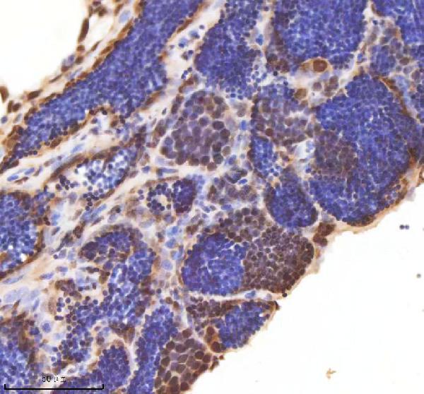
IHC analysis of ASH2L using anti-ASH2L antibody (AZA0A8M2B2E1).
ASH2L was detected in a paraffin-embedded section of zebrafish testis tissue. Heat mediated antigen retrieval was performed in EDTA buffer (pH 8.0, epitope retrieval solution). The tissue section was blocked with 10% goat serum. The tissue section was then incubated with 2 μg/ml rabbit anti-ASH2L Antibody (AZA0A8M2B2E1) overnight at 4°C. Peroxidase Conjugated Goat Anti-rabbit IgG was used as secondary antibody and incubated for 30 minutes at 37°C. The tissue section was developed using HRP Conjugated Rabbit IgG Super Vision Assay Kit (Catalog # SV0002) with DAB as the chromogen.

Click image to see more details
IF analysis of ASH2L using anti-ASH2L antibody (AZA0A8M2B2E1).
ASH2L was detected in a paraffin-embedded section of zebrafish embryo tissue. Heat mediated antigen retrieval was performed in EDTA buffer (pH 8.0, epitope retrieval solution). The tissue section was blocked with 10% goat serum. The tissue section was then incubated with 5 μg/mL rabbit anti-ASH2L Antibody (AZA0A8M2B2E1) overnight at 4°C. Biotin conjugated goat anti-rabbit IgG (BA1003) was used as secondary antibody and incubated for 30 minutes at 37°C. The tissue section was developed using DyLight®594-conjugated Anti-rabbit IgG Secondary Antibody (red) (Catalog # BA1142). The section was counterstained with DAPI. Visualize using a fluorescence microscope and filter sets appropriate for the label used.
Specific Publications For Anti-Zebrafish ASH2L Antibody (AZA0A8M2B2E1)
Loading publications
Recommended Resources
Here are featured tools and databases that you might find useful.
- Boster's Pathways Library
- Protein Databases
- Bioscience Research Protocol Resources
- Data Processing & Analysis Software
- Photo Editing Software
- Scientific Literature Resources
- Research Paper Management Tools
- Molecular Biology Software
- Primer Design Tools
- Bioinformatics Tools
- Phylogenetic Tree Analysis
Customer Reviews
Have you used Anti-Zebrafish ASH2L Antibody?
Share your experimental results or join a short interview to earn up to $1,000 in product credits or other rewards.
0 Reviews For Anti-Zebrafish ASH2L Antibody
Customer Q&As
Have a question?
Find answers in Q&As, reviews.
Can't find your answer?
Submit your question
