Click image to see more details
Product Info Summary
| SKU: | AZE9QB71 |
|---|---|
| Size: | 100 μg/vial |
| Reactive Species: | Zebrafish |
| Host: | Rabbit |
| Application: | WB |
Customers Who Bought This Also Bought

Product info
Product Name
Anti-Zebrafish KIF3A Antibody Picoband®
SKU/Catalog Number
AZE9QB71
Size
100 μg/vial
Form
Lyophilized
Description
Boster Bio Anti-Zebrafish KIF3A Antibody Picoband® catalog # AZE9QB71. Tested in WB applications. This antibody reacts with Zebrafish. The brand Picoband indicates this is a premium antibody that guarantees superior quality, high affinity, and strong signals with minimal background in Western blot applications. Only our best-performing antibodies are designated as Picoband, ensuring unmatched performance.
Storage & Handling
At -20°C for one year from date of receipt. After reconstitution, at 4°C for one month. It can also be aliquotted and stored frozen at -20°C for six months. Avoid repeated freezing and thawing.
Cite This Product
Anti-Zebrafish KIF3A Antibody Picoband® (Boster Biological Technology, Pleasanton CA, USA, Catalog # AZE9QB71)
Host
Rabbit
Contents
Each vial contains 4 mg Trehalose, 0.9 mg NaCl, 0.2 mg Na2HPO4.
Clonality
Polyclonal
Immunogen
E.coli-derived zebrafish KIF3A recombinant protein (Position: D486-Q701).
Reactive Species
AZE9QB71 is reactive to KIF3A in Zebrafish
Observed Molecular Weight
70 kDa
Background of KIF3A
Kinesin-like protein KIF3A is a protein that in humans is encoded by the KIF3A gene. KIF3A is one subunit of the heterotrimeric motor protein, kinesin-2, that was initially isolated from sea urchin egg/embryo cytosol using microtubule affinity purification. This motor consists of two kinesin-related subunits (called KIF3A and KIF3B or 3C in vertebrates) and an associated protein (KAP3), and it transports protein complexes, nucleic acids and organelles towards the "plus" ends of microtubule tracks within cells. Work done in a broad range of eukaryotic cells has revealed that heterotrimeric kinesin-2 is the primary motor protein driving the intra-flagellar transport of tubulins and other axonemal building blocks from the base of the ciliary/flagellar axoneme to their site of assembly at the distal tips. This process is required for cilium assembly/maintenance and cilium-based signalling which play key roles in various cell and developmental processes. For example, in vertebrate embryos, kinesin-2 function is required for cilia-dependent nodal flow and the development of left-right asymmetry.
Antibody Validation
Boster validates all antibodies on WB, IHC, ICC, Immunofluorescence, and ELISA with known positive control and negative samples to ensure specificity and high affinity, including thorough antibody incubations.
Application & Images
Applications
AZE9QB71 is guaranteed for WB Boster Guarantee
Recommend Dilution
| Application | Dilution | Species |
|---|---|---|
| Western blot | 0.25-0.5 μg/ml | Zebrafish |
Tested application
Suggested blocking solution with 5% non-fat milk or BSA; (*)Recommended protein loading: 20-40 µg per lane
Validation Images & Assay Conditions
Click image to see more details
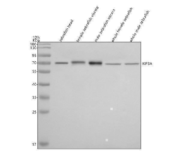
Western blot analysis of KIF3A using anti-KIF3A antibody (AZE9QB71).
Electrophoresis was performed on a 10% SDS-PAGE gel at 80V (Stacking gel) / 120V (Resolving gel) for 2 hours. The sample well of each lane was loaded with 30 ug of sample under reducing conditions.
Lane 1: zebrafish head tissue lysates,
Lane 2: female zebrafish viscera tissue lysates,
Lane 3: male zebrafish viscera tissue lysates,
Lane 4: whole zebrafish tissue lysates,
Lane 5: zebrafish embryo tissue lysates.
After electrophoresis, proteins were transferred to a nitrocellulose membrane at 150 mA for 50-90 minutes. Blocked the membrane with 5% non-fat milk/TBS for 1.5 hour at RT. The membrane was incubated with rabbit anti-KIF3A antigen affinity purified polyclonal antibody (AZE9QB71) at 0.5 μg/mL overnight at 4°C, then washed with TBS-0.1%Tween 3 times with 5 minutes each and probed with a goat anti-rabbit IgG-HRP secondary antibody at a dilution of 1:5000 for 1.5 hour at RT. The signal is developed using an ECL Plus Western Blotting Substrate (Catalog # AR1196-200) with Tanon 5200 system. A specific band was detected for KIF3A at approximately 70 kDa. The expected band size for KIF3A is at 80 kDa.
Specific Publications For Anti-Zebrafish KIF3A Antibody Picoband® (AZE9QB71)
Loading publications
Recommended Resources
Here are featured tools and databases that you might find useful.
- Boster's Pathways Library
- Protein Databases
- Bioscience Research Protocol Resources
- Data Processing & Analysis Software
- Photo Editing Software
- Scientific Literature Resources
- Research Paper Management Tools
- Molecular Biology Software
- Primer Design Tools
- Bioinformatics Tools
- Phylogenetic Tree Analysis
Customer Reviews
Have you used Anti-Zebrafish KIF3A Antibody Picoband®?
Share your experimental results or join a short interview to earn up to $1,000 in product credits or other rewards.
0 Reviews For Anti-Zebrafish KIF3A Antibody Picoband®
Customer Q&As
Have a question?
Find answers in Q&As, reviews.
Can't find your answer?
Submit your question
