Click image to see more details
Product Info Summary
| SKU: | AZA0A0R4ILA8 |
|---|---|
| Size: | 100 μg/vial |
| Reactive Species: | Zebrafish |
| Host: | Rabbit |
| Application: | IHC, WB |
Customers Who Bought This Also Bought

Product info
Product Name
Anti-Zebrafish TBP-1/PSMC3 Antibody Picoband®
SKU/Catalog Number
AZA0A0R4ILA8
Size
100 μg/vial
Form
Lyophilized
Description
Boster Bio Anti-Zebrafish TBP-1/PSMC3 Antibody Picoband® catalog # AZA0A0R4ILA8. Tested in WB, IHC applications. This antibody reacts with Zebrafish. The brand Picoband indicates this is a premium antibody that guarantees superior quality, high affinity, and strong signals with minimal background in Western blot applications. Only our best-performing antibodies are designated as Picoband, ensuring unmatched performance.
Storage & Handling
At -20°C for one year from date of receipt. After reconstitution, at 4°C for one month. It can also be aliquotted and stored frozen at -20°C for six months. Avoid repeated freezing and thawing.
Cite This Product
Anti-Zebrafish TBP-1/PSMC3 Antibody Picoband® (Boster Biological Technology, Pleasanton CA, USA, Catalog # AZA0A0R4ILA8)
Host
Rabbit
Contents
Each vial contains 4 mg Trehalose, 0.9 mg NaCl, 0.2 mg Na2HPO4.
Clonality
Polyclonal
Immunogen
E.coli-derived zebrafish TBP-1/PSMC3 recombinant protein (Position: M1-A427).
Reactive Species
AZA0A0R4ILA8 is reactive to PSMC3 in Zebrafish
Background of PSMC3
26S protease regulatory subunit 6A, also known as 26S proteasome AAA-ATPase subunit Rpt5, is an enzyme that in humans is encoded by the PSMC3 gene. The 26S proteasome is a multicatalytic proteinase complex with a highly ordered structure composed of 2 complexes, a 20S core and a 19S regulator. The 20S core is composed of 4 rings of 28 non-identical subunits; 2 rings are composed of 7 alpha subunits and 2 rings are composed of 7 beta subunits. The 19S regulator is composed of a base, which contains 6 ATPase subunits and 2 non-ATPase subunits, and a lid, which contains up to 10 non-ATPase subunits. Proteasomes are distributed throughout eukaryotic cells at a high concentration and cleave peptides in an ATP/ubiquitin-dependent process in a non-lysosomal pathway. An essential function of a modified proteasome, the immunoproteasome, is the processing of class I MHC peptides. This gene encodes one of the ATPase subunits, a member of the triple-A family of ATPases that have chaperone-like activity. This subunit may compete with PSMC2 for binding to the HIV tat protein to regulate the interaction between the viral protein and the transcription complex. A pseudogene has been identified on chromosome 9.
Antibody Validation
Boster validates all antibodies on WB, IHC, ICC, Immunofluorescence, and ELISA with known positive control and negative samples to ensure specificity and high affinity, including thorough antibody incubations.
Application & Images
Applications
AZA0A0R4ILA8 is guaranteed for IHC, WB Boster Guarantee
Recommend Dilution
| Application | Dilution | Species |
|---|---|---|
| Western blot | 0.25-0.5 μg/ml | Zebrafish |
| Immunohistochemistry(Paraffin-embedded Section) | 2-5 μg/ml | Zebrafish |
Validation Images & Assay Conditions
Click image to see more details
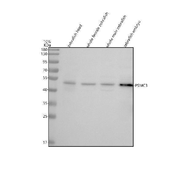
Western blot analysis of PSMC3 using anti-PSMC3 antibody (AZA0A0R4ILA8).
Electrophoresis was performed on a 10% SDS-PAGE gel at 80V (Stacking gel) / 120V (Resolving gel) for 2 hours. The sample well of each lane was loaded with 30 ug of sample under reducing conditions.
Lane 1: zebrafish head tissue lysates,
Lane 2: whole female zebrafish tissue lysates,
Lane 3: whole male zebrafish tissue lysates,
Lane 4: zebrafish embryo tissue lysates.
After electrophoresis, proteins were transferred to a nitrocellulose membrane at 150 mA for 50-90 minutes. Blocked the membrane with 5% non-fat milk/TBS for 1.5 hour at RT. The membrane was incubated with rabbit anti-PSMC3 antigen affinity purified polyclonal antibody (AZA0A0R4ILA8) at 0.5 μg/mL overnight at 4°C, then washed with TBS-0.1%Tween 3 times with 5 minutes each and probed with a goat anti-rabbit IgG-HRP secondary antibody at a dilution of 1:5000 for 1.5 hour at RT. The signal is developed using an ECL Plus Western Blotting Substrate (Catalog # AR1196-200) with Tanon 5200 system. A specific band was detected for PSMC3 at approximately 49 kDa. The expected band size for PSMC3 is at 49 kDa.

Click image to see more details
IHC analysis of PSMC3 using anti-PSMC3 antibody (AZA0A0R4ILA8).
PSMC3 was detected in a paraffin-embedded section of zebrafish spinal tissue. Heat mediated antigen retrieval was performed in EDTA buffer (pH 8.0, epitope retrieval solution). The tissue section was blocked with 10% goat serum. The tissue section was then incubated with 2 μg/ml rabbit anti-PSMC3 Antibody (AZA0A0R4ILA8) overnight at 4°C. Peroxidase Conjugated Goat Anti-rabbit IgG was used as secondary antibody and incubated for 30 minutes at 37°C. The tissue section was developed using HRP Conjugated Rabbit IgG Super Vision Assay Kit (Catalog # SV0002) with DAB as the chromogen.

Click image to see more details
IHC analysis of PSMC3 using anti-PSMC3 antibody (AZA0A0R4ILA8).
PSMC3 was detected in a paraffin-embedded section of zebrafish brain tissue. Heat mediated antigen retrieval was performed in EDTA buffer (pH 8.0, epitope retrieval solution). The tissue section was blocked with 10% goat serum. The tissue section was then incubated with 2 μg/ml rabbit anti-PSMC3 Antibody (AZA0A0R4ILA8) overnight at 4°C. Peroxidase Conjugated Goat Anti-rabbit IgG was used as secondary antibody and incubated for 30 minutes at 37°C. The tissue section was developed using HRP Conjugated Rabbit IgG Super Vision Assay Kit (Catalog # SV0002) with DAB as the chromogen.

Click image to see more details
IHC analysis of PSMC3 using anti-PSMC3 antibody (AZA0A0R4ILA8).
PSMC3 was detected in a paraffin-embedded section of zebrafish heart tissue. Heat mediated antigen retrieval was performed in EDTA buffer (pH 8.0, epitope retrieval solution). The tissue section was blocked with 10% goat serum. The tissue section was then incubated with 2 μg/ml rabbit anti-PSMC3 Antibody (AZA0A0R4ILA8) overnight at 4°C. Peroxidase Conjugated Goat Anti-rabbit IgG was used as secondary antibody and incubated for 30 minutes at 37°C. The tissue section was developed using HRP Conjugated Rabbit IgG Super Vision Assay Kit (Catalog # SV0002) with DAB as the chromogen.
Specific Publications For Anti-Zebrafish TBP-1/PSMC3 Antibody Picoband® (AZA0A0R4ILA8)
Loading publications
Recommended Resources
Here are featured tools and databases that you might find useful.
- Boster's Pathways Library
- Protein Databases
- Bioscience Research Protocol Resources
- Data Processing & Analysis Software
- Photo Editing Software
- Scientific Literature Resources
- Research Paper Management Tools
- Molecular Biology Software
- Primer Design Tools
- Bioinformatics Tools
- Phylogenetic Tree Analysis
Customer Reviews
Have you used Anti-Zebrafish TBP-1/PSMC3 Antibody Picoband®?
Share your experimental results or join a short interview to earn up to $1,000 in product credits or other rewards.
0 Reviews For Anti-Zebrafish TBP-1/PSMC3 Antibody Picoband®
Customer Q&As
Have a question?
Find answers in Q&As, reviews.
Can't find your answer?
Submit your question
