AKT1 / RAC-alpha serine/threonine-protein kinase · IHC design guide

Design Immunohistochemistry for AKT1

Plan AKT1 staining in paraffin sections using the observed widespread nuclear tissue pattern (HPA tissue IHC). Compare staining intensity across consistently fixed sections, and assess localisation in the context of activation-dependent nuclear translocation (UniProt).

Evidence assembled Oct 2026 · For research use; verify linked source records and product datasheet before use
Immunohistochemistry protocol sheet for AKT1 (IHC for AKT1): expected localisation Nuclear staining observed in tissue IHC (HPA tissue IHC), antibody M00024-1, validated IHC image, and IHC protocol steps
Printable AKT1 IHC protocol sheet — expected localisation Nuclear staining observed in tissue IHC (HPA tissue IHC), antibody M00024-1, controls and protocol steps. Open the full AKT1 IHC guide →

AKT1 Immunohistochemistry Experimental Design Guide

Expected localisation, validated protocols, controls and antibodies — the at-a-glance facts below, then the full design guide.

Must know before staining
Expected localisation Nuclear staining observed in tissue IHC (HPA tissue IHC)
Staining pattern Widespread nuclear staining across tissue cell types (HPA tissue IHC)
Antigen retrieval Tris-EDTA pH 9.0 HIER, 95–98 °C, 20 min (rule: nuclear antigen)
Positive control ⓘ Adrenal gland+4 more · see all
Negative control ⓘ None in HPA (detected in all 45 tissues); use no-primary + isotype controls
Important caveats
Reasons your staining may differ from the expected pattern.
Fixation Keep fixation consistent across sections. (standard IHC practice; not target-specific)
Caveat Activation can alter nuclear localisation (UniProt)
Regulation Protein levels rise in prostate cancer (UniProt)
Isoform / epitope Two isoforms; one 1–480 chain is annotated (UniProt)
Section 1

Recommended AKT1 IHC & IF Protocols

Compare the catalog antibody’s IHC-P protocol with published phospho-AKT1 melanoma and AKT1 tissue-section methods (PMC6324666; PMC3369856; PMC9600081).

Recommended immunohistochemistry (IHC-P) protocol parameters
SampleParaffin-embedded human colon tissue; fixative not specified (datasheet M00024-1)
FixationImage fixative and duration unreported (datasheet M00024-1); verify before use.
Sectioning4–5 µm sections on charged slides (standard)
DeparaffinisationXylene, graded ethanol series to water (standard)
Antigen retrievalHeat-induced epitope retrieval in Tris-EDTA buffer, pH 9.0, 20 min at 95–98 °C (standard rule: nuclear antigen)
Peroxidase block3% H2O2, 10 min, room temperature (standard)
Blocking10% normal serum of the secondary host, 30 min, room temperature (standard)
Primary antibodyRabbit monoclonal (clone EBG-1) anti-AKT1, 1:50 (datasheet M00024-1)
Primary incubationOvernight at 4 °C (standard)
DetectionHRP-polymer secondary, DAB chromogen 5–10 min (standard)
CounterstainHematoxylin, blue, dehydrate and mount (standard)
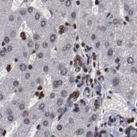
Expected resultAKT1-positive staining in glandular cells of adrenal gland (HPA tissue IHC: High). HPA tissue profile: Ubiquitous nuclear expression. No signal in the no-primary control.
💡Decision noteStart with Tris-EDTA pH 9.0 retrieval at 95–98 °C for 20 min (page retrieval rule); compare the published 100 °C, 30 min condition if needed (PMC9600081).
Section 2

What Is the Expected AKT1 Staining Pattern?

AKT1 is a non-transmembrane protein found in the nucleus, cytoplasm and, under some conditions, at the cell membrane (UniProt P31749 topology and location). In IHC, expect widespread nuclear staining, including strong staining in breast glandular cells; HPA rates the tissue pattern Supported (HPA: ubiquitous nuclear expression; High in breast glandular cells; Supported).

What am I looking at on my slide?
Widespread nuclear stain, strong in breast glands (HPA: ubiquitous nuclear; High in breast glands)Consistent with the supported tissue pattern (HPA: Supported).
Extracellular-only stain (UniProt P31749 topology)Unexpected for non-transmembrane AKT1; assess artefact (UniProt P31749 topology).
Myocytes stain strongly while breast glands are blank (HPA: Low in myocytes; High in breast glands)Consider cross-reactivity or endogenous detection activity (general IHC practice).
Diffuse stain obscures nuclei (HPA: ubiquitous nuclear expression)Background may prevent compartment scoring (general IHC practice).
No stain in breast glandular cells (HPA: High in breast glandular cells)Investigate assay failure before calling the sample negative (general IHC practice).
💡Expected AKT1 appearanceCall a positive when nuclei stain across cells, strongly in breast glands; extracellular-only colour is suspect (HPA: ubiquitous nuclear; High in breast glands; UniProt P31749 topology).
How each factor affects the staining
Antibody validation (HPA: IHC Supported for HPA002891, CAB003765, CAB080436)The reported tissue pattern is Supported (HPA: high staining–RNA consistency).
Compartment (UniProt P31749 location)Cytoplasmic or membrane signal can occur; assess it alongside nuclear staining (UniProt P31749 location).
IF/ICC?Nucleoplasm is supported; microtubules are uncertain (HPA: ICC-IF subcellular).
Why is my staining missing, weak or wrong?
SituationLikely causeNext action
Breast glands are blank (HPA: High in breast glandular cells)Possible assay failure (general IHC practice).Check a known-positive section and detection reagents (general IHC practice).
Diffuse colour obscures nuclei (HPA: ubiquitous nuclear expression)Possible nonspecific background (general IHC practice).Review blocking, antibody dilution and washes (general IHC practice).
Colour persists without primary antibody (general IHC practice)Possible endogenous detection activity (general IHC practice).Check the no-primary control and appropriate blocking (general IHC practice).
Extracellular-only colour (UniProt P31749 topology)Possible artefact (UniProt P31749 topology).Compare with a no-primary control (general IHC practice).
Nuclear stain is weak across the section (HPA: ubiquitous nuclear expression)Possible weak assay signal (general IHC practice).Check retrieval and detection steps with a positive control (general IHC practice).

Sample controls for AKT1 IHC & IF

🧪Run bone marrow first: hematopoietic cells should stain because HPA scores them High (HPA: bone marrow, hematopoietic cells, High). HPA detects AKT1 in all 45 scored tissues, so there is no supported negative tissue; use no-primary and isotype controls, and treat any unstained cells within the positive section as internal background only if they show counterstain without chromogen (HPA: no negative rows; standard IHC practice).
Positive control tissue: Adrenal gland (Glandular cells, HPA High)
Negative control tissue: None in HPA: AKT1 is detected in all 45 scored tissues. Use a no-primary (secondary-only) and an isotype control instead.
ICC-IF cell lines (HPA subcellular resource): HPA ICC-IF images show AKT1 in A-431, U-251MG, U2OS, ASC52telo, RPTEC/TERT1, hTERT-RPE1 (serum starved), Sperm, with annotated localisation: Nucleoplasm (supported), Microtubules (uncertain), Principal piece (approved), End piece (approved) (HPA subcellular).
Technical controls: Include a no-primary, secondary-only section and a nonimmune control matched to the primary antibody’s host species and immunoglobulin class; use an AKT1 knockout specimen, if available, as a biological specificity control (standard IHC practice). Quench endogenous peroxidase and inspect bone marrow for signal from peroxidase-rich hematopoietic cells before scoring chromogenic staining (standard IHC practice).
⚠️Feasibility: A target-specific fixation window and retrieval effect are unreported in the supplied evidence, so optimize antigen retrieval empirically for the paraffin-section assay (supplied evidence: no fixation window or retrieval comparison; standard IHC practice). The selected M00024-1 caption shows paraffin-embedded colon IHC but does not report a fixative (selected-SKU tissue-IHC caption: fixative not stated). The supplied evidence does not establish that frozen sections or IF are easier for AKT1; in bone marrow, endogenous peroxidase can complicate chromogenic interpretation (supplied evidence: no frozen-section or IF comparison; standard IHC practice).

HPA tissue IHC evidence for AKT1

Comprehensive Human Protein Atlas IHC scoring per tissue (reliability: Supported — High consistency between antibody staining and RNA expression data.). Rows are taken directly from the HPA tissue chart — click any row's HPA link to view the source.

Positive expression · recommended positive controls

TissueCell typeLevelEvidenceSource
Adrenal gland Glandular cells High Protein (IHC) HPA →
Appendix Lymphoid tissue High Protein (IHC) HPA →
Bone marrow Hematopoietic cells High Protein (IHC) HPA →
Breast Glandular cells High Protein (IHC) HPA →
Bronchus Respiratory epithelial cells High Protein (IHC) HPA →

Undetected expression · recommended negative controls

TissueCell typeLevelEvidenceSource
None in HPA: AKT1 is detected in all 45 scored tissues. Use a no-primary (secondary-only) and an isotype control instead.
Section 3

Advanced AKT1 IHC Tips

Troubleshoot chromogenic AKT1 staining in paraffin sections by checking retrieval, cellular distribution, antibody specificity, controls, and scoring.

How should I retrieve AKT1 in paraffin sections with weak nuclear staining?
Start with Tris-EDTA at pH 9.0, heated to 95–98 °C for 20 min (page retrieval rule: nuclear antigen). If staining remains weak, compare a small retrieval time series on matched sections while keeping antibody dilution and chromogen development constant (standard IHC practice). Include a known positive section and a no-primary control in each run so a gain in nuclear signal can be judged against background (standard IHC practice; HPA: ubiquitous nuclear expression). Because AKT1 also occupies cytoplasm and cell membrane, assess those compartments separately when deciding whether retrieval improved the result (UniProt P31749 subcellular location).
Could fixation explain variable AKT1 staining between paraffin blocks?
The selected AKT1 image documents staining in paraffin-embedded human colon but does not state a fixative, so target-specific fixation sensitivity is unknown (catalog caption: M00024-1). Compare blocks with recorded fixation and processing histories before attributing a staining difference to AKT1 abundance (standard IHC practice). On adjacent sections, hold retrieval at Tris-EDTA pH 9.0, 95–98 °C for 20 min and keep detection conditions identical (page retrieval rule: nuclear antigen; standard IHC practice). If one block remains weak, use a positive control from the same staining run and examine tissue preservation; neither a topology annotation nor a tissue staining pattern establishes a fixation effect (standard IHC practice).
Is nuclear AKT1 staining expected, and how should membrane staining be assessed?
Nuclear staining is plausible: the tissue profile describes ubiquitous nuclear expression, and the subcellular profile supports nucleoplasmic localisation (HPA: tissue IHC profile; HPA: supported nucleoplasm). AKT1 is also annotated in cytoplasm and at the cell membrane, with nuclear translocation linked to activation in the UniProt record (UniProt P31749 subcellular location). Score nuclear, cytoplasmic, and membranous signal separately, and compare cells within the same section under one chromogen development time (standard IHC practice). A change in compartment alone does not prove kinase activation; total AKT1 staining cannot identify a particular phosphorylation state without a separately validated phospho-specific assay (UniProt P31749 phosphorylation and localisation; standard IHC practice).
Can this stain distinguish AKT1 isoforms or activation states?
AKT1 has 2 annotated isoforms, while its PH domain spans residues 5–108 and kinase domain spans 150–408 (UniProt P31749 isoforms and domains). The supplied caption does not identify the catalog antibody’s epitope or establish isoform selectivity, so report staining as antibody-detected AKT1 unless separate validation resolves either question (catalog caption: M00024-1). Likewise, total staining cannot assign phosphorylation at Thr-308 or Ser-473, which are linked to activation in the record (UniProt P31749 subcellular location). For an isoform or phospho-specific claim, check the antibody’s stated immunogen and validation, then use an appropriate specificity control on sections (standard IHC practice).
How can IF help investigate an ambiguous AKT1 IHC pattern?
Use IF as a separate validation experiment, pairing AKT1 with an independently validated marker for the cell type being scored in IHC (standard IF practice). Choose fluorophores after inspecting unstained tissue autofluorescence, favoring channels with clear separation from that signal and including single-color controls for multiplex imaging (standard IF practice). AKT1 has no annotated transmembrane segment and is reported in the nucleoplasm, cytoplasm, and at the cell membrane; select permeabilisation according to the antibody’s mapped epitope and intended intracellular compartment (UniProt P31749 topology and subcellular location; standard IF practice). Because the supplied IHC caption gives no IF fixation or permeabilisation conditions, validate those settings in IF rather than transferring them from the paraffin image (catalog caption: M00024-1; standard IF practice).
What should I check when AKT1 DAB staining looks diffuse?
Inspect the no-primary control first, then compare it with the AKT1 section under the same DAB development time (standard IHC practice). If both show color, check peroxidase blocking, secondary reagent background, and residual chromogen before changing the primary antibody concentration (standard chromogenic IHC practice). If only the AKT1 section is diffuse, compare antibody dilution and wash conditions on matched sections while retaining the same retrieval at pH 9.0 for 20 min (page retrieval rule: nuclear antigen; standard IHC practice). Do not discard all cytoplasmic staining as background: cytoplasm is an annotated AKT1 location, alongside nucleus and cell membrane (UniProt P31749 subcellular location).
How should I quantify AKT1 staining across paraffin sections? ⚠ ANSWER MARKED FOR VERIFICATION
Define the cell population and compartment before scoring, since AKT1 may appear in nucleus, cytoplasm, or at the cell membrane (UniProt P31749 subcellular location). For a chromogenic readout, record the percentage of positive cells and staining intensity, or calculate a compartment-specific H-score as the sum of intensity category multiplied by its percentage of cells (standard IHC practice). Normalize counts to eligible, preserved cells or sampled tissue area in mm², and use matched regions and one scoring threshold across sections (standard IHC practice). Report nuclear and nonnuclear scores separately; ubiquitous nuclear expression in the tissue profile makes an unspecified whole-section positive fraction hard to interpret (HPA: tissue IHC profile; standard IHC practice).
How do I distinguish genuine AKT1 signal from tissue artefact?
Interpret staining against the expected cell population and compartment: AKT1 is broadly expressed, with nuclear tissue staining reported and cytoplasmic and membrane locations annotated (HPA: tissue IHC profile; UniProt P31749 subcellular location). Review matched positive and no-primary controls before accepting a new pattern as specific (standard IHC practice). Flag signal confined to section edges, necrotic areas, or visibly damaged tissue, and investigate color in the no-primary control as possible endogenous enzyme or detection background (standard chromogenic IHC practice). A compartment shift may be biologically plausible, but a total-AKT1 stain alone does not establish activation or a specific phosphorylated form (UniProt P31749 phosphorylation and localisation; standard IHC practice).
Boster reagents

Best AKT1 / RAC-alpha serine/threonine-protein kinase IHC Antibodies

Three catalog antibodies have IHC images from human paraffin-embedded tissues; IF/ICC is also listed for each, and catalog reactivity covers human, mouse, and rat (catalog: IHC captions, applications, reactivity).

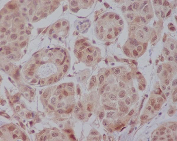
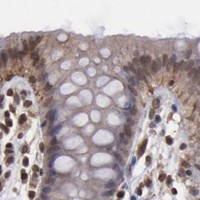
Real IHC data Immunohistochemical analysis of paraffin-embedded human colon, using AKT1 Antibody.
Anti-AKT1 Monoclonal Antibody
Cat # M00024-1
Real IHC data IHC analysis of AKT1 using anti-AKT1 antibody (P00024-2). AKT1 was detected in a paraffin-embedded section of human colorectal adenocarcinoma tissue. Heat mediated antigen retrieval was performed in EDTA buffer (pH 8.0, epitope retrieval solution). The tissue section was blocked with 10% goat serum. The tissue section was then incubated with 1:50 rabbit anti-AKT1 Antibody (P00024-2) overnight at 4°C. Peroxidase Conjugated Goat Anti-rabbit IgG was used as secondary antibody and incubated for 30 minutes at 37°C. The tissue section was developed using HRP Conjugated Rabbit IgG Super Vision Assay Kit (Catalog # SV0002) with DAB as the chromogen.
Anti-Phospho-AKT1 (T450) Rabbit Monoclonal Antibody
Cat # P00024-2
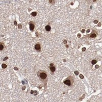
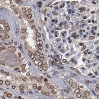
Real IHC data Immunohistochemical analysis of paraffin-embedded human kidney, using Phospho-Akt(Ser473) Antibody.
Anti-Phospho-Akt (Ser473) AKT1 Rabbit Monoclonal Antibody
Cat # P00024-5

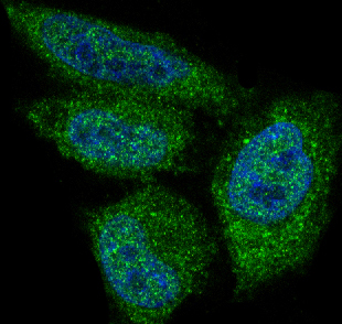
M00024-1 shows IHC in paraffin-embedded human colon and IF in HeLa cells (catalog: image captions); P00024-2 shows IHC in paraffin-embedded human colorectal adenocarcinoma and IF in A549 cells (catalog: image captions). P00024-5 shows IHC in paraffin-embedded human kidney and IF in PDGF-treated NIH/3T3 cells (catalog: image captions).

Which to pick: For tissue IHC, choose M00024-1 for AKT1, P00024-2 for phospho-AKT1 T450, or P00024-5 for phospho-AKT Ser473 according to the listed antibody targets; each has its own paraffin-section IHC image, with fixative unreported (catalog: titles, IHC captions). For IF/ICC, all three list those applications, but the illustrated samples differ: HeLa for M00024-1, A549 for P00024-2, and NIH/3T3 for P00024-5 (catalog: applications, IF captions). All three are rabbit monoclonals listed as reactive with human, mouse, and rat; the cited tissue IHC images are human, so mouse and rat tissue performance still needs verification (catalog: host, clone, reactivity, IHC captions).

Each figure is that product's own IHC / IF validation image from its datasheet.

References

  1. UniProt Consortium. UniProt entry P31749 (AKT1_HUMAN, RAC-alpha serine/threonine-protein kinase).
  2. Human Protein Atlas. AKT1 tissue IHC expression (reliability: Supported).
  3. Human Protein Atlas. AKT1 subcellular location (ICC-IF): Mainly localized to the nucleoplasm, microtubules, principal piece and end piece. In addition localized to the primary cilium, basal body, perinuclear theca and calyx..
  4. Human Protein Atlas. AKT1 antibody validation summary (3 antibodies).
  5. Network pharmacology and bioinformatics analysis-based strategy for exploring the mechanisms of luteolin in the treatment of five types of cancer: A retrospective computational study. The Journal of international medical research 2026 — PMC13309665.
  6. Phosphorylated Akt1 expression is associated with poor prognosis in cutaneous, oral and sinonasal melanomas. Oncotarget 2018 — PMC6324666.
  7. AKT1 loss correlates with episomal HPV16 in vulval intraepithelial neoplasia. PloS one 2012 — PMC3369856.
  8. Expression of AKT1 Related with Clinicopathological Parameters in Clear Cell Renal Cell Carcinoma. Current issues in molecular biology 2022 — PMC9600081.
  9. PubMed PMID:1851997 — UniProt-cited evidence.
  10. PubMed PMID:11508278 — UniProt-cited evidence.
  11. PubMed PMID:14702039 — UniProt-cited evidence.