Click image to see more details
-

-
-
-
-
+15
Product Info Summary
| SKU: | M00024-1 |
|---|---|
| Size: | 100 μl |
| Reactive Species: | Human, Mouse, Rat |
| Host: | Rabbit |
| Application: | Flow Cytometry, IP, IF, IHC, ICC, WB |
Customers Who Bought This Also Bought

Product info
Product Name
Anti-AKT1 Monoclonal Antibody
SKU/Catalog Number
M00024-1
BM4390 is an alternative SKU for this antibody, used in previous lots.
Size
100 μl
Form
Liquid
Description
Boster Bio Anti-AKT1 Monoclonal Antibody catalog # M00024-1. Tested in WB, IHC, ICC/IF, IP, Flow Cytometry applications. This antibody reacts with Human, Mouse, Rat.
Storage & Handling
Store at -20°C for one year. For short term storage and frequent use, store at 4°C for up to one month. Avoid repeated freeze-thaw cycles.
Cite This Product
Anti-AKT1 Monoclonal Antibody (Boster Biological Technology, Pleasanton CA, USA, Catalog # M00024-1)
Host
Rabbit
Contents
Rabbit IgG in stabilizing components, phosphate buffered saline, pH 7.4, 150mM NaCl, 0.02% sodium azide and 50% glycerol.
*This antibody is supplied in a stabilized formulation.
Compatibility with conjugation reactions depends on the chemistry of the conjugation method used.
For conjugation methods that are not compatible with the stabilizing components present in this formulation, a carrier-free antibody format is required.
Clonality
Monoclonal
Clone Number
EBG-1
Isotype
Rabbit IgG
Immunogen
A synthesized peptide derived from human AKT1 Akt, also referred to as PKB or Rac, plays a critical role in controlling survival and apoptosis. This protein kinase is activated by insulin and various growth and survival factors to function in a wortmannin-sensitive pathway involving PI3 kinase. Akt is activated by phospholipid binding and activation loop phosphorylation at Thr308 by PDK1 and by phosphorylation within the carboxy terminus at Ser473.
Reactive Species
M00024-1 is reactive to AKT1 in Human, Mouse, Rat
Observed Molecular Weight
56 kDa
Calculated molecular weight
55.7 kDa
Antibody Validation
Boster validates all antibodies on WB, IHC, ICC, Immunofluorescence, and ELISA with known positive control and negative samples to ensure specificity and high affinity, including thorough antibody incubations.
Application & Images
Applications
M00024-1 is guaranteed for Flow Cytometry, IP, IF, IHC, ICC, WB Boster Guarantee
Recommend Dilution
WB 1:500-2000
IHC 1:50-200
ICC/IF 1:50-200
IP 1:50
FC 1:50
Tested application
Suggested blocking solution with 5% non-fat milk or BSA; (*)Recommended protein loading: 20-40 µg per lane
Use TE buffer pH 9.0 for antigen retrieval; (*) citrate buffer pH 6.0 is an alternative.
Validation Images & Assay Conditions
Click image to see more details
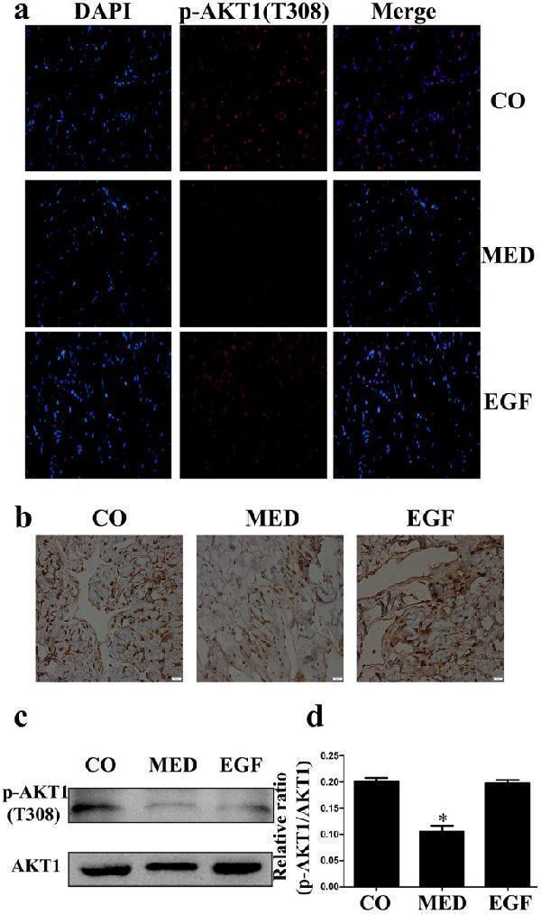
( a ) Immunofluorescence staining of cavernous tissue using an antibody against p-Akt1(Tyr308) in the CO, MED and EGF groups. ( b ) Immunohistochemistry staining of cavernous tissue was performed with an antibody against Akt1 in three groups (magnification: ×400 scale bar: 20 μm). ( c,d ) Western blot analysis of p-Akt1 (Tyr308) and Akt1 expression in the three groups. Data in the bar graphs are expressed as the means ± SD of 5~7 rats. *p < 0.05 vs the CO group. p-Akt, phosphor-protein kinase B; CO, normal control rats; MED, metabolic syndrome-related erectile dysfunction rats; EGF, MED rats treated with epithelial growth factor; SD, standard deviation.
Index in PubMed under a CC BY license. PMID: 29044143

Click image to see more details
Western blot analysis of AKT1 using anti-AKT1 antibody (M00024-1).
Electrophoresis was performed on a 5-20% SDS-PAGE gel at 70V (Stacking gel) / 90V (Resolving gel) for 2-3 hours. The sample well of each lane was loaded with 30 ug of sample under reducing conditions.
Lane 1: human HepG2 whole cell lysates,
Lane 2: human A549 whole cell lysates,
Lane 3: human Hela whole cell lysates,
Lane 4: human Raji whole cell lysates,
Lane 5: human MCF-7 whole cell lysates,
Lane 6: human SiHa whole cell lysates,
Lane 7: human 293T whole cell lysates.
After electrophoresis, proteins were transferred to a nitrocellulose membrane at 150 mA for 50-90 minutes. Blocked the membrane with 5% non-fat milk/TBS for 1.5 hour at RT. The membrane was incubated with rabbit anti-AKT1 antigen affinity purified monoclonal antibody (Catalog # M00024-1) at 1:500 overnight at 4°C, then washed with TBS-0.1%Tween 3 times with 5 minutes each and probed with a goat anti-rabbit IgG-HRP secondary antibody at a dilution of 1:5000 for 1.5 hour at RT. The signal is developed using an Enhanced Chemiluminescent detection (ECL) kit (Catalog # EK1002) with Tanon 5200 system. A specific band was detected for AKT1 at approximately 56 kDa. The expected band size for AKT1 is at 56 kDa.

Click image to see more details
Treatment with quercetin in PASMC proliferation and antioxidation under hypoxia. (A, B) Quercetin in cell proliferation were assessed using Ki67 immunofluorescence and quantitative evaluation in hypoxia-induced PASMCs (n = 3, scale bar = 100 μm). (C–H) Primitive Western blots and quantitative densities of PCNA, p-PI3K, PI3K, p-AKT1 Ser473, AKT1 with or without 740Y-P(10 μM), LY294002(10 μM), or quercetin (18 μM) in PASMCs under 3% O 2 for 24 h (I–L) Quantitative evaluation of SOD and GSH-Px activities and GSH and MDA contents in 3% O 2 -induced PASMCs. n = 3. All data represent mean ± SD. * p < 0.05 vs. control group, # p < 0.05 vs. 3% O 2 group, and p < 0.05 vs. 3% O 2 + QCT-18 μM.
Index in PubMed under a CC BY license. PMID: 40385484

Click image to see more details
Western blot analysis of AKT1 using anti-AKT1 antibody (M00024-1).
Electrophoresis was performed on a 5-20% SDS-PAGE gel at 70V (Stacking gel) / 90V (Resolving gel) for 2-3 hours. The sample well of each lane was loaded with 30 ug of sample under reducing conditions.
Lane 1: rat liver tissue lysates,
Lane 2: rat brain tissue lysates,
Lane 3: rat C6 whole cell lysates,
Lane 4: rat RH35 whole cell lysates,
Lane 5: mouse liver tissue lysates,
Lane 6: mouse brain tissue lysates,
Lane 7: Mouse Neuro-2a whole cell lysates,
Lane 8: Mouse HEPA1-6 whole cell lysates.
After electrophoresis, proteins were transferred to a nitrocellulose membrane at 150 mA for 50-90 minutes. Blocked the membrane with 5% non-fat milk/TBS for 1.5 hour at RT. The membrane was incubated with rabbit anti-AKT1 antigen affinity purified monoclonal antibody (Catalog # M00024-1) at 1:500 overnight at 4°C, then washed with TBS-0.1%Tween 3 times with 5 minutes each and probed with a goat anti-rabbit IgG-HRP secondary antibody at a dilution of 1:5000 for 1.5 hour at RT. The signal is developed using an Enhanced Chemiluminescent detection (ECL) kit (Catalog # EK1002) with Tanon 5200 system. A specific band was detected for AKT1 at approximately 56 kDa. The expected band size for AKT1 is at 56 kDa.

Click image to see more details
Effect of Lactucin on the activation of signaling pathways. (A) The whole-cell lysates were extracted for immunoblotting to determine the level of iNOS, COX-2. (B, C) The whole-cell lysates were extracted for immunoblotting to determine the levels of phospho- or total MAPKs (ERK, p38, and JNK) and AKT identified based on their antibodies. Data are shown as mean ± SD for each group (* p < 0.05 with the LPS Group, n = 3. Normal Group: RAW264.7 cells without LPS activation).
Index in PubMed under a CC BY license. PMID: 33995112

Click image to see more details
Immunohistochemical analysis of paraffin-embedded human colon, using AKT1 Antibody.

Click image to see more details
Treatment with eriocitrin in PASMC proliferation and antioxidation under hypoxic conditions. (A, B) Eriocitrin in cell proliferation were assessed using Ki67 immunofluorescence and quantitative evaluation in hypoxia-induced PASMCs (n = 3, scale bar = 100 μm). (C–H) Primitive Western blots and quantitative densities of PCNA, p-PI3K, PI3K, p-AKT1 (Ser473), AKT1 with or without 740Y-P (10 μM), LY294002 (10 μM), or eriocitrin (11 μM) in PASMCs under 3% O 2 for 24 h (I–L) Quantitative evaluation of SOD and GSH-Px activities and GSH and MDA contents in 3% O 2 -induced PASMCs. n = 3. All data are represented as the mean ± SD. * p < 0.05 vs. control group, # p < 0.05 vs. 3% O 2 group, and p < 0.05 vs. 3% O 2 + ERI-11 μM.
Index in PubMed under a CC BY license. PMID: 40385484

Click image to see more details
Immunofluorescent analysis of Hela cells, using AKT1 Antibody.

Click image to see more details
Eriocitrin and quercetin are responsible for anti-proliferation by targeting the PI3K protein in PASMCs under hypoxic conditions. ERI, eriocitrin; QCT, quercetin. (A, B) Primitive bands and quantitative evaluation of p-mTOR, mTOR, p-AKT1 (Ser473), and AKT1 with or without PS210 (2 μM) by Western blotting in PASMCs under 3% O 2 . n = 3. All data are represented as the mean ± SD. * p < 0.05 vs. control group, # p < 0.05 vs. 3% O 2 group, and p < 0.05 vs. 3% O 2 + FLA-50 μg/ml group. (C–G) Primitive bands and quantitative densities of p-PI3K and PI3K by Western blots. n = 3. All data are represented as the mean ± SD. * p < 0.05 vs. control group and # p < 0.05 vs. 3% O 2 group. (H–N) BECC, ERI, and QCT treatment increased the stability of PI3K in PASMC protease lysates by the DARTS experiment. (H–K) Primitive Western blots of PI3K. (L–N) Quantitative evaluation of PI3K levels. n = 3. All data are represented as the mean ± SD. * p < 0.05 vs. DMSO group.
Index in PubMed under a CC BY license. PMID: 40385484

Click image to see more details
Immunofluorescent analysis using the Antibody at 1:50 dilution.

Click image to see more details
Flavonoids inhibit the proliferation of PASMCs under hypoxic conditions by inhibiting the PI3K/AKT axis. (A, B) Primitive bands and quantitative densities of p-AKT1 Ser473 and AKT1 with or without Sc79 (20 μM) by Western blots in PASMCs under 3% O 2 . (C, D) Primitive bands and quantitative densities of p-PI3K, PI3K, p-AKT1 Ser473, and AKT1 with or without 740Y-P (10 μM) by Western blots in PASMCs under 3% O 2 . (E, F) Primitive bands and quantitative densities of p-PDPK1, PDPK, p-AKT1 Ser473, and AKT1 with or without MYH1485 (2 μM) in PASMCs by Western blots under 3% O 2 . n = 3. All data are represented as the mean ± SD. * p < 0.05 vs. control group, # p < 0.05 vs. 3% O 2 group, and p < 0.05 vs. 3% O 2 + Fla-50 μg/ml group.
Index in PubMed under a CC BY license. PMID: 40385484

Click image to see more details
Immunofluorescent analysis using the Antibody at 1:50 dilution.

Click image to see more details
BECCs regulate the AKT/GSK3β/CDK/cyclin signaling pathway in HAPH rats. (A–C) Primitive bands of p-AKT1 S473, AKT1, p-GSK3β, GSK3β, CDK4, cyclin D1, CDK2, cyclin A, and P27 by Western blots in lung tissues. (D–F) Quantitative evaluation of p-AKT1 (S473), AKT1, p-GSK3β, GSK3β, CDK4, cyclin D1, CDK2, cyclin A, and P27 in the lung tissues. n = 5. All data are represented as the mean ± SD. * p < 0.05 vs. control group and # p < 0.05 vs. hypoxia group.
Index in PubMed under a CC BY license. PMID: 40385484

Click image to see more details
Immunofluorescent analysis using the Antibody at 1:150 dilution.

Click image to see more details
Immunofluorescent analysis using the Antibody at 1:50 dilution.

Click image to see more details
Immunoprecipitate (IP) analysis using the Antibody at 1:50 dilution. (wb at 1:3K dilution)

Click image to see more details
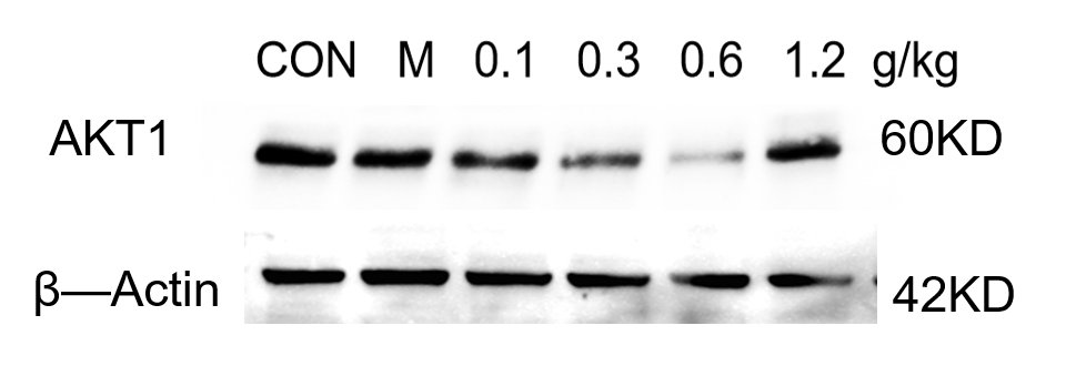
Western blot analysis of AKT1 using anti-AKT1 antibody (M00024-1).
Electrophoresis was performed on a 5-20% SDS-PAGE gel at 80V (Stacking gel) / 120V (Resolving gel) for 2 hours. The sample well of each lane was loaded with 30 ug of sample under reducing conditions.
Lane 1: Control group-mouse hippocampus tissue,
Lane 2: Model group-mouse hippocampus tissue,
Lane 3: Drug treatment (0.1 g/kg) – Mouse hippocampus tissue,
Lane 4: Drug treatment (0.3 g/kg) – Mouse hippocampus tissue,
Lane 5: Drug treatment (0.6 g/kg) – Mouse hippocampus tissue,
Lane 5: Drug treatment (1.2 g/kg) – Mouse hippocampus tissue.
After electrophoresis, proteins were transferred to a nitrocellulose membrane at 150 mA for 50-90 minutes. Blocked the membrane with 5% non-fat milk/TBS for 1.5 hour at RT. The membrane was incubated with rabbit anti-AKT1 antigen affinity purified monolonal antibody (A04887-1) overnight at 4°C, then washed with TBS-0.1%Tween 3 times with 5 minutes each and probed with a goat anti-rabbit IgG-HRP secondary antibody (Catalog # BA1054) at a dilution of 1:5000 for 1.5 hour at RT. The signal is developed using an ECL Plus Western Blotting Substrate with ChemiDoc MP system. A specific band was detected for AKT1 at approximately 60 kDa. The expected band size for AKT1 is at 56 kDa.

Click image to see more details
Western blot analysis of AKT1 using anti-AKT1 antibody (M00024-1).
Electrophoresis was performed on a 5-20% SDS-PAGE gel at 80V (Stacking gel) / 120V (Resolving gel) for 2 hours. The sample well of each lane was loaded with 30 ug of sample under reducing conditions.
Lane 1: Normal group-Rat colon tissue lysates,
Lane 2: Control group-Rat colon tissue lysates,
Lane 3: Drug treatment (low) –Rat colon tissue lysates,
Lane 4: Drug treatment (medium) –Rat colon tissue lysates,
Lane 5: Drug treatment (high) –Rat colon tissue lysates,
Lane 5: Drug treatment (positive) –Rat colon tissue lysates.
After electrophoresis, proteins were transferred to a nitrocellulose membrane at 150 mA for 50-90 minutes. Blocked the membrane with 5% non-fat milk/TBS for 1.5 hour at RT. The membrane was incubated with rabbit anti-AKT1 antigen affinity purified monolonal antibody (A04887-1) overnight at 4°C, then washed with TBS-0.1%Tween 3 times with 5 minutes each and probed with a goat anti-rabbit IgG-HRP secondary antibody (Catalog # BA1054) at a dilution of 1:5000 for 1.5 hour at RT. The signal is developed using an ECL Plus Western Blotting Substrate with ChemiDoc MP system. A specific band was detected for AKT1 at approximately 60 kDa. The expected band size for AKT1 is at 56 kDa.

Click image to see more details
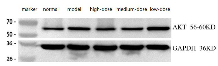
Western blot analysis of AKT1 using anti-AKT1 antibody (M00024-1).
Electrophoresis was performed on a 5-20% SDS-PAGE gel at 70V (Stacking gel) / 90V (Resolving gel) for 2-3 hours. The sample well of each lane was loaded with 30 ug of sample under reducing conditions.
Lane 1: normal group-Rat skeletal muscle tissue lysates,
Lane 2: model group-Rat skeletal muscle tissue lysates,
Lane 3: high-dose group-Rat skeletal muscle tissue lysates,
Lane 4: medium-dose group-Rat skeletal muscle tissue lysates,
Lane 5: low-dose group-Rat skeletal muscle tissue lysates.
After electrophoresis, proteins were transferred to a nitrocellulose membrane at 150 mA for 50-90 minutes. Blocked the membrane with 5% non-fat milk/TBS for 1.5 hour at RT. The membrane was incubated with rabbit anti-AKT1 antigen affinity purified monoclonal antibody (Catalog # M00024-1) at 1:2000 overnight at 4°C, then washed with TBS-0.1%Tween 3 times with 5 minutes each and probed with a goat anti-rabbit IgG-HRP secondary antibody at a dilution of 1:10000 for 1 hour at RT. The signal is developed using an Enhanced Chemiluminescent detection (ECL) kit (Catalog # EK1002) with Tanon 5200 system. A specific band was detected for AKT1 at approximately 56-60 kDa. The expected band size for AKT1 is at 56 kDa.
Specific Publications For Anti-AKT1 Monoclonal Antibody (M00024-1)
Loading publications
Recommended Resources
Here are featured tools and databases that you might find useful.
- Boster's Pathways Library
- Protein Databases
- Bioscience Research Protocol Resources
- Data Processing & Analysis Software
- Photo Editing Software
- Scientific Literature Resources
- Research Paper Management Tools
- Molecular Biology Software
- Primer Design Tools
- Bioinformatics Tools
- Phylogenetic Tree Analysis
Customer Reviews
Have you used Anti-AKT1 Monoclonal Antibody?
Share your experimental results or join a short interview to earn up to $1,000 in product credits or other rewards.
3 Reviews For Anti-AKT1 Monoclonal Antibody
The AKT1 Antibody (M00024-1) showed clear, specific bands in WB of rat skeletal muscle, with expression changes consistent with expectations.
Excellent
| SKU | M00024-1 |
|---|---|
| Application | Western Blot |
| Sample | Rat skeletal muscle tissue |
| Sample Processing Description | Normal rat skeletal muscle, injury model, and high-, medium-, and low-dose treatment groups, with total protein extracted. |
| Other Reagents | RIPA lysis buffer, Protease inhibitor, Running buffer, Transfer buffer, Blocking buffer |
| Primary Antibody | AKT1 Monoclonal Antibody |
| Primary Incubation | 1:2000, overnight at 4 ℃ |
| Secondary Antibody | HRP Conjugated AffiniPure Goat Anti-Rabbit IgG (H+L) (BA1054) |
| Secondary Incubation | 1:10000, 1 h in RT |
| Detection | Substrate: ECL substrate, Image system: ChemiDoc MP |
| Results Summary | The AKT1 bands are clear and correctly positioned; expression is significantly higher in the model group than in the control, and decreases with increasing drug dose in the treatment groups, consistent with expectations. |
Guangtian Yu, Ningxia Medical University
Verified customer
Submitted 2026-03-25
This antibody is highly specific and efficient, suitable for detecting AKT1 protein in rat colon by Western blot, with only minor nonspecific bands.
Excellent
| SKU | M00024-1 |
|---|---|
| Application | Western Blot |
| Sample | rat colon tissue |
| Sample Processing Description | RIPA lysis buffer: Add protease inhibitor PMSF (100:1), lyse for 10 min, centrifuge at 12,000 rpm for 15 min, transfer the supernatant and mix with 5× loading buffer, denature at 100 °C for 10 min, and load onto SDS-PAGE. |
| Other Reagents | Blocking buffer |
| Primary Antibody | AKT1 Monoclonal Antibody |
| Primary Incubation | 1:1000, overnight at 4 ℃ |
| Secondary Antibody | HRP Conjugated AffiniPure Goat Anti-Rabbit IgG (H+L) |
| Secondary Incubation | 1:1000, 1 hour in room temperature |
| Detection | Substrate: ECL, Imaging system:ChemiDoc MP |
| Results Summary | The figure shows a schematic of Western blot results for the target protein AKT1 and the internal control Actin in rat colon across different groups. No significant differences were observed between groups; the target bands are clear and distinct, and the experimental results are satisfactory. |
Shiyu Zhang, LUTCM
Verified customer
Submitted 2025-12-29
Western blot analysis of AKT1 protein in mouse hippocampus using the AKT1 antibody showed clear bands. The antibody is suitable for mouse hippocampal tissue samples.
Excellent
| SKU | M00024-1 |
|---|---|
| Application | Western Blot |
| Sample | Mouse hippocampus tissue |
| Sample Processing Description | The mouse hippocampus was lysed with RIPA buffer containing a protease inhibitor cocktail. After protein quantification, samples were mixed with 5× protein loading buffer and heated for 10 minutes to denature. Load 5 μL of protein per lane and apply to SDS-PAGE. |
| Primary Antibody | Anti-AKT1 Monoclonal Antibody |
| Primary Incubation | overnight at 4 ℃ |
| Secondary Antibody | HRP-conjugated Anti-Rabbit IgG Secondary Antibody |
| Secondary Incubation | 1 hour in room temperature |
| Detection | Substrate: Ultra-sensitive ECL luminescent reagent (Cat# AR1191), Imaging system: ChemiDoc MP (Bio-Rad) |
| Results Summary | Western blot analysis of AKT1 protein in the mouse hippocampus using the AKT1 antibody showed clear bands. This antibody is suitable for mouse hippocampal tissue samples. |
Changbin Yuan, LNUTCM
Verified customer
Submitted 2025-11-05
Customer Q&As
Have a question?
Find answers in Q&As, reviews.
Can't find your answer?
Submit your question
16 Customer Q&As for Anti-AKT1 Monoclonal Antibody
Question
See below the WB image, lot number and protocol we used for adrenal gland using anti-AKT1 Monoclonal antibody M00024-1. Please let me know if you require anything else.
A. Krishna
Verified customer
Asked: 2020-03-10
Answer
Thank you very much for the data. Our lab team are working to resolve this as quickly as possible, and we appreciate your patience and understanding! You have provided everything we needed. Please let me know if there is anything you need in the meantime.
Boster Scientific Support
Answered: 2020-03-10
Question
My boss were content with the WB result of your anti-AKT1 Monoclonal antibody. However we have observed positive staining in cervix carcinoma cytoplasm. using this antibody. Is that expected? Could you tell me where is AKT1 supposed to be expressed?
Verified Customer
Verified customer
Asked: 2020-02-14
Answer
From literature, cervix carcinoma does express AKT1. Generally AKT1 expresses in cytoplasm. Regarding which tissues have AKT1 expression, here are a few articles citing expression in various tissues:
Adrenal gland, Pubmed ID: 14702039
Cervix carcinoma, Pubmed ID: 17081983, 18669648
Cervix carcinoma, and Erythroleukemia, Pubmed ID: 23186163
Foreskin, Pubmed ID: 1718748
Liver, Pubmed ID: 24275569
Muscle, and Ovary, Pubmed ID: 15489334
Boster Scientific Support
Answered: 2020-02-14
Question
Is this M00024-1 anti-AKT1 Monoclonal antibody reactive to the isotypes of AKT1?
Verified Customer
Verified customer
Asked: 2019-09-23
Answer
The immunogen of M00024-1 anti-AKT1 Monoclonal antibody is A synthesized peptide derived from human AKT1. Could you tell me which isotype you are interested in so I can help see if the immunogen is part of this isotype?
Boster Scientific Support
Answered: 2019-09-23
Question
Is a blocking peptide available for product anti-AKT1 Monoclonal antibody (M00024-1)?
A. Moore
Verified customer
Asked: 2019-09-12
Answer
We do provide the blocking peptide for product anti-AKT1 Monoclonal antibody (M00024-1). If you would like to place an order for it please contact support@bosterbio.com and make a special request.
Boster Scientific Support
Answered: 2019-09-12
Question
We have observed staining in human cervix carcinoma. What should we do? Is anti-AKT1 Monoclonal antibody supposed to stain cervix carcinoma positively?
G. Bhatt
Verified customer
Asked: 2019-06-04
Answer
According to literature cervix carcinoma does express AKT1. According to Uniprot.org, AKT1 is expressed in left adrenal gland, adrenal gland, muscle ovary, foreskin, cervix carcinoma, cervix carcinoma erythroleukemia, liver, among other tissues. Regarding which tissues have AKT1 expression, here are a few articles citing expression in various tissues:
Adrenal gland, Pubmed ID: 14702039
Cervix carcinoma, Pubmed ID: 17081983, 18669648
Cervix carcinoma, and Erythroleukemia, Pubmed ID: 23186163
Foreskin, Pubmed ID: 1718748
Liver, Pubmed ID: 24275569
Muscle, and Ovary, Pubmed ID: 15489334
Boster Scientific Support
Answered: 2019-06-04
Question
I see that the anti-AKT1 Monoclonal antibody M00024-1 works with ICC, what is the protocol used to produce the result images on the product page?
Verified Customer
Verified customer
Asked: 2018-07-05
Answer
You can find protocols for ICC on the "support/technical resources" section of our navigation menu. If you have any further questions, please send an email to support@bosterbio.com
Boster Scientific Support
Answered: 2018-07-05
Question
Our lab used your anti-AKT1 Monoclonal antibody for WB on muscle ovary a few years ago. I am using mouse, and I plan to use the antibody for IHC next. Our lab want to know about examining muscle ovary as well as cervix carcinoma in our next experiment. Could you please give me some suggestion on which antibody would work the best for IHC?
Verified Customer
Verified customer
Asked: 2018-06-20
Answer
I viewed the website and datasheets of our anti-AKT1 Monoclonal antibody and it seems that M00024-1 has been tested on mouse in both WB and IHC. Thus M00024-1 should work for your application. Our Boster satisfaction guarantee will cover this product for IHC in mouse even if the specific tissue type has not been validated. We do have a comprehensive range of products for IHC detection and you can check out our website bosterbio.com to find out more information about them.
Boster Scientific Support
Answered: 2018-06-20
Question
Thank you for helping with my inquiry over the phone. Here are the WB image, lot number and protocol we used for adrenal gland using anti-AKT1 Monoclonal antibody M00024-1. Let me know if you need anything else.
Verified Customer
Verified customer
Asked: 2018-05-04
Answer
Thanks for the data. You have provided everything we needed. Our lab team are working to resolve your inquiry as quickly as possible, and we appreciate your patience and understanding! Please let me know if there is anything you need in the meantime.
Boster Scientific Support
Answered: 2018-05-04
Question
My question regarding product M00024-1, anti-AKT1 Monoclonal antibody. I was wondering if it would be possible to conjugate this antibody with biotin. I would need it to be without BSA or sodium azide. I am planning on using a buffer exchange of sodium azide with PBS only. Would there be problems for me to conjugate the antibody and store it in -20 degrees in small aliquots?
B. Jones
Verified customer
Asked: 2017-07-03
Answer
We suggest not storing this antibody with PBS buffer only in -20 degrees. If you want to store it in -20 degrees it is best to add some cryoprotectant like glycerol. If you want carrier free M00024-1 anti-AKT1 Monoclonal antibody, we can provide it to you in a special formula with trehalose and/or glycerol. These molecules will not interfere with conjugation chemistry and provide a good level of protection for the antibody from degradation. Please be sure to specify this in your purchase order.
Boster Scientific Support
Answered: 2017-07-03
Question
Would anti-AKT1 Monoclonal antibody M00024-1 work for ICC with adrenal gland?
N. Krishna
Verified customer
Asked: 2016-11-21
Answer
According to the expression profile of adrenal gland, AKT1 is highly expressed in adrenal gland. So, it is likely that anti-AKT1 Monoclonal antibody M00024-1 will work for ICC with adrenal gland.
Boster Scientific Support
Answered: 2016-11-21
Question
I would like to test anti-AKT1 Monoclonal antibody M00024-1 on mouse adrenal gland for research purposes, then I may be interested in using anti-AKT1 Monoclonal antibody M00024-1 for diagnostic purposes as well. Is the antibody suitable for diagnostic purposes?
P. Rodriguez
Verified customer
Asked: 2016-09-28
Answer
The products we sell, including anti-AKT1 Monoclonal antibody M00024-1, are only intended for research use. They would not be suitable for use in diagnostic work. If you have the means to develop a product into diagnostic use, and are interested in collaborating with us and develop our product into an IVD product, please contact us for more discussions.
Boster Scientific Support
Answered: 2016-09-28
Question
Do you have a BSA free version of anti-AKT1 Monoclonal antibody M00024-1 available?
S. Li
Verified customer
Asked: 2015-05-15
Answer
We appreciate your recent telephone inquiry. I can confirm that some lots of this anti-AKT1 Monoclonal antibody M00024-1 are BSA free. For now, these lots are available and we can make a BSA free formula for you free of charge. It will take 3 extra days to prepare. If you require this antibody BSA free again in future, please do not hesitate to contact me and I will be pleased to check which lots we have in stock that are BSA free.
Boster Scientific Support
Answered: 2015-05-15
Question
Would M00024-1 anti-AKT1 Monoclonal antibody work on parafin embedded sections? If so, which fixation method do you recommend we use (PFA, paraformaldehyde, other)?
A. Evans
Verified customer
Asked: 2015-04-10
Answer
It shows on the product datasheet, M00024-1 anti-AKT1 Monoclonal antibody as been tested on ICC. It is best to use PFA for fixation because it has better tissue penetration ability. PFA needs to be prepared fresh before use. Long term stored PFA turns into formalin, as the PFA molecules congregate and become formalin.
Boster Scientific Support
Answered: 2015-04-10
Question
We are interested in using your anti-AKT1 Monoclonal antibody for cellular response to dna damage stimulus studies. Has this antibody been tested with western blotting on hela cells? We would like to see some validation images before ordering.
G. Jones
Verified customer
Asked: 2015-04-03
Answer
We appreciate your inquiry. This M00024-1 anti-AKT1 Monoclonal antibody is validated on hela cells. It is guaranteed to work for Flow Cytometry, IP, IF, IHC, ICC, WB in human, mouse, rat. Our Boster guarantee will cover your intended experiment even if the sample type has not been be directly tested.
Boster Scientific Support
Answered: 2015-04-03
Question
We are currently using anti-AKT1 Monoclonal antibody M00024-1 for human tissue, and we are happy with the IP results. The species of reactivity given in the datasheet says human, mouse, rat. Is it possible that the antibody can work on feline tissues as well?
R. Singh
Verified customer
Asked: 2013-10-01
Answer
The anti-AKT1 Monoclonal antibody (M00024-1) has not been validated for cross reactivity specifically with feline tissues, though there is a good chance of cross reactivity. We have an innovator award program that if you test this antibody and show it works in feline you can get your next antibody for free. Please contact me if I can help you with anything.
Boster Scientific Support
Answered: 2013-10-01
Question
I was wanting to use your anti-AKT1 Monoclonal antibody for ICC for mouse adrenal gland on frozen tissues, but I want to know if it has been validated for this particular application. Has this antibody been validated and is this antibody a good choice for mouse adrenal gland identification?
K. Moore
Verified customer
Asked: 2013-09-13
Answer
As indicated on the product datasheet, M00024-1 anti-AKT1 Monoclonal antibody has been validated for Flow Cytometry, IP, IF, IHC, ICC, WB on human, mouse, rat tissues. We have an innovator award program that if you test this antibody and show it works in mouse adrenal gland in IHC-frozen, you can get your next antibody for free.
Boster Scientific Support
Answered: 2013-09-13
