Click image to see more details
-

-
-
-
-
+6
Product Info Summary
| SKU: | P00024-2 |
|---|---|
| Size: | 100 μl |
| Reactive Species: | Human, Mouse, Rat |
| Host: | Rabbit |
| Application: | IP, IF, IHC, ICC, WB |
Customers Who Bought This Also Bought

Product info
Product Name
Anti-Phospho-AKT1 (T450) Rabbit Monoclonal Antibody
SKU/Catalog Number
P00024-2
BM4721 is an alternative SKU for this antibody, used in previous lots.
Size
100 μl
Form
Liquid
Description
Boster Bio Anti-Phospho-AKT1 (T450) Rabbit Monoclonal Antibody catalog # P00024-2. Tested in WB, IHC, ICC/IF, IP applications. This antibody reacts with Human, Mouse, Rat.
Storage & Handling
Store at -20°C for one year. For short term storage and frequent use, store at 4°C for up to one month. Avoid repeated freeze-thaw cycles.
Cite This Product
Anti-Phospho-AKT1 (T450) Rabbit Monoclonal Antibody (Boster Biological Technology, Pleasanton CA, USA, Catalog # P00024-2)
Host
Rabbit
Contents
Rabbit IgG in stabilizing components, phosphate buffered saline, pH 7.4, 150mM NaCl, 0.02% sodium azide and 50% glycerol.
*This antibody is supplied in a stabilized formulation.
Compatibility with conjugation reactions depends on the chemistry of the conjugation method used.
For conjugation methods that are not compatible with the stabilizing components present in this formulation, a carrier-free antibody format is required.
Clonality
Monoclonal
Clone Number
HEA-1
Isotype
Rabbit IgG
Immunogen
A synthesized peptide derived from human AKT1
Reactive Species
P00024-2 is reactive to AKT1 in Human, Mouse, Rat
Observed Molecular Weight
65 kDa
Calculated molecular weight
55.7 kDa
Antibody Validation
Boster validates all antibodies on WB, IHC, ICC, Immunofluorescence, and ELISA with known positive control and negative samples to ensure specificity and high affinity, including thorough antibody incubations.
Application & Images
Applications
P00024-2 is guaranteed for IP, IF, IHC, ICC, WB Boster Guarantee
Recommend Dilution
WB 1:500-2000
IHC 1:50-200
ICC/IF 1:50-200
IP 1:20
Tested application
Suggested blocking solution with 5% non-fat milk or BSA; (*)Recommended protein loading: 20-40 µg per lane
Use TE buffer pH 9.0 for antigen retrieval; (*) citrate buffer pH 6.0 is an alternative.
Validation Images & Assay Conditions
Click image to see more details
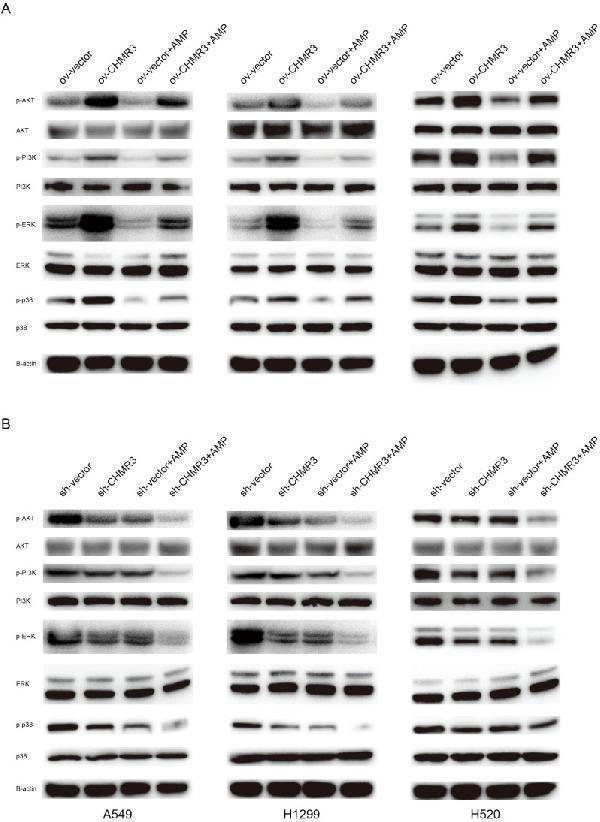
AMP treatment regulates the PI3K/AKT and MAPK signaling pathways. Lung cells (A549, NCI-H1299, and NCI-H520) with stable CHRM3 overexpression (A) or knockdown (B) were treated with or without 100 μg/mL AMPs for 48 h. The expression levels of PI3K/AKT pathway proteins and MAPK pathway proteins were detected using western blot.
Index in PubMed under a CC BY license. PMID: 40718823

Click image to see more details
a Western blotting was used to examine mitochondrial apoptotic pathway-related proteins after treatment with control (i), PANI-PEG-CS (ii), Ir(III) complex (iii), and Ir(III)@PANI-PEG-CS (iv). Key proteins involved in apoptosis such as Bax, Bcl-2, cytochrome c, and cleaved caspase-3 were examined to better understand how each treatment affects cell death at the mitochondrial level. b To further investigate the underlying molecular mechanisms, western blotting was used to investigate the PI3K/AKT/mTOR pathway (i), PANI-PEG-CS (ii), Ir(III) complex (iii), and Ir(III)@PANI-PEG-CS (iv). Expression levels of PI3K, AKT (total and phosphorylated), and mTOR were evaluated to assess whether this survival pathway was activated or suppressed. Protein levels were quantified and compared to the control group to determine statistical significance. * p < 0.05, ** p < 0.01, *** p < 0.001 in comparison to the respective control
Index in Springer Nature under a CC BY license. DOI: 10.1186/s12645-025-00336-z

Click image to see more details
Western blot analysis of AKT1 using anti-AKT1 antibody (P00024-2).
Electrophoresis was performed on a 5-20% SDS-PAGE gel at 70V (Stacking gel) / 90V (Resolving gel) for 2-3 hours. The sample well of each lane was loaded with 30 ug of sample under reducing conditions.
Lane 1: human Hela whole cell lysates,
Lane 2: human MCF-7 whole cell lysates,
Lane 3: rat brain tissue lysates,
Lane 4: rat PC-12 whole cell lysates,
Lane 5: mouse brain tissue lysates,
Lane 6: mouse NIH/3T3 whole cell lysates.
After electrophoresis, proteins were transferred to a nitrocellulose membrane at 150 mA for 50-90 minutes. Blocked the membrane with 5% non-fat milk/TBS for 1.5 hour at RT. The membrane was incubated with rabbit anti-AKT1 antigen affinity purified monoclonal antibody (Catalog # P00024-2) at 1:1000 overnight at 4°C, then washed with TBS-0.1%Tween 3 times with 5 minutes each and probed with a goat anti-rabbit IgG-HRP secondary antibody at a dilution of 1:1000 for 1.5 hour at RT. The signal is developed using an Enhanced Chemiluminescent detection (ECL) kit (Catalog # EK1002) with Tanon 5200 system. A specific band was detected for AKT1 at approximately 65 kDa. The expected band size for AKT1 is at 56 kDa.

Click image to see more details
Effect of Lactucin on the activation of signaling pathways. (A) The whole-cell lysates were extracted for immunoblotting to determine the level of iNOS, COX-2. (B, C) The whole-cell lysates were extracted for immunoblotting to determine the levels of phospho- or total MAPKs (ERK, p38, and JNK) and AKT identified based on their antibodies. Data are shown as mean ± SD for each group (* p < 0.05 with the LPS Group, n = 3. Normal Group: RAW264.7 cells without LPS activation).
Index in PubMed under a CC BY license. PMID: 33995112

Click image to see more details
IHC analysis of AKT1 using anti-AKT1 antibody (P00024-2).
AKT1 was detected in a paraffin-embedded section of human colorectal adenocarcinoma tissue. Heat mediated antigen retrieval was performed in EDTA buffer (pH 8.0, epitope retrieval solution). The tissue section was blocked with 10% goat serum. The tissue section was then incubated with 1:50 rabbit anti-AKT1 Antibody (P00024-2) overnight at 4°C. Peroxidase Conjugated Goat Anti-rabbit IgG was used as secondary antibody and incubated for 30 minutes at 37°C. The tissue section was developed using HRP Conjugated Rabbit IgG Super Vision Assay Kit (Catalog # SV0002) with DAB as the chromogen.

Click image to see more details
IHC analysis of AKT1 using anti-AKT1 antibody (P00024-2).
AKT1 was detected in a paraffin-embedded section of human prostate cancer tissue. Heat mediated antigen retrieval was performed in EDTA buffer (pH 8.0, epitope retrieval solution). The tissue section was blocked with 10% goat serum. The tissue section was then incubated with 1:50 rabbit anti-AKT1 Antibody (P00024-2) overnight at 4°C. Peroxidase Conjugated Goat Anti-rabbit IgG was used as secondary antibody and incubated for 30 minutes at 37°C. The tissue section was developed using HRP Conjugated Rabbit IgG Super Vision Assay Kit (Catalog # SV0002) with DAB as the chromogen.

Click image to see more details
IHC analysis of AKT1 using anti-AKT1 antibody (P00024-2).
AKT1 was detected in a paraffin-embedded section of human lung adenocarcinoma tissue. Heat mediated antigen retrieval was performed in EDTA buffer (pH 8.0, epitope retrieval solution). The tissue section was blocked with 10% goat serum. The tissue section was then incubated with 1:50 rabbit anti-AKT1 Antibody (P00024-2) overnight at 4°C. Peroxidase Conjugated Goat Anti-rabbit IgG was used as secondary antibody and incubated for 30 minutes at 37°C. The tissue section was developed using HRP Conjugated Rabbit IgG Super Vision Assay Kit (Catalog # SV0002) with DAB as the chromogen.

Click image to see more details
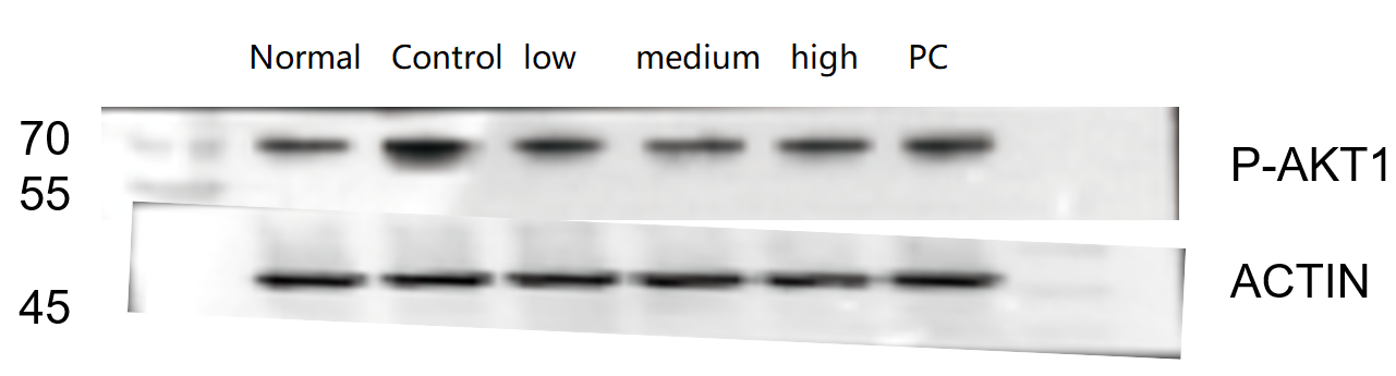
Western blot analysis of AKT1 using anti-AKT1 antibody (P00024-2).
Electrophoresis was performed on a 5-20% SDS-PAGE gel at 70V (Stacking gel) / 90V (Resolving gel) for 2-3 hours. The sample well of each lane was loaded with 30 ug of sample under reducing conditions.
Lane 1: Normal group-rat colon tissue lysates,
Lane 2: Control group-rat colon tissue lysates,
Lane 3: Low-dose drug treatment-rat colon tissue lysates,
Lane 4: Medium-dose drug treatment-rat colon tissue lysates,
Lane 5: Positive drug treatment-rat colon tissue lysates.
After electrophoresis, proteins were transferred to a nitrocellulose membrane at 150 mA for 50-90 minutes. Blocked the membrane with 5% non-fat milk/TBS for 1.5 hour at RT. The membrane was incubated with rabbit anti-AKT1 antigen affinity purified monoclonal antibody (Catalog # P00024-2) at 1:1000 overnight at 4°C, then washed with TBS-0.1%Tween 3 times with 5 minutes each and probed with a goat anti-rabbit IgG-HRP secondary antibody at a dilution of 1:1000 for 1 hour at RT. The signal is developed using an Enhanced Chemiluminescent detection (ECL) kit (Catalog # EK1002) with ChemiDoc MP system. A specific band was detected for AKT1 at approximately 70 kDa. The expected band size for AKT1 is at 56 kDa.

Click image to see more details
IHC analysis of AKT1 using anti-AKT1 antibody (P00024-2).
AKT1 was detected in a paraffin-embedded section of intestinal diffuse large B-cell lymphoma tissue. Heat mediated antigen retrieval was performed in EDTA buffer (pH 8.0, epitope retrieval solution). The tissue section was blocked with 10% goat serum. The tissue section was then incubated with 1:50 rabbit anti-AKT1 Antibody (P00024-2) overnight at 4°C. Peroxidase Conjugated Goat Anti-rabbit IgG was used as secondary antibody and incubated for 30 minutes at 37°C. The tissue section was developed using HRP Conjugated Rabbit IgG Super Vision Assay Kit (Catalog # SV0002) with DAB as the chromogen.

Click image to see more details
IF analysis of AKT1 using anti-AKT1 antibody (P00024-2) and anti-Beta Tubulin antibody (M01857-3).
AKT1 was detected in immunocytochemical section of A549 cell. Enzyme antigen retrieval was performed using IHC enzyme antigen retrieval reagent (AR0022) for 15 mins. The cells were blocked with 10% goat serum. And then incubated at 1:50 with rabbit anti-AKT1 Antibody (P00024-2) and mouse anti-Beta Tubulin antibody (M01857-3) overnight at 4°C. DyLight®488 Conjugated Goat Anti-Rabbit IgG (BA1127) and Cy3 Conjugated Goat Anti-Mouse IgG (BA1031) were used as secondary antibody at 1:500 dilution and incubated for 30 minutes at 37°C. Visualize using a fluorescence microscope and filter sets appropriate for the label used.
Specific Publications For Anti-Phospho-AKT1 (T450) Rabbit Monoclonal Antibody (P00024-2)
Loading publications
Recommended Resources
Here are featured tools and databases that you might find useful.
- Boster's Pathways Library
- Protein Databases
- Bioscience Research Protocol Resources
- Data Processing & Analysis Software
- Photo Editing Software
- Scientific Literature Resources
- Research Paper Management Tools
- Molecular Biology Software
- Primer Design Tools
- Bioinformatics Tools
- Phylogenetic Tree Analysis
Customer Reviews
Have you used Anti-Phospho-AKT1 (T450) Rabbit Monoclonal Antibody?
Share your experimental results or join a short interview to earn up to $1,000 in product credits or other rewards.
1 Reviews For Anti-Phospho-AKT1 (T450) Rabbit Monoclonal Antibody
This antibody is highly specific and efficient, suitable for Western blot detection of AKT1 (Phospho-T450) protein in rat colon, with only minor nonspecific bands observed.
Excellent
| SKU | P00024-2 |
|---|---|
| Application | Western Blot |
| Sample | rat colon tissue |
| Sample Processing Description | RIPA lysis buffer supplemented with PMSF (100:1) was used to lyse the samples for 10 min. The lysates were centrifuged at 12,000 rpm for 15 min, and the supernatants were collected. Fivefold loading buffer was added, and the samples were denatured at 100 °C for 10 min before loading onto SDS-PAGE. |
| Other Reagents | Blocking buffer |
| Primary Antibody | Phospho-AKT1 (T450) Rabbit Monoclonal Antibody |
| Primary Incubation | 1:1000, overnight at 4 ℃ |
| Secondary Antibody | HRP Conjugated AffiniPure Goat Anti-Rabbit IgG (H+L) |
| Secondary Incubation | 1:2000, 1 hour in room temperature |
| Detection | Substrate: ECL, Imaging system:ChemiDoc MP |
| Results Summary | The figure shows representative Western blot results of AKT1 (Phospho-T450) and the internal control Actin in rat colon tissues from different groups. Among the low, medium, and high doses of the traditional Chinese medicine, the high-dose group exhibits the best therapeutic effect. The target bands are clear and well defined, and the experimental results are satisfactory. |
Shiyu Zhang, LUTCM
Verified customer
Submitted 2025-12-26
Customer Q&As
Have a question?
Find answers in Q&As, reviews.
Can't find your answer?
Submit your question
4 Customer Q&As for Anti-Phospho-AKT1 (T450) Rabbit Monoclonal Antibody
Question
We are currently using anti-Phospho-AKT1 (T450) Rabbit Monoclonal antibody P00024-2 for mouse tissue, and we are well pleased with the IP results. The species of reactivity given in the datasheet says human, mouse, rat. Is it likely that the antibody can work on dog tissues as well?
Verified Customer
Verified customer
Asked: 2020-01-30
Answer
The anti-Phospho-AKT1 (T450) Rabbit Monoclonal antibody (P00024-2) has not been tested for cross reactivity specifically with dog tissues, though there is a good chance of cross reactivity. We have an innovator award program that if you test this antibody and show it works in dog you can get your next antibody for free. Please contact me if I can help you with anything.
Boster Scientific Support
Answered: 2020-01-30
Question
We ordered your anti-Phospho-AKT1 (T450) Rabbit Monoclonal antibody for WB on adrenal gland in a previous project. I am using human, and We are going to use the antibody for IP next. I am interested in examining adrenal gland as well as foreskin in our next experiment. Could give a recommendation on which antibody would work the best for IP?
Verified Customer
Verified customer
Asked: 2019-05-31
Answer
I looked at the website and datasheets of our anti-Phospho-AKT1 (T450) Rabbit Monoclonal antibody and it appears that P00024-2 has been validated on human in both WB and IP. Thus P00024-2 should work for your application. Our Boster satisfaction guarantee will cover this product for IP in human even if the specific tissue type has not been validated. We do have a comprehensive range of products for IP detection and you can check out our website bosterbio.com to find out more information about them.
Boster Scientific Support
Answered: 2019-05-31
Question
My team were content with the WB result of your anti-Phospho-AKT1 (T450) Rabbit Monoclonal antibody. However we have been able to see positive staining in left adrenal gland cytoplasm. using this antibody. Is that expected? Could you tell me where is AKT1 supposed to be expressed?
Verified Customer
Verified customer
Asked: 2019-02-19
Answer
Based on literature, left adrenal gland does express AKT1. Generally AKT1 expresses in cytoplasm. Regarding which tissues have AKT1 expression, here are a few articles citing expression in various tissues:
Adrenal gland, Pubmed ID: 14702039
Cervix carcinoma, Pubmed ID: 17081983, 18669648
Cervix carcinoma, and Erythroleukemia, Pubmed ID: 23186163
Foreskin, Pubmed ID: 1718748
Liver, Pubmed ID: 24275569
Muscle, and Ovary, Pubmed ID: 15489334
Boster Scientific Support
Answered: 2019-02-19
Question
We have been able to see staining in mouse cervix carcinoma erythroleukemia. What should we do? Is anti-Phospho-AKT1 (T450) Rabbit Monoclonal antibody supposed to stain cervix carcinoma erythroleukemia positively?
Verified Customer
Verified customer
Asked: 2018-05-22
Answer
From literature cervix carcinoma erythroleukemia does express AKT1. From Uniprot.org, AKT1 is expressed in left adrenal gland, adrenal gland, muscle ovary, foreskin, cervix carcinoma, cervix carcinoma erythroleukemia, liver, among other tissues. Regarding which tissues have AKT1 expression, here are a few articles citing expression in various tissues:
Adrenal gland, Pubmed ID: 14702039
Cervix carcinoma, Pubmed ID: 17081983, 18669648
Cervix carcinoma, and Erythroleukemia, Pubmed ID: 23186163
Foreskin, Pubmed ID: 1718748
Liver, Pubmed ID: 24275569
Muscle, and Ovary, Pubmed ID: 15489334
Boster Scientific Support
Answered: 2018-05-22
