This website uses cookies to ensure you get the best experience on our website.
- Table of Contents
2 Citations 7 Q&As
1 Citations
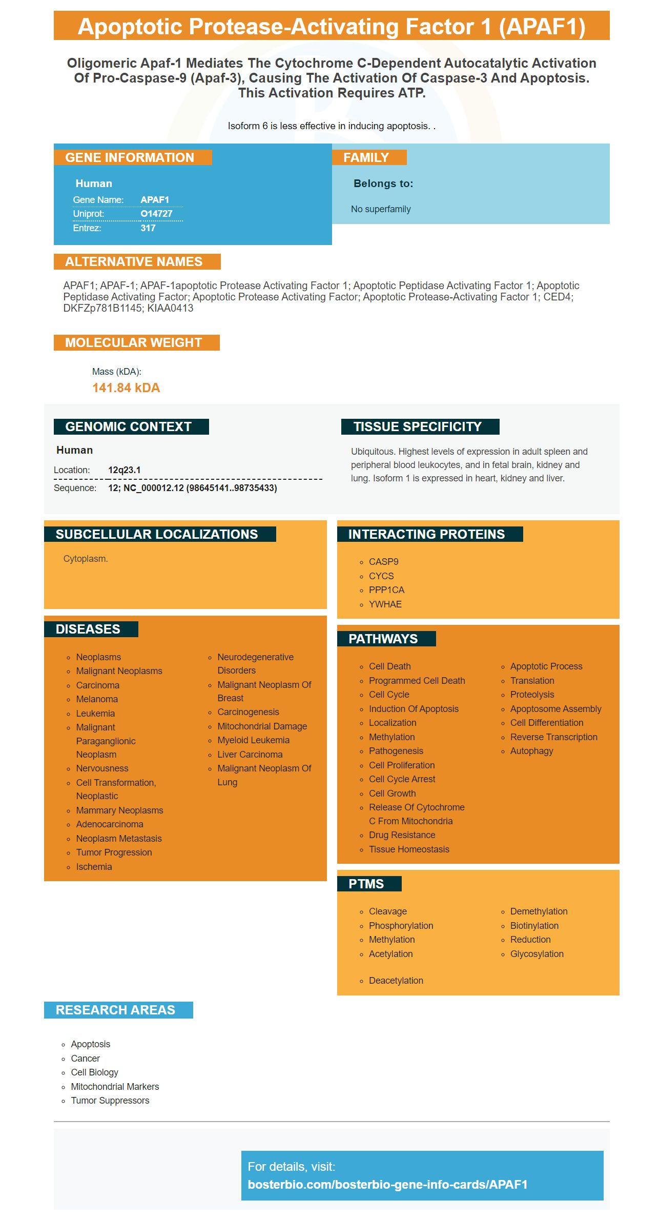
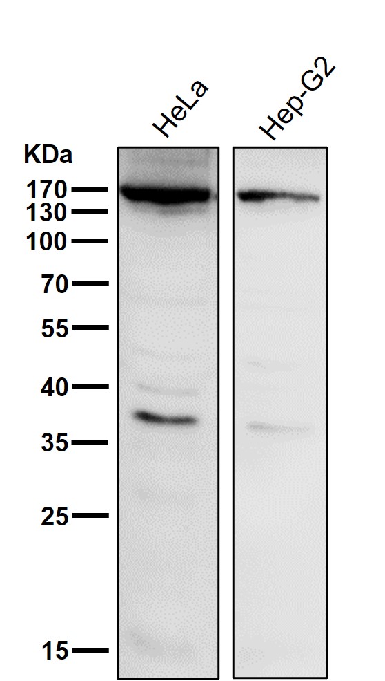
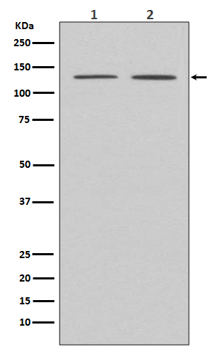
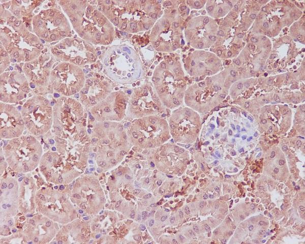
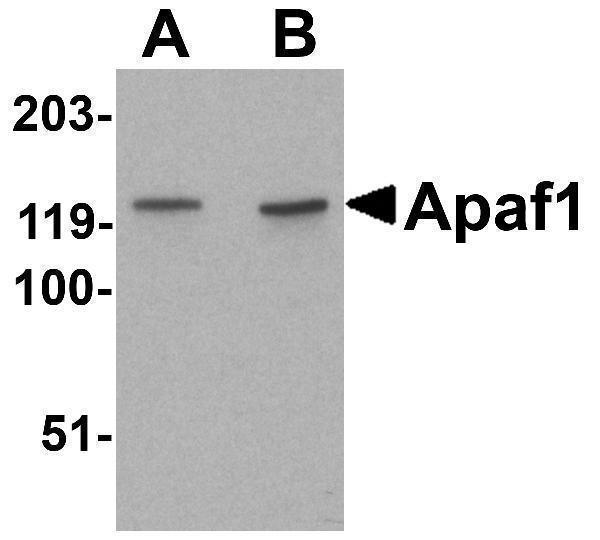
Facts about Apoptotic protease-activating factor 1.
Isoform 6 is less effective in inducing apoptosis. .
Human | |
---|---|
Gene Name: | APAF1 |
Uniprot: | O14727 |
Entrez: | 317 |
Belongs to: |
---|
No superfamily |
APAF1; APAF-1; APAF-1apoptotic protease activating factor 1; apoptotic peptidase activating factor 1; apoptotic peptidase activating factor; apoptotic protease activating factor; apoptotic protease-activating factor 1; CED4; DKFZp781B1145; KIAA0413
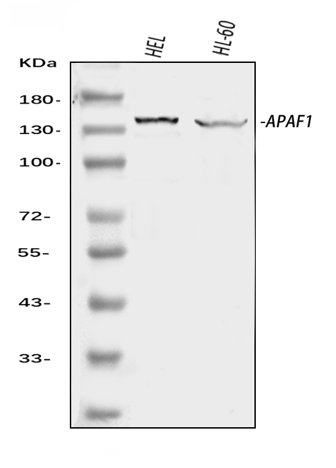
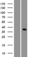
Mass (kDA):
141.84 kDA
Human | |
---|---|
Location: | 12q23.1 |
Sequence: | 12; NC_000012.12 (98645141..98735433) |
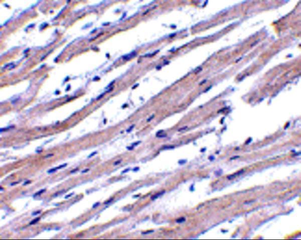
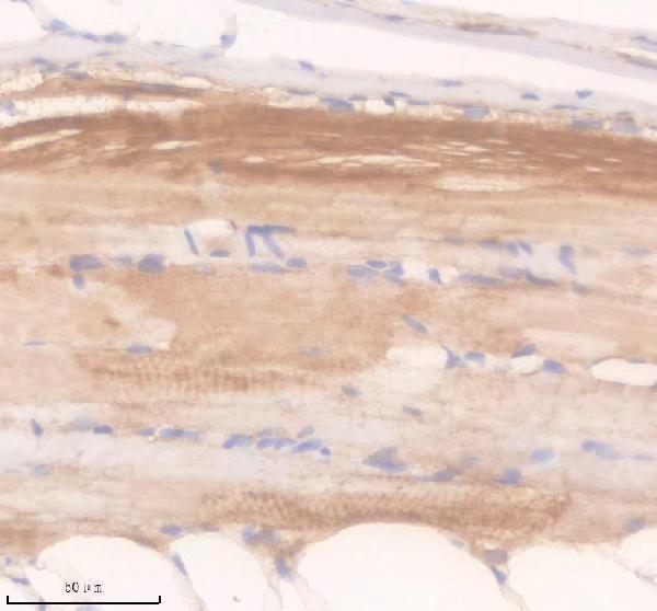
Ubiquitous. Highest levels of expression in adult spleen and peripheral blood leukocytes, and in fetal brain, kidney and lung. Isoform 1 is expressed in heart, kidney and liver.
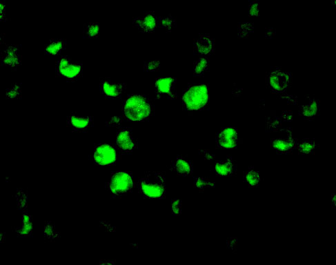
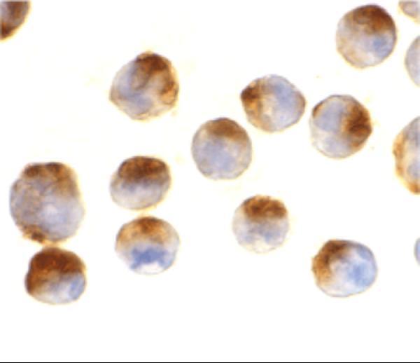
Cytoplasm.
In this article we will discuss the cSCC and ATP binding to Apaf1 markers. We will also discuss the ELISA platform and the assay kits for aspartate aminotransferase (AST).
cSCC can be a deadly form of cancer. However the mutational landscape of APAF1 has been linked to both immunocompetent patients and immunosuppressed ones. The APAF1 marker promotes the growth of prostate cancer through its Plexin B1 receptor, which has intrinsic GAP activity. A variety of tumors show unfavorable outcomes when Semaphorin-3C levels are high.
cSCC research is dominated by the APAF1 target gene. MiR186 inhibits APAF1 mRNA expression. A luciferase reporters assay revealed a direct target genes. Cell proliferation is facilitated when the APAF1 protein or mRNA are suppressed. Overexpression of SRCIN1 reverses miR-346's effects on A431 cells.
Researchers are currently investigating the mechanism of cancer cell progression and growth. These mutations are related to the RAS family which is made up small guanosinetriphosphate protein. RAS proteins regulate downstream proteins and gene expression to promote cell growth and differentiation.
Additional benefits may accrue from using DMBT1 as a therapeutic target for CSCC. It can inhibit apoptosis, migration, and invasion of CSCC cells. This is a new strategy to treat the illness. These findings are a step towards identifying new therapeutic options. While DMBT1 does not directly inhibit apoptosis, it is still an effective method of treatment for the disease.
This study investigated ATP-binding to Apaf-1 in rat's brain using surface plasmon resonance (SPR), a non-cell-free system that can also be used to study biomolecular interactions. The binding affinity of ZYZ-488 for Apaf-1 was assessed using a kinetics parameter called response unite (RU). The curve for cycle 6 was almost the same as the curve for cycle 7, which indicates good reproducibility. The kinetic parameters were calculated by subtracting the baseline response unit from AbsResp.
ATP binding is observed in Apaf-1-bound brain cells, both in mice and humans. Apaf-1 binds to cytochrome c in a Y-like configuration that promotes apoptosome assembly. However, Apaf-1 is also seen to be bound between WD40 b propellers, rendering them inactive.
This study also showed ZYZ-488 a novel ATP binder to Apaf-1 inhibitor, reduces the infarct size in MI mice. ZYZ-488 can also improve cardiac function, reduce the size of infarcts and attenuate histopathology changes that occur in MI. These results suggest a new therapeutic target for the treatment of myocardial infarction.
The Hsp70 mutants interfere with the activity of the caspases in a mitochondrial-initiated pathway. The Hsp70C-terminal mutant interacts with Apaf-1 for longer periods than Hsp70WT. The Hsp70 DXXD mutation has a higher cell protection than Hsp70WT. These mutants could prove to be useful antiapoptotic therapeutics.
Using an ELISA platform for the APAF-1 marker is a simple, reliable, and accurate way to determine the protein expression levels of this gene. Like other ELISA technologies the sensitivity of an ELISA test depends on the detection platform and the plate coating conditions. Sandwich ELISAs capture the target proteins by using an antibody precoated onto a liquid phase.
The APAF1 procaspase marker is a specific protein that is present throughout the body. It is also widely expressed in human tissues, but its role is not yet completely understood. It is pro-survival but has a complex role in the development and maintenance of the nervous system. This important protein can be studied using the APAF1 ELISA platform.
2E12 is an APAF1 mAb that recognizes a single 130-kD hematopoietic or epithelial protein. Apaf-1's HA-tagged protein is a transiently expressing Gene in 293T Cells. Apaf-1 can be recognized by the 2E12 mAb from many species. It is suitable for a wide variety applications.
Apaf-1 belongs to the caspase clan. It is an evolutionary-conserved process that removes undesirable cells from tissues. It can also lead to many other diseases. Caspases are involved in triggering apoptosis. Caspases-3, in particular, initiate a cascade involving proteolytic enzymes. Caspases then act as downstream regulators. The Apaf-1 proteins acts as adapters by activating caspases-9.
Cayman Aspartate Aminotransferase Colorimetric Activity Assay Kit provides a convenient means to measure the AST's activity. The method measures the rate that NADH oxidation occurs in a sample. This is accompanied by a decrease at 340 nm in absorbance. This decrease in absorbance corresponds to the activity of AST within the sample. The kit also contains lactate-dehydrogenase, which is used to prevent interference from endogenous Pyruvate found in serum.
The APAF1 marker, a biomarker for cancer, is widely used. It recruits caspase-9 to initiate cell death. This protein is also known to be Apoptosis-Protein (APO).
The APAF1 protein, a potent antigen, is highly expressed in cancer cells. This protein is controlled through the APAF1 genome. BosterBio: Best Uses of The APAF1Marker
PMID: 9267021 by Zou H., et al. Apaf-1, a human protein homologous to C. elegans CED-4, participates in cytochrome c-dependent activation of caspase-3.
PMID: 10441496 by Hahn C., et al. Three new types of Apaf-1 in mammalian cells.
*More publications can be found for each product on its corresponding product page