This website uses cookies to ensure you get the best experience on our website.


- Table of Contents
Use this guide to plan ATM staining in paraffin sections around its general nuclear tissue profile (HPA tissue IHC). Interpret goblet cell staining cautiously because HPA disregarded that signal (HPA tissue IHC).

Expected localisation, validated protocols, controls and antibodies — the at-a-glance facts below, then the full design guide.
| Expected localisation | Predominantly nuclear (HPA tissue IHC; UniProt) | |
| Staining pattern | General nuclear staining across cell types (HPA tissue IHC) | |
| Antigen retrieval | Tris-EDTA pH 9.0 HIER, 95–98 °C, 20 min (rule: nuclear antigen) | |
| Positive control | Adrenal gland+4 more · see all | |
| Negative control | None in HPA (detected in all 44 tissues); use no-primary + isotype controls |
| Fixation | Keep fixation consistent across paraffin sections. (standard IHC practice; not target-specific) | |
| Caveat | Goblet cell staining was disregarded (HPA tissue IHC) | |
| Regulation | Damage induces ATM autophosphorylation (UniProt) | |
| Isoform / epitope | No isoforms listed; chain spans residues 2–3056 (UniProt) |
The catalog antibody’s IHC-P protocol is followed by three published protocols for ATM staining in paraffin sections, including one that measures phosphorylated ATM.
| Sample | Tissue sections; selected-image fixative not specified (standard IHC workflow) |
| Fixation | Image fixative and duration unreported (datasheet A00014); verify before use. |
| Sectioning | 4–5 µm sections on charged slides (standard) |
| Deparaffinisation | Xylene, graded ethanol series to water (standard) |
| Antigen retrieval | Heat-induced epitope retrieval in Tris-EDTA buffer, pH 9.0, 20 min at 95–98 °C (standard rule: nuclear antigen) |
| Peroxidase block | 3% H2O2, 10 min, room temperature (standard) |
| Blocking | 10% normal serum of the secondary host, 30 min, room temperature (standard) |
| Primary antibody | Rabbit anti-ATM, 2.5 μg/mL (datasheet A00014) |
| Primary incubation | Overnight at 4 °C (standard) |
| Detection | HRP-polymer secondary, DAB chromogen 5–10 min (standard) |
| Counterstain | Hematoxylin, blue, dehydrate and mount (standard) |
| Expected result | ATM-positive staining in glandular cells of adrenal gland (HPA tissue IHC: High). HPA tissue profile: General nuclear expression. No signal in the no-primary control. |
ATM is primarily nuclear and has no transmembrane segment (UniProt Q13315: localization, topology). In paraffin sections, expect a general nuclear pattern, including high staining reported in adrenal glandular cells, bronchial respiratory epithelial cells and bone marrow hematopoietic cells (HPA: tissue IHC). HPA rates the tissue profile Supported, with medium consistency between antibody staining and RNA expression; its reported goblet cell staining was disregarded (HPA: tissue IHC reliability).
| Distinct nuclear staining in glandular, respiratory epithelial or hematopoietic cells. | This fits ATM's primary nuclear localization and HPA's high staining calls for adrenal glandular, bronchial epithelial and bone marrow hematopoietic cells (UniProt Q13315: localization; HPA: tissue IHC). Judge localization within the identified cells, alongside a negative detection control (standard IHC practice). |
| Predominantly membrane-bound staining, with little or no nuclear signal. | A membrane-dominant pattern conflicts with ATM's primary nuclear location and lack of a transmembrane segment, so assess it as possible nonspecific staining (UniProt Q13315: localization, topology). Cytoplasmic signal alone is less decisive: UniProt also records vesicle, centrosome and peroxisome locations (UniProt Q13315: localization). |
| Strong staining in an unexpected cell population, especially if nuclei are unstained. | Consider cross-reactivity or endogenous chromogenic detection activity and check a negative detection control (standard IHC practice). ATM is broadly distributed, so an unlisted cell type is not automatically wrong; HPA reports low tissue specificity and provides no negative tissue call here (HPA: tissue IHC). |
| Diffuse color across tissue, extracellular spaces or slide background. | Poorly localized color cannot establish ATM expression (standard IHC practice). Compare with a negative detection control, then review blocking, washes and detection conditions (standard IHC practice); do not score diffuse background as HPA's general nuclear pattern (HPA: tissue IHC). |
| No nuclear signal in a section selected from an HPA high-staining tissue. | First check cell identity, tissue preservation and run controls, antibody application, detection and counterstain (standard IHC practice). Absence in one section does not establish biological loss: HPA's tissue profile has medium antibody–RNA consistency, and its high call applies to specified cells rather than every cell in that tissue (HPA: tissue IHC). |
| Tissue and cell selection | HPA reports high staining in adrenal glandular cells, bronchial respiratory epithelial cells and bone marrow hematopoietic cells, while caudate glial cells and ovarian stroma cells are listed as low (HPA: tissue IHC). Select and score the named cells; these calls do not define a universal negative control (HPA: tissue IHC). |
| Goblet cell signal | HPA lists high goblet cell staining in appendix, colon and duodenum but explicitly says goblet cell staining was observed and disregarded (HPA: tissue IHC). Do not use goblet cell color alone to validate an ATM positive result (HPA: tissue IHC reliability). |
| Antibody evidence | CAB000102 and CAB080442 have Supported IHC status; HPA067142 has no IHC status in the supplied antibody record (HPA: antibodies). An ICC Enhanced designation for HPA067142 does not establish its performance in paraffin IHC (HPA: antibodies). |
| Subcellular exceptions | UniProt describes ATM as primarily nuclear, with vesicle, centrosome and ROS-associated peroxisome localization also recorded (UniProt Q13315: localization). These exceptions warrant context when assessing cytoplasmic staining; they do not make a diffuse chromogenic haze diagnostic of ATM (UniProt Q13315: localization; standard IHC practice). |
| IF/ICC Q&A: where should ATM appear? | Mainly in nucleoplasm; HPA also reports vesicles and Golgi localization as uncertain (HPA: subcellular ICC-IF). This IF observation is context for localization, not an IHC staining or antibody-validation claim (HPA: subcellular ICC-IF; HPA: antibodies). |
| Situation | Likely cause | Next action |
|---|---|---|
| Expected high-staining cell population has no nuclear signal. | The selected cells may be absent from the section, or the staining run may have failed (HPA: tissue IHC; standard IHC practice). | Confirm cell identity, then check the positive run control, antibody application and chromogenic detection (standard IHC practice). Use HPA's high call as a reference for its specified cells, not for every cell in the tissue (HPA: tissue IHC). |
| Signal is predominantly at cell borders or membranes. | This conflicts with primary nuclear ATM localization and its lack of a transmembrane segment (UniProt Q13315: localization, topology). | Check whether nuclei stain in the same cells; compare with a negative detection control and reassess antibody specificity before scoring membrane color (standard IHC practice). |
| Color appears broadly in an unexpected cell type. | Cross-reactivity or endogenous detection activity is possible (standard IHC practice), but HPA reports low tissue specificity and supplies no negative tissue category here (HPA: tissue IHC). | Identify the stained cells, compare their compartment with the expected nuclear pattern, and inspect a negative detection control (HPA: tissue IHC; standard IHC practice). Avoid calling the cell type ATM-negative from this record alone (HPA: tissue IHC). |
| Goblet cells show strong color but nearby nuclei do not. | HPA explicitly disregarded the observed goblet cell staining, despite listing high levels in appendix, colon and duodenum (HPA: tissue IHC). | Do not treat goblet cell color as confirmation; evaluate nuclear staining in identified cells and check detection controls (HPA: tissue IHC; standard IHC practice). |
| The whole section has weak, diffuse chromogenic color. | Background from blocking, washing or detection can obscure cell boundaries (standard IHC practice). Such color does not resolve HPA's general nuclear pattern (HPA: tissue IHC). | Review blocking, wash steps and detection conditions, then compare with a negative detection control before assigning a cell-level score (standard IHC practice). |
| Nuclear staining varies between sections or antibody choices. | HPA reports only medium consistency between antibody staining and RNA expression; the supplied antibody records also have different application statuses (HPA: tissue IHC reliability; HPA: antibodies). | Check that the antibody has IHC support, compare the same identified cell population across sections, and interpret discordance with tissue controls (HPA: antibodies; standard IHC practice). Target-specific fixation sensitivity is unreported in the supplied sources (HPA: tissue IHC; UniProt Q13315). |


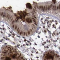

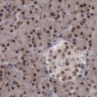


Comprehensive Human Protein Atlas IHC scoring per tissue (reliability: Supported — Medium consistency between antibody staining and RNA expression data. Goblet cell staining observed and disregarded.). Rows are taken directly from the HPA tissue chart — click any row's HPA link to view the source.
| Tissue | Cell type | Level | Evidence | Source |
|---|---|---|---|---|
| Adrenal gland | Glandular cells | High | Protein (IHC) | HPA → |
| Appendix | Goblet cells | High | Protein (IHC) | HPA → |
| Bone marrow | Hematopoietic cells | High | Protein (IHC) | HPA → |
| Breast | Glandular cells | High | Protein (IHC) | HPA → |
| Bronchus | Respiratory epithelial cells | High | Protein (IHC) | HPA → |
| Tissue | Cell type | Level | Evidence | Source |
|---|---|---|---|---|
| None in HPA: ATM is detected in all 44 scored tissues. Use a no-primary (secondary-only) and an isotype control instead. | ||||
Troubleshoot ATM staining in paraffin sections by checking retrieval, nuclear localisation, controls and cell-based scoring before interpreting chromogenic signal.
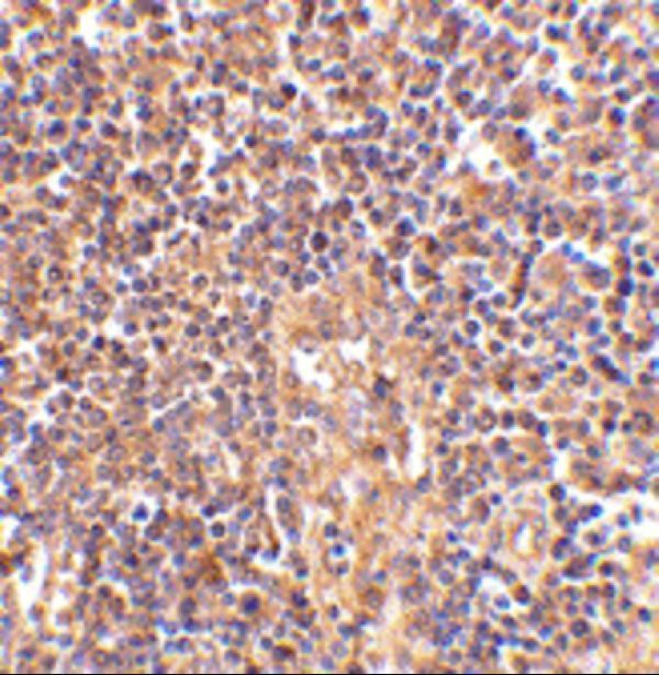
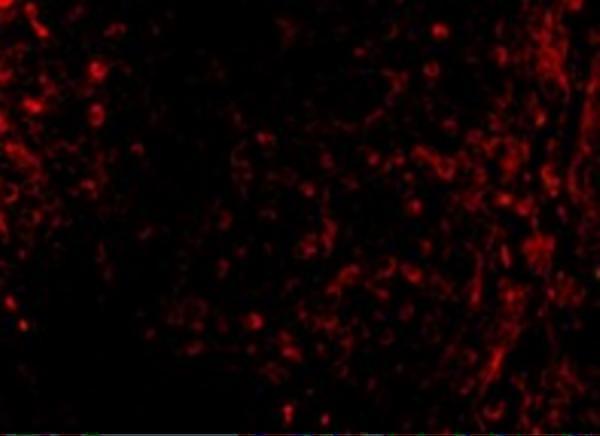
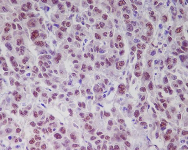
Human-reactive ATM antibodies have IHC images from lymph node, breast cancer, and liver (catalog IHC image captions); IF images show human lymph node cells and HeLa cells (catalog IF image captions).
A00014 has pictured IHC in human lymph node and IF in human lymph node cells; M00014 has pictured IHC in paraffin-embedded human breast cancer and IF in HeLa cells (catalog image captions). P00014-1 has pictured IHC in paraffin-embedded human liver and is listed for IF without an IF image (catalog image caption and applications).
Which to pick: For tissue IHC, choose A00014 for human lymph node (A00014 IHC image caption), M00014 for paraffin-embedded human breast cancer (M00014 IHC image caption), or P00014-1 when detecting phospho-ATM S1981 in paraffin-embedded human liver (P00014-1 title and IHC image caption); the captions do not report a fixative. For IF/ICC, M00014 is a monoclonal antibody listed for both applications, with pictured IF in HeLa cells (catalog clone, applications, and IF image caption); A00014 is listed for IF and has pictured IF in human lymph node cells (catalog applications and IF image caption). For human, mouse, or rat IF/ICC, consider monoclonal M00014-1; it is listed for those species and applications but has no IHC application or IHC image (catalog reactivity, clone, applications, and image list).