This website uses cookies to ensure you get the best experience on our website.


- Table of Contents
Real validated CASP9 Western blot protocols, expected-band and isoform facts, troubleshooting for weak or shifted signal, and recommended anti-CASP9 WB antibodies. Everything you need to plan the experiment before you commit precious samples.
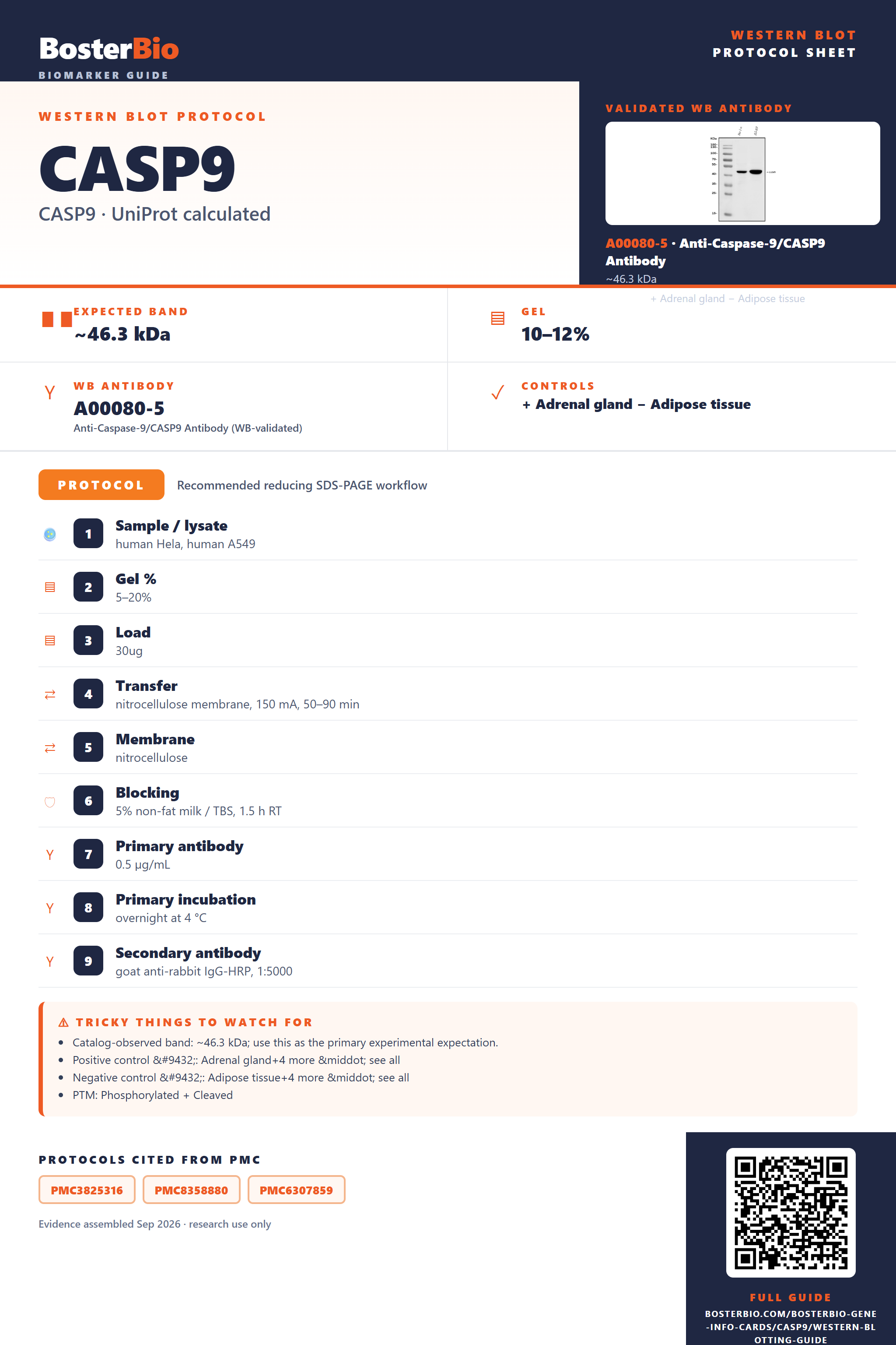
Expected bands, validated protocols, controls and antibodies — the at-a-glance facts below, then the full design guide.
| Expected band | ~46.3 kDa | |
| Observed band | 46 kDa | |
| Gel | 10–12% | |
| Positive control | Adrenal gland+4 more · see all | |
| Negative control | Adipose tissue+4 more · see all |
| PTM | Phosphorylated + Cleaved | |
| Caveat | Proteolytic cleavage | |
| Regulation | Apoptosis | |
| Isoform | 4 isoform(s) |
Literature-validated Western blot parameters for CASP9 — gel percentage, transfer, blocking, antibody incubation and detection, extracted from published methods.
| Sample / lysate | human Hela, human A549 |
| Gel % | 5–20% |
| Load | 30ug |
| Transfer | nitrocellulose membrane, 150 mA, 50–90 min |
| Membrane | nitrocellulose |
| Blocking | 5% non-fat milk / TBS, 1.5 h RT |
| Primary antibody | 0.5 µg/mL |
| Primary incubation | overnight at 4 °C |
| Secondary antibody | goat anti-rabbit IgG-HRP, 1:5000 |
| Wash | TBS-0.1% Tween, 3 × 5 min |
| Detection | ECL |
| Exposure / imaging | Tanon 5200 |
| Observed band | 46 kDa |
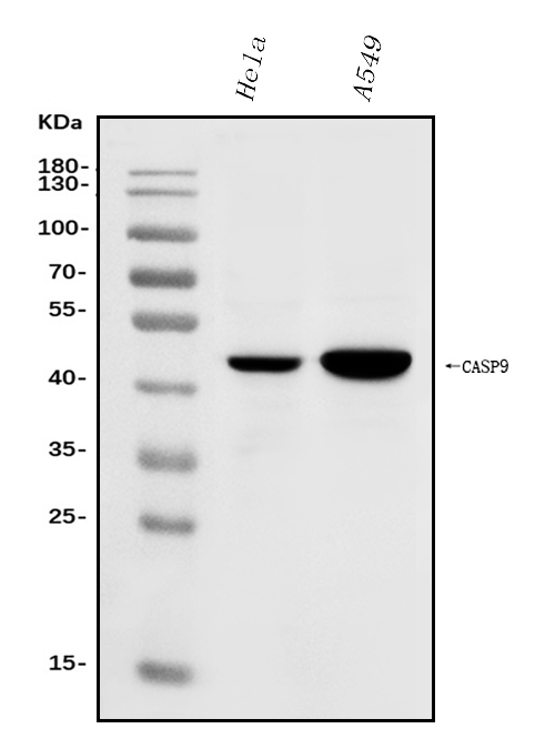
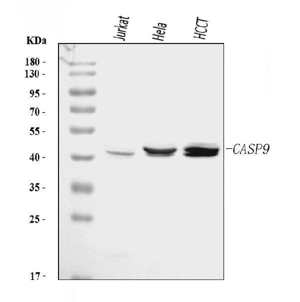
CASP9 has a 46.3 kDa backbone and typically runs at 46 kDa as the zymogen, with lower bands arising from proteolytic processing to p35 and p10.
| Single band at 46 kDa | full-length caspase-9 zymogen close to the 46.3 kDa predicted backbone |
| Band near 35 kDa | processed caspase-9 p35 cleavage product from zymogen maturation |
| Smaller fragment near 10 kDa | processed caspase-9 p10 small subunit |
| Multiple bands between full-length and smaller fragments | coexistence of precursor, processed subunits, and splice isoforms 1 to 4 |
| 46.3 kDa predicted backbone | supports a full-length band around 46 kDa |
| zymogen propeptide and proteolytic maturation | creates smaller mature cleavage products than the precursor |
| p35 subunit formation | produces a prominent lower band near 35 kDa after processing |
| p10 subunit formation | produces a much smaller fragment band |
| alternative splicing isoforms 1, 2, 3, and 4 | can generate additional bands of different relative sizes |
| Situation | Likely cause | Next action |
|---|---|---|
| Band higher than expected | full-length zymogen is being compared with a processed p35 expectation | distinguish whether your antibody should detect precursor 46 kDa, processed p35, or both |
| Band lower than expected | caspase-9 has undergone proteolytic processing from the zymogen into smaller subunits | look for the accompanying full-length 46 kDa band and expected processed fragments such as p35 |
| Multiple bands | mixture of precursor, processed products, and splice isoforms 1 to 4 | interpret bands in the context of activation state and confirm which isoforms or cleavage forms your antibody recognizes |
| Weak or no signal | the detected epitope may be absent from the dominant processed form or from some isoforms | check the antibody immunogen region and use a lysate expected to contain full-length CASP9 if targeting the 46 kDa species |
| Fragments below expected size | normal caspase-9 maturation generates smaller subunits including p35 and p10 | treat lower bands as plausible processing products rather than nonspecific bands if they accompany CASP9 precursor loss |


Comprehensive Human Protein Atlas IHC scoring per tissue. Rows are taken directly from the HPA tissue chart — click any row's HPA link to view the source.
| Tissue | Cell type | Level | Evidence | Source |
|---|---|---|---|---|
| Adrenal gland | glandular cells | Medium | Protein (IHC) | HPA → |
| Appendix | glandular cells | Medium | Protein (IHC) | HPA → |
| Bone marrow | hematopoietic cells | Medium | Protein (IHC) | HPA → |
| Breast | glandular cells | Medium | Protein (IHC) | HPA → |
| Bronchus | respiratory epithelial cells | Medium | Protein (IHC) | HPA → |
| Tissue | Cell type | Level | Evidence | Source |
|---|---|---|---|---|
| Adipose tissue | adipocytes | Low | Protein (IHC) | HPA → |
| Esophagus | squamous epithelial cells | Low | Protein (IHC) | HPA → |
| Liver | cholangiocytes | Low | Protein (IHC) | HPA → |
| Lung | alveolar cells | Low | Protein (IHC) | HPA → |
| Oral mucosa | squamous epithelial cells | Low | Protein (IHC) | HPA → |
Deeper troubleshooting and optimisation questions for CASP9, answered from its protein features.
BosterBio's CASP9 antibodies are among the best-performing WB antibodies on the market — well cited, thoroughly validated, and orthogonally cross-validated against negative tissues and complementary methods.
These recommended anti-CASP9 Western blot antibodies represent Boster’s best-performing, well-cited, thoroughly validated options, with orthogonal cross-validation supported by negative-tissue and complementary-method evidence for confident band assignment.
Which to pick: For CASP9, all listed SKUs include WB validation images. Start with A00080-5, A00080-6, or A00080-8 for direct CASP9 WB examples; consider PA1595 or Picoband PB9332 if you want additional lysate-panel validation.