Click image to see more details
-

-
-
-
-
+1
Product Info Summary
| SKU: | PA1595 |
|---|---|
| Size: | 100 μg/vial |
| Reactive Species: | Human |
| Host: | Rabbit |
| Application: | IHC, WB |
Customers Who Bought This Also Bought

Product info
Product Name
Anti-Caspase 9/CASP9 Antibody Picoband®
SKU/Catalog Number
PA1595
BA3974 is an alternative SKU for this antibody, used in previous lots.
Size
100 μg/vial
Form
Lyophilized
Description
Boster Bio Anti-Caspase 9/CASP9 Antibody catalog # PA1595. Tested in IHC, WB applications. This antibody reacts with Human. The brand Picoband indicates this is a premium antibody that guarantees superior quality, high affinity, and strong signals with minimal background in Western blot applications. Only our best-performing antibodies are designated as Picoband, ensuring unmatched performance.
Storage & Handling
Store at -20˚C for one year from date of receipt. After reconstitution, at 4˚C for one month. It can also be aliquotted and stored frozen at -20˚C for six months. Avoid repeated freeze-thaw cycles.
Cite This Product
Anti-Caspase 9/CASP9 Antibody Picoband® (Boster Biological Technology, Pleasanton CA, USA, Catalog # PA1595)
Host
Rabbit
Contents
Each vial contains antibody formulated with stabilizing components, 0.9mg NaCl, 0.2mg Na2HPO4, 0.05mg Thimerosal, 0.05mg NaN3.
*This antibody is supplied in a stabilized formulation.
Compatibility with conjugation reactions depends on the chemistry of the conjugation method used.
For conjugation methods that are not compatible with the stabilizing components present in this formulation, a carrier-free antibody format is required.
Clonality
Polyclonal
Isotype
Rabbit IgG
Immunogen
A synthetic peptide corresponding to a sequence in the middle region of human Caspase-9.
Cross-reactivity
No cross-reactivity with other proteins
Reactive Species
PA1595 is reactive to CASP9 in Human
Observed Molecular Weight
35 kDa
Calculated molecular weight
46.3 kDa
Background of CASP9
CASP9 (CASPASE9), also called APAF3, is an initiator caspase, encoded by the CASP9 gene. The CASP9 gene is mapped to chromosome 1p36.3-p36.1 by FISH. CASP9 is identified as a member of the caspase family that participates in caspase-3 activation in vitro. And it also regarded as the most upstream member of the apoptotic protease cascade that is triggered by cytochrome c and dATP. Its genomic coordinates (GRCh37) is 1:15,818,768-15,851,284. The crystal structure of CASP9 is complex with the BIR3 in an inhibitory domain of XIAP at 2.4-angstrom resolution and the CASP9 gene contains 9 exons and spans approximately 35 kb of genomic DNA. Caspase-9 and APAF1 bind to each other via their respective NH2-terminal CED-3 homologous domains in the presence of cytochrome c and dATP, an event that leads to caspase-9 activation. CASP9 activity increases dramatically upon association with the apoptosome complex. And the majority of Casp9 knockout mice died perinatally with a markedly enlarged and malformed cerebrum caused by reduced apoptosis during brain development.
Antibody Validation
Boster validates all antibodies on WB, IHC, ICC, Immunofluorescence, and ELISA with known positive control and negative samples to ensure specificity and high affinity, including thorough antibody incubations.
Application & Images
Applications
PA1595 is guaranteed for IHC, WB Boster Guarantee
Recommend Dilution
| Application | Dilution | Species |
|---|---|---|
| Immunohistochemistry (Paraffin-embedded Section) | 0.5-1μg/ml | Human |
| Western blot | 0.1-0.5μg/ml | Human |
Tested application
Suggested blocking solution with 5% non-fat milk or BSA; (*)Recommended protein loading: 20-40 µg per lane
Use TE buffer pH 9.0 for antigen retrieval; (*) citrate buffer pH 6.0 is an alternative.
Validation Images & Assay Conditions
Click image to see more details
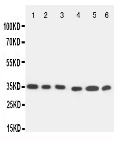
Anti-Caspase-9 antibody, PA1595, Western blotting
All lanes: Anti Caspase-9 (PA1595) at 0.5ug/ml
Lane 1: SMMC Whole Cell Lysate at 40ug
Lane 2: MCF-7 Whole Cell Lysate at 40ug
Lane 3: CEM Whole Cell Lysate at 40ug
Lane 4: JURKAT Whole Cell Lysate at 40ug
Lane 5: RAJI Whole Cell Lysate at 40ug
Lane 6: HELA Whole Cell Lysate at 40ug
Predicted bind size: 35KD
Observed bind size: 35KD

Click image to see more details
Anti-Caspase-9 antibody, PA1595, IHC(P)
IHC(P): Human Tonsil Tissue

Click image to see more details
PPM-18 triggers apoptotic cell death in bladder cancer cells. (A) T24 and EJ cells were treated with the indicated concentration of PPM-18, and stained with annexin V-FITC and PI. The apoptotic effect of PPM-18 on T24 and EJ cells was measured by flow cytometry. (B) Western blotting showed the effect of PPM-18 on the expression of cleaved caspase-9, 3, and PARP in T24 and EJ cells. (C) T24 and EJ cells were exposed to 10 μM PPM-18 for 24 h, and stained with JC-1 fluorescent probe. The effect of PPM-18 on the alteration of mitochondria membrane potential was analyzed by flow cytometry. (D) Western blotting displayed the effect of PPM-18 on expression of BAX and BCL-2 in T24 and EJ cells. (E) and (F) The effect of Z-VAD-FMK on PPM-18 reduced the viability of T24 and EJ cells, or PPM-18 triggered apoptosis in T24 cells. Cells treated with 15 μM PPM-18 combined with or without 20 μM Z-VAD-FMK for 24 h, and cell viability and apoptosis were respectively measured by MTS assay and flow cytometry. Data are presented as the mean ± SD of at least three independent experiments. * p < 0.05, ** p < 0.01, and *** p < 0.001 vs. the control group, or vs. PPM-18+Z-VAD-FMK.
Index in PubMed under a CC BY license. PMID: 34305598

Click image to see more details
PPM-18 induces AMPK-dependent autophagy and apoptotic cell death in bladder cancer cells. (A) and (B) T24 and EJ cells were exposed to the indicated concentration of PPM-18 for 24 h, and the expression of phospho-AMPK, AMPK, phospho-mTORC1, phospho-P70S6K, phospho-PI3K, PI3K, phospho-AKT, and AKT were analyzed by western blot. (C) and (D) Western blot showed the effect of AMPK siRNA or 5 μM Compound C on the phosphorylation of AMPK, mTORC1, and P70S6K, and the expression of LC3B and cleaved caspase-9, 3 in PPM-18–treated T24 cells. (E) and (F) MTS assay and flow cytometry demonstrated that treatment with Compound C affected PPM-18–reduced viability of T24 cells and PPM-18–induced apoptosis in T24 cells. Data are presented as the mean ± SD of at least three independent experiments. *** p < 0.001 vs. the control group, or vs. PPM-18+Compund C.
Index in PubMed under a CC BY license. PMID: 34305598

Click image to see more details
PPM-18 stimulates autophagy that promotes apoptosis in bladder cancer cells. (A) T24 and EJ cells were treated with various concentrations of PPM-18 for 24 h. Then cells were harvested for western blotting to detect the expression of p62 and LC3B, two autophagy indicators. (B) The formation of autophagosomes in T24 and EJ cells treated with or without 10 μM PPM-18 for 18 h under the transmission electron microscope. Arrows indicated the autophagosomes containing intact and degraded cellular debris. Scale bar: left: 5.0 μm, middle: 2.0 μm, and right: 1.0 μm. (C) Western blot showed the effect of 5 mM 3-MA, a typical autophagy inhibitor, on the expression of p62, LC3B, and cleaved caspase-9, 3 in PPM-18–treated T24 and EJ cells. (D) and (E) T24 and EJ cells were treated with 15 μM PPM-18 combined with or without 5 mM 3-MA, and the effect of 3-MA on PPM-18–reduced viability or PPM-18–triggered apoptosis in T24 cells and EJ cells were measured by MTS assay and flow cytometry, respectively. (F) T24 and EJ cells were treated with 15 μM PPM-18 combined with or without 10 μM rapamycin, a typical autophagy agonist, and the effect of rapamycin on PPM-18–induced autophagy and apoptosis were evaluated by western blot. (G) The MTS assay demonstrated that rapamycin affected PPM-18–reduced viability in T24 and EJ cells. (H) Flow cytometry displayed the effect of rapamycin on PPM-18–triggered apoptosis in T24 and EJ cells. Data are presented as the mean ± SD of at least three independent experiments. ** p < 0.01 and *** p < 0.001 vs. the control group, or vs. PPM-18+3-MA. * p < 0.05 and *** p < 0.001 vs. PPM-18+Rapa.
Index in PubMed under a CC BY license. PMID: 34305598
Specific Publications For Anti-Caspase 9/CASP9 Antibody Picoband® (PA1595)
Loading publications
Recommended Resources
Here are featured tools and databases that you might find useful.
- Boster's Pathways Library
- Protein Databases
- Bioscience Research Protocol Resources
- Data Processing & Analysis Software
- Photo Editing Software
- Scientific Literature Resources
- Research Paper Management Tools
- Molecular Biology Software
- Primer Design Tools
- Bioinformatics Tools
- Phylogenetic Tree Analysis
Customer Reviews
Have you used Anti-Caspase 9/CASP9 Antibody Picoband®?
Share your experimental results or join a short interview to earn up to $1,000 in product credits or other rewards.
0 Reviews For Anti-Caspase 9/CASP9 Antibody Picoband®
Customer Q&As
Have a question?
Find answers in Q&As, reviews.
Can't find your answer?
Submit your question
5 Customer Q&As for Anti-Caspase 9/CASP9 Antibody Picoband®
Question
Do you have any data about PA1595 for use in IF with PFA fixed cells?
Verified customer
Asked: 2020-06-19
Answer
Our lab hasn't performed IF test with PFA fixed cells using the Anti-Caspase 9/CASP9 Antibody (PA1595). Please run a pilot test to see if it would work.
Boster Scientific Support
Answered: 2020-06-22
Question
We have observed staining in human adrenal tissue. Are there any suggestions? Is anti-Caspase 9/CASP9 antibody supposed to stain adrenal tissue positively?
Verified Customer
Verified customer
Asked: 2019-09-24
Answer
From literature adrenal tissue does express CASP9. From Uniprot.org, CASP9 is expressed in adrenal tissue, t-cell, stomach cancer, kidney, eye lymph, cervix carcinoma, cervix carcinoma erythroleukemia, among other tissues. Regarding which tissues have CASP9 expression, here are a few articles citing expression in various tissues:
Cervix carcinoma, Pubmed ID: 18669648, 20068231
Cervix carcinoma, and Erythroleukemia, Pubmed ID: 23186163
Eye, and Lymph, Pubmed ID: 15489334
Kidney, Pubmed ID: 14702039
Stomach cancer, Pubmed ID: 9890966
T-cell, Pubmed ID: 8900201
Boster Scientific Support
Answered: 2019-09-24
Question
We are currently using anti-Caspase 9/CASP9 antibody PA1595 for human tissue, and we are content with the WB results. The species of reactivity given in the datasheet says human. Is it true that the antibody can work on dog tissues as well?
Verified Customer
Verified customer
Asked: 2018-12-31
Answer
The anti-Caspase 9/CASP9 antibody (PA1595) has not been validated for cross reactivity specifically with dog tissues, though there is a good chance of cross reactivity. We have an innovator award program that if you test this antibody and show it works in dog you can get your next antibody for free. Please contact me if I can help you with anything.
Boster Scientific Support
Answered: 2018-12-31
Question
We ordered your anti-Caspase 9/CASP9 antibody for IHC on kidney in the past. I am using human, and We are going to use the antibody for WB next. I was wanting to use examining kidney as well as cervix carcinoma in our next experiment. Could you please give me some suggestion on which antibody would work the best for WB?
Verified Customer
Verified customer
Asked: 2017-12-29
Answer
I have checked the website and datasheets of our anti-Caspase 9/CASP9 antibody and it seems that PA1595 has been validated on human in both IHC and WB. Thus PA1595 should work for your application. Our Boster satisfaction guarantee will cover this product for WB in human even if the specific tissue type has not been validated. We do have a comprehensive range of products for WB detection and you can check out our website bosterbio.com to find out more information about them.
Boster Scientific Support
Answered: 2017-12-29
Question
I am interested in using your anti-Caspase 9/CASP9 antibody for activation of caspases through apoptosome-mediated cleavage studies. Has this antibody been tested with western blotting on smmc whole cell lysate? We would like to see some validation images before ordering.
C. Bhatt
Verified customer
Asked: 2014-10-16
Answer
We appreciate your inquiry. This PA1595 anti-Caspase 9/CASP9 antibody is tested on smmc whole cell lysate, cem whole cell lysate, jurkat whole cell lysate, raji whole cell lysate, hela whole cell lysate, human tonsil tissue. It is guaranteed to work for IHC, WB in human. Our Boster guarantee will cover your intended experiment even if the sample type has not been be directly tested.
Boster Scientific Support
Answered: 2014-10-16
