CDK6 / Cyclin-dependent kinase 6 · IHC design guide

Design Immunohistochemistry for CDK6

Plan chromogenic CDK6 IHC around cytoplasmic staining that can also include nuclei (HPA tissue IHC). Use the tissue profile to select controls and score staining by cell type and compartment.

Evidence assembled Oct 2026 · For research use; verify linked source records and product datasheet before use
Immunohistochemistry protocol sheet for CDK6 (IHC for CDK6): expected localisation Cytoplasmic, sometimes nuclear (HPA tissue IHC), antibody PB9995, validated IHC image, and IHC protocol steps
Printable CDK6 IHC protocol sheet — expected localisation Cytoplasmic, sometimes nuclear (HPA tissue IHC), antibody PB9995, controls and protocol steps. Open the full CDK6 IHC guide →

CDK6 Immunohistochemistry Experimental Design Guide

Expected localisation, validated protocols, controls and antibodies — the at-a-glance facts below, then the full design guide.

Must know before staining
Expected localisation Cytoplasmic, sometimes nuclear (HPA tissue IHC)
Staining pattern Marrow cells highest; cytoplasmic, sometimes nuclear (HPA tissue IHC)
Antigen retrieval Citrate pH 6 HIER, heat-mediated (datasheet PB9995)
Positive control ⓘ Duodenum+4 more · see all
Negative control ⓘ Caudate+4 more · see all
Important caveats
Reasons your staining may differ from the expected pattern.
Fixation Keep fixation consistent across sections (standard IHC practice; not target-specific)
Caveat Staining and RNA show medium consistency (HPA tissue IHC)
Regulation Reduced during differentiation (UniProt)
Isoform / epitope No isoforms; one 1–326 chain, no transmembrane segment (UniProt)
Section 1

Recommended CDK6 IHC & IF Protocols

The catalog antibody’s IHC-P protocol (datasheet PB9995) is accompanied by two published CDK6 IHC protocols (PMC10878183; PMC4144705).

Recommended immunohistochemistry (IHC-P) protocol parameters
SampleParaffin-embedded human intestinal cancer tissues; fixative not specified (datasheet PB9995)
FixationImage fixative and duration unreported (datasheet PB9995); verify before use.
Sectioning4–5 µm sections on charged slides (standard)
DeparaffinisationXylene, graded ethanol series to water (standard)
Antigen retrievalHeat retrieval: Citrate pH 6, 20 min (datasheet PB9995)
Peroxidase block3% H2O2, 10 min, room temperature (standard)
Blocking10% goat serum (datasheet PB9995)
Primary antibodyRabbit anti-CDK6, 0.5-1μg/ml recommended; image 2μg/ml (datasheet PB9995)
Primary incubationOvernight at 4 °C (datasheet PB9995)
DetectionStreptavidin-biotin complex (SABC), DAB chromogen (datasheet PB9995)
CounterstainHematoxylin, blue, dehydrate and mount (standard)
Expected resultCDK6-positive staining in paneth cells of duodenum (HPA tissue IHC: High). HPA tissue profile: Cytoplasmic expression in several tissues, which in cases was combined with nuclear expression. Highest expression in bone marrow cells. No signal in the no-primary control.
💡Decision noteStart with heat-mediated citrate retrieval at pH 6 (datasheet PB9995); one published protocol specifies microwave heating at 800 W for 25 min (PMC10878183).
Section 2

What Is the Expected CDK6 Staining Pattern?

CDK6 is a non-membrane protein found in the nucleus and cytoplasm (UniProt Q00534 topology and subcellular location). In tissue IHC, expect cytoplasmic staining in several tissues, sometimes with nuclear staining; bone marrow cells show the highest expression in HPA’s profile (HPA tissue IHC). HPA rates tissue staining “Enhanced,” with medium consistency between antibody staining and RNA expression (HPA tissue IHC).

What am I looking at on my slide?
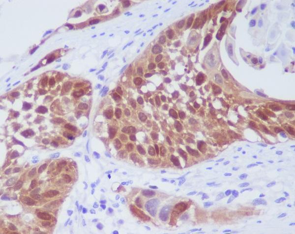
Cytoplasmic staining, sometimes accompanied by nuclear staining, in expected cell populations (HPA tissue IHC).This fits HPA’s tissue pattern (HPA tissue IHC). High staining in duodenal Paneth cells, placental cytotrophoblasts or small-intestinal enterocytes offers a useful positive reference (HPA tissue IHC). Score the two compartments separately; nuclear staining is plausible because CDK6 also localizes to the nucleus (UniProt Q00534 subcellular location).
Predominantly cell-surface or extracellular staining, without a convincing intracellular pattern.That distribution warrants an artefact check: CDK6 has no transmembrane segment, and its reported locations are intracellular (UniProt Q00534 topology and subcellular location). Recheck tissue morphology and the detection control before interpreting a sharp outline or extracellular deposit as CDK6 (general IHC practice).
Strong staining in an HPA “Not detected” population, such as heart cardiomyocytes (HPA tissue IHC).Investigate antibody cross-reactivity or endogenous detection activity (general IHC practice). “Not detected” describes HPA’s observation; it does not prove that every specimen must be negative (HPA tissue IHC). Compare a tissue-matched detection control and a known-positive population before assigning specificity (general IHC practice).
Uniform color over cells, stroma and empty areas, with poorly defined boundaries.A diffuse deposit weakens compartment and cell-type interpretation (general IHC practice). Compare a no-primary control, then assess blocking, washes and chromogen development (general IHC practice). HPA’s reported CDK6 pattern is cellular, chiefly cytoplasmic and sometimes nuclear (HPA tissue IHC).
No staining in a known-positive Paneth-cell, cytotrophoblast or enterocyte sample (HPA tissue IHC).Target-specific fixation effects are not established by the supplied assay evidence. Verify with a matched IHC source before attributing a result to fixation.
💡Expected CDK6 appearanceA convincing positive shows defined intracellular, chiefly cytoplasmic staining, sometimes nuclear, in the expected cells; Paneth cells, cytotrophoblasts and small-intestinal enterocytes are reported High by HPA, whereas broad surface outlines or diffuse deposits warrant control checks (HPA tissue IHC; UniProt Q00534 topology; general IHC practice).
How each factor affects the staining
Cell population and intensityHPA reports High staining in duodenal Paneth cells, placental cytotrophoblasts and small-intestinal enterocytes; bone-marrow hematopoietic cells are listed as Medium despite the profile’s “highest expression” description (HPA tissue IHC). Identify the cell population before comparing intensities (general IHC practice).
Antibody evidenceBoth listed antibodies, HPA002637 and CAB004363, have “Enhanced” IHC validation, a category supported by independent antibodies or orthogonal data (HPA antibody validation). This supports pattern assessment but does not make every stained cell or compartment specific; interpret the slide with controls (general IHC practice).
Protein location and processingCDK6 has no transmembrane segment, signal peptide or propeptide, and the recorded chain spans residues 1–326 (UniProt Q00534 topology and processing). These features support an intracellular expectation; they do not identify an antibody epitope or establish how retrieval affects staining (UniProt Q00534; general IHC practice).
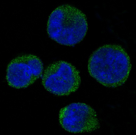
IF/ICC Q: Where should signal appear?A: HPA’s cell-image summary places CDK6 mainly in the nucleoplasm, with additional cytosolic signal; both locations are supported (HPA subcellular ICC-IF). Use that as a localization cross-check, while interpreting paraffin tissue against HPA’s chiefly cytoplasmic IHC profile (HPA tissue IHC).
Why is my staining missing, weak or wrong?
SituationLikely causeNext action
A positive-reference section shows no signal.The run may have failed, or the expected cell population may be absent from the section (general IHC practice).Confirm Paneth cells, cytotrophoblasts or small-intestinal enterocytes are present, then check primary-antibody application, retrieval and detection controls (HPA tissue IHC; general IHC practice). No CDK6-specific retrieval condition is supplied.
Nuclear signal appears alongside cytoplasmic signal.Both compartments are reported for CDK6; mixed staining can be expected (HPA tissue IHC; UniProt Q00534 subcellular location).Record nuclear and cytoplasmic staining separately and compare their cell distribution with morphology and controls (general IHC practice). Do not reject an otherwise credible result solely for nuclear signal (HPA tissue IHC).
A nominally negative cell population stains strongly.Possible cross-reactivity or endogenous detection activity requires investigation (general IHC practice); HPA’s “Not detected” category is an observed reference, not an absolute biological exclusion (HPA tissue IHC).Run a tissue-matched no-primary detection control and compare a known-positive cell population in the same run (general IHC practice; HPA tissue IHC).
Brown signal blankets the section and obscures cell boundaries.Background from detection, inadequate washing or excessive chromogen development may obscure localization (general IHC practice).Inspect a no-primary control; review blocking, washes and development time, then reassess whether signal follows cells and intracellular compartments (general IHC practice; HPA tissue IHC).
Only membrane-like rims or extracellular material stain.That pattern conflicts with the reported intracellular locations and lack of a transmembrane segment (UniProt Q00534 subcellular location and topology).Check morphology and detection controls, then compare an expected positive tissue before scoring the rim as specific CDK6 (general IHC practice; HPA tissue IHC).
Tissue IHC and an IF/ICC image emphasize different compartments.HPA summarizes tissue IHC as chiefly cytoplasmic, sometimes nuclear, and ICC-IF as mainly nucleoplasmic with additional cytosolic signal (HPA tissue IHC; HPA subcellular ICC-IF).Interpret each preparation against its own HPA reference and record both compartments; use controls to assess unexpected signal (HPA tissue IHC; HPA subcellular ICC-IF; general IHC practice).

Sample controls for CDK6 IHC & IF

🧪Run duodenum first and score Paneth cells for CDK6 staining (HPA: High in duodenal Paneth cells). Use heart muscle cardiomyocytes as the negative tissue (HPA: Not detected in cardiomyocytes); on the duodenal slide, internal background cells should show counterstain without convincing CDK6 signal, but their negative status requires validation.
Positive control tissue: Duodenum (Paneth cells, HPA High)
Negative control tissue: Caudate (HPA Not detected)
ICC-IF cell lines (HPA subcellular resource): HPA ICC-IF images show CDK6 in A-431, U-251MG, U2OS, ASC52telo, hTERT-RPE1 (serum starved), with annotated localisation: Nucleoplasm (supported) (HPA subcellular).
Technical controls: Include a no-primary (secondary-only) slide, a rabbit host- and clonality-matched isotype control, and a CDK6 knockout specimen where available. Quench endogenous peroxidase and check for endogenous biotin in duodenum because the reported detection uses an avidin–biotin complex with DAB (PB9995 tissue-IHC caption).
⚠️Feasibility: A target-specific fixation window or fixation effect is unreported, and the selected caption does not state the fixative (PB9995 tissue-IHC caption). The reported paraffin-section staining used citrate retrieval at pH 6 for 20 minutes; treat this as a starting condition, not proof that retrieval is required (PB9995 tissue-IHC caption). The supplied evidence does not establish whether frozen sections or IF are easier; IF/ICC images show mainly nucleoplasmic and additional cytosolic localization (HPA: subcellular ICC-IF).

HPA tissue IHC evidence for CDK6

Comprehensive Human Protein Atlas IHC scoring per tissue (reliability: Enhanced — Medium consistency between antibody staining and RNA expression data.). Rows are taken directly from the HPA tissue chart — click any row's HPA link to view the source.

Positive expression · recommended positive controls

TissueCell typeLevelEvidenceSource
Duodenum Paneth cells High Protein (IHC) HPA →
Placenta Cytotrophoblasts High Protein (IHC) HPA →
Small intestine Enterocytes High Protein (IHC) HPA →
Appendix Enterocytes Medium Protein (IHC) HPA →
Bone marrow Hematopoietic cells Medium Protein (IHC) HPA →

Undetected expression · recommended negative controls

TissueCell typeLevelEvidenceSource
Caudate Glial cells Not detected Protein (IHC) HPA →
Heart muscle Cardiomyocytes Not detected Protein (IHC) HPA →
Hippocampus Glial cells Not detected Protein (IHC) HPA →
Liver Cholangiocytes Not detected Protein (IHC) HPA →
Nasopharynx Basal cells Not detected Protein (IHC) HPA →
Section 3

Advanced CDK6 IHC Tips

Troubleshoot CDK6 staining by checking retrieval, tissue preservation, cellular location, controls, and scoring in paraffin-section chromogenic IHC.

How should I retrieve CDK6 in paraffin sections when staining is weak?
Start with heat-mediated antigen retrieval in citrate buffer at pH 6 (datasheet PB9995). The selected PB9995 tissue image used 20 minutes of citrate retrieval before staining a paraffin-embedded human intestinal cancer section; its fixative was not reported (PB9995 caption). If signal remains weak, vary heating or cooling conditions on adjacent sections while holding antibody concentration and detection constant, because retrieval intensity can change both signal and tissue integrity (standard IHC practice). Include a known positive section and a no-primary control in each comparison, then select conditions that preserve morphology and give cell-associated staining above background (standard IHC practice).
Could fixation explain weak or uneven CDK6 staining?
CDK6-specific sensitivity to fixation is unknown from the supplied evidence, and the PB9995 paraffin-section caption does not state its fixative (PB9995 caption). Record the fixative, fixation duration, section age, and processing history for each specimen before comparing staining intensity across cases (standard IHC practice). Use adjacent sections from the same block to test retrieval and detection changes, so differences in processing do not masquerade as antibody effects (standard IHC practice). Examine tissue morphology and a positive control processed alongside the test section; patchy loss near damaged or poorly preserved areas warrants a processing review before a biological interpretation (standard IHC practice).
Is cytoplasmic CDK6 staining plausible when I expected a nuclear signal?
Yes: CDK6 is reported in both nucleus and cytoplasm, with kinase activity confined to the nucleus (UniProt Q00534 localisation). Tissue IHC shows cytoplasmic staining in several tissues, sometimes with nuclear staining, while cell-based imaging supports nucleoplasmic and cytosolic locations (HPA tissue IHC; HPA subcellular). Score nuclear and cytoplasmic staining separately against the counterstain, and compare cells of the same type and morphology rather than calling every cytoplasmic signal background (standard IHC practice). A sharp rim limited to folds, cut edges, or damaged tissue needs control-section review before it is assigned to CDK6, despite reported localisation at cell projections and centrosomes (UniProt Q00534 localisation; standard IHC practice).
How do isoforms and epitope location affect CDK6 IHC interpretation?
The supplied CDK6 record lists one 326-amino-acid chain and zero isoforms, so it provides no basis for attributing different staining patterns to splice isoforms (UniProt Q00534 processing and isoforms). Its protein kinase domain spans residues 13–300, and several modified residues are reported, but the PB9995 caption does not identify the antibody epitope (UniProt Q00534 domains and modified residues; PB9995 caption). Check the antibody's documented immunogen or epitope before proposing a modification-sensitive explanation for lost staining (standard IHC practice). Compare adjacent positive and negative control sections under identical retrieval and detection conditions; compartment differences alone cannot locate the recognized epitope (standard IHC practice).
How can IF help check a disputed CDK6 IHC pattern?
Use IF/ICC as a separate localisation check: cell-based imaging places CDK6 mainly in the nucleoplasm and additionally in the cytosol, while tissue IHC can show both compartments (HPA subcellular; HPA tissue IHC). Multiplex CDK6 with a validated marker for the expected cell type and a nuclear counterstain, selecting fluorophores after measuring autofluorescence in an unstained specimen (standard IF practice). CDK6 has no transmembrane segment and occupies intracellular compartments, so permeabilise sufficiently for antibody access while preserving nuclear and cytoplasmic structure (UniProt Q00534 topology and localisation; standard IF practice). Evaluate single-stain and no-primary controls for spectral bleed-through and background before comparing the IF pattern with chromogenic IHC (standard IF practice).
What should I change when CDK6 DAB staining covers most of the section?
First inspect the no-primary control for detection-system staining and apply a peroxidase block before DAB development when using enzyme-based chromogenic IHC (standard IHC practice). The selected PB9995 image used 10% goat serum blocking, 2 µg/mL primary antibody overnight at 4°C, and a biotin-based DAB workflow (PB9995 caption). If background persists, titrate the primary and secondary reagents on adjacent sections, shorten chromogen development, and check whether biotin-based detection contributes to staining in the tissue (standard IHC practice). Compare cell-associated signal with folds, edges, and necrotic regions before changing retrieval, since those regions can distort apparent staining (standard IHC practice).
How should I quantify CDK6 across heterogeneous IHC sections? ⚠ ANSWER MARKED FOR VERIFICATION
Define the cell population and analysis area before scoring, then record nuclear and cytoplasmic staining separately because both patterns are supported for CDK6 (HPA tissue IHC; UniProt Q00534 localisation). An H-score combines the percentage of cells at each intensity with its intensity grade; report it alongside percent-positive cells, or use positive-cell density per mm² when cell abundance varies (standard IHC practice). Normalise scores to the number of evaluable cells or viable tissue area within the same annotated compartment, and exclude folds, necrosis, and blank spaces (standard IHC practice). Keep retrieval, detection, exposure, and scoring thresholds consistent across specimens and include control sections in each staining run (standard IHC practice).
When is a CDK6-positive cell credible rather than an artefact?
A credible signal is cell-associated and reproducible, with nuclear or cytoplasmic localisation that fits reported CDK6 patterns (UniProt Q00534 localisation; HPA tissue IHC). Compare the stained cells with the tissue's morphology and a suitable positive control; HPA reports high staining in duodenal Paneth cells and placental cytotrophoblasts, but its tissue IHC has only medium consistency with RNA data (HPA tissue IHC). Question staining restricted to section edges, folds, necrosis, or compartments unsupported by the localisation record, and inspect a no-primary control for endogenous enzyme or detection background (UniProt Q00534 localisation; standard IHC practice). Interpret staining as protein detection, since chromogenic intensity alone does not establish nuclear CDK6 kinase activity (UniProt Q00534 localisation; standard IHC practice).
Boster reagents

Best CDK6 / Cyclin-dependent kinase 6 IHC Antibodies

The catalog shows human tissue IHC images for four anti-CDK6 antibodies (IHC image captions) and a K562 cell IF image for M00358 (IF image caption).

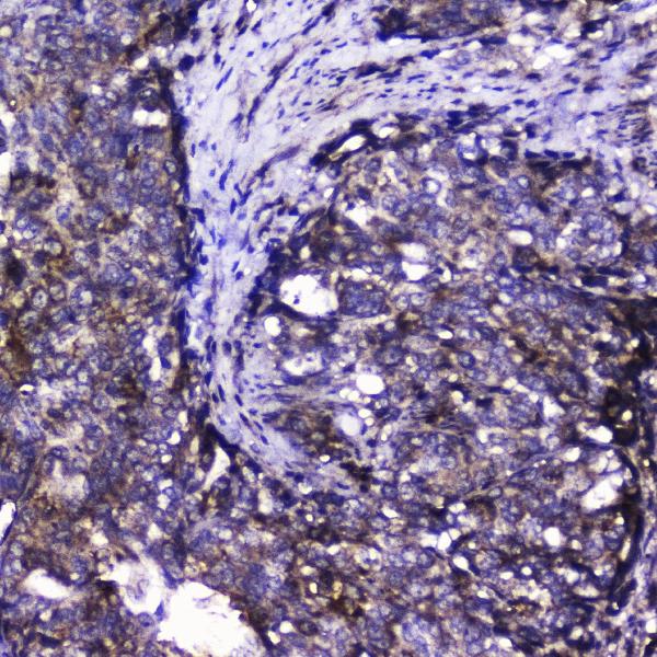
Real IHC data IHC analysis of Cdk6 using anti-Cdk6 antibody (PB9995). Cdk6 was detected in paraffin-embedded section of human intestinal cancer tissues. Heat mediated antigen retrieval was performed in citrate buffer (pH6, epitope retrieval solution) for 20 mins. The tissue section was blocked with 10% goat serum. The tissue section was then incubated with 2μg/ml rabbit anti-Cdk6 Antibody (PB9995) overnight at 4°C. Biotinylated goat anti-rabbit IgG was used as secondary antibody and incubated for 30 minutes at 37°C. The tissue section was developed using Strepavidin-Biotin-Complex (SABC)(Catalog # SA1022) with DAB as the chromogen.
Anti-Cdk6 Antibody ®
Cat # PB9995
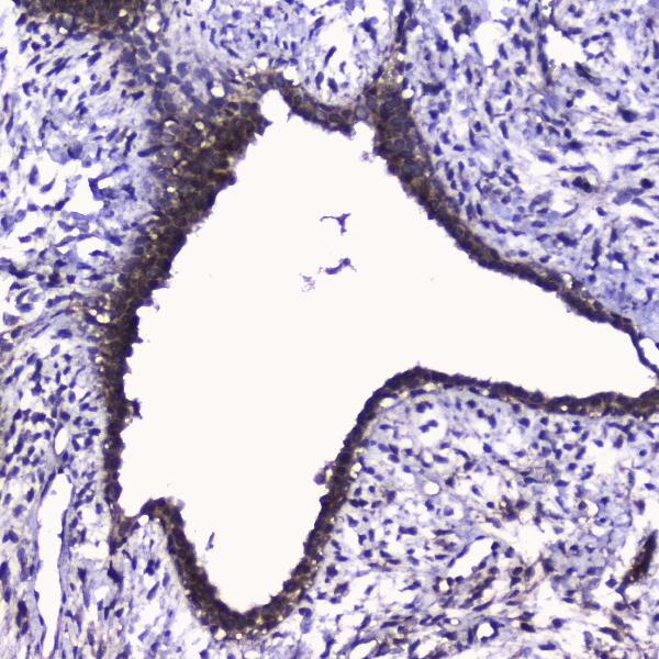
Real IHC data IHC analysis of Cdk6 using anti-Cdk6 antibody (A00358). Cdk6 was detected in paraffin-embedded section of human intestinal cancer tissue. Heat mediated antigen retrieval was performed in citrate buffer (pH6, epitope retrieval solution) for 20 mins. The tissue section was blocked with 10% goat serum. The tissue section was then incubated with 2μg/ml rabbit anti-Cdk6 Antibody (A00358) overnight at 4℃. Biotinylated goat anti-rabbit IgG was used as secondary antibody and incubated for 30 minutes at 37℃. The tissue section was developed using Strepavidin-Biotin-Complex (SABC)(Catalog # SA1022) with DAB as the chromogen.
Anti-Cdk6 Antibody ®
Cat # A00358
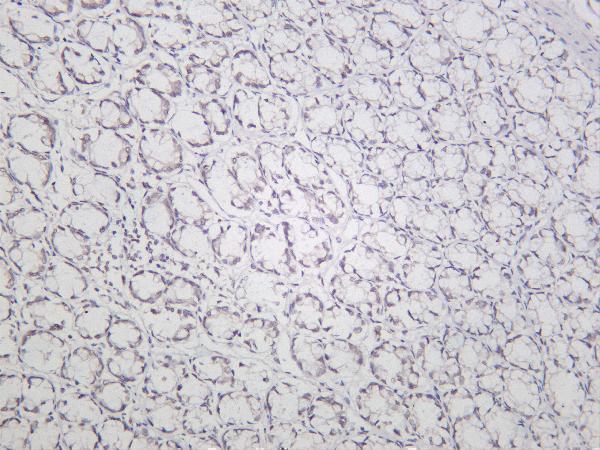
Real IHC data Rat colon tonsil was stained with Anti-CDK6 rabbit antibody
Anti-CDK6 Rabbit Monoclonal Antibody
Cat # M00358-2
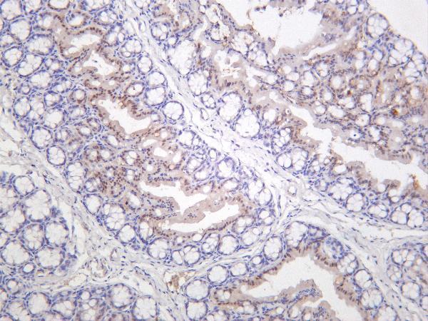
Real IHC data Immunohistochemical analysis of paraffin-embedded human lung cancer, using CDK6 Antibody.
Anti-CDK6 Rabbit Monoclonal Antibody
Cat # M00358

PB9995 and A00358 show CDK6 staining in paraffin sections of human intestinal cancer tissue (respective IHC image captions). M00358-2 shows human tonsil staining (IHC image caption), while M00358 shows paraffin section staining of human lung cancer and IF staining of K562 cells (respective image captions).

Which to pick: For human tissue IHC, PB9995 or A00358 offers a documented starting workflow: citrate retrieval at pH 6 for 20 minutes, followed by serum blocking and chromogenic detection (respective IHC image captions); the fixative is unreported (respective IHC image captions). For IF/ICC, M00358 lists both applications and has a K562 IF image (catalog applications; IF image caption). For cross-species planning, A00358 lists human and mouse reactivity, while PB9995 lists human and rat reactivity; their illustrated IHC sections are human, so verify staining in the intended nonhuman tissue (catalog reactivity; respective IHC image captions).

Each figure is that product's own IHC / IF validation image from its datasheet.

References

  1. UniProt Consortium. UniProt entry Q00534 (CDK6_HUMAN, Cyclin-dependent kinase 6).
  2. Human Protein Atlas. CDK6 tissue IHC expression (reliability: Enhanced).
  3. Human Protein Atlas. CDK6 subcellular location (ICC-IF): Mainly localized to the nucleoplasm. In addition localized to the cytosol..
  4. Human Protein Atlas. CDK6 antibody validation summary (2 antibodies).
  5. CDK6 protein expression is associated with disease progression and treatment resistance in multiple myeloma. HemaSphere 2024 — PMC10878183.
  6. Cyclin-dependent kinase 6 (CDK6) is a candidate diagnostic biomarker for early non-small cell lung cancer. Translational cancer research 2020 — PMC8798208.
  7. Expression and Significance of Cyclin-Dependent Protein Kinase 6 in Diffuse Large B-Cell Lymphoma. International journal of general medicine 2022 — PMC9483138.
  8. MiR-506 suppresses proliferation and induces senescence by directly targeting the CDK4/6-FOXM1 axis in ovarian cancer. The Journal of pathology 2014 — PMC4144705.
  9. PubMed PMID:1639063 — UniProt-cited evidence.
  10. PubMed PMID:14702039 — UniProt-cited evidence.
  11. PubMed PMID:12853948 — UniProt-cited evidence.