This website uses cookies to ensure you get the best experience on our website.
- Table of Contents
1 Citations 15 Q&As
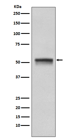


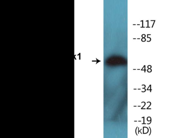

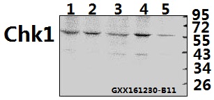
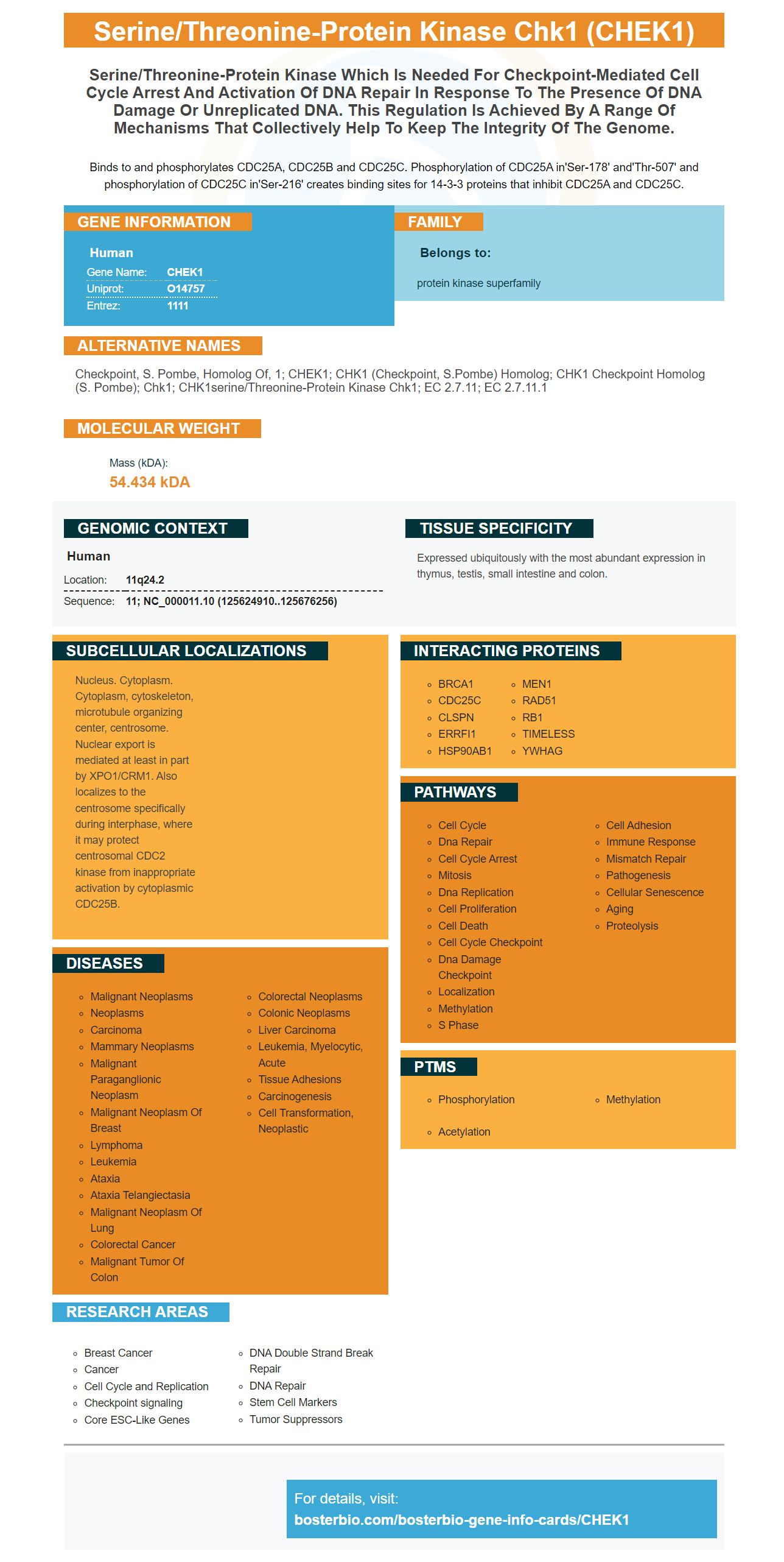
Facts about Serine/threonine-protein kinase Chk1.

Binds to and phosphorylates CDC25A, CDC25B and CDC25C. Phosphorylation of CDC25A in'Ser-178' and'Thr-507' and phosphorylation of CDC25C in'Ser-216' creates binding sites for 14-3-3 proteins that inhibit CDC25A and CDC25C.
| Human | |
|---|---|
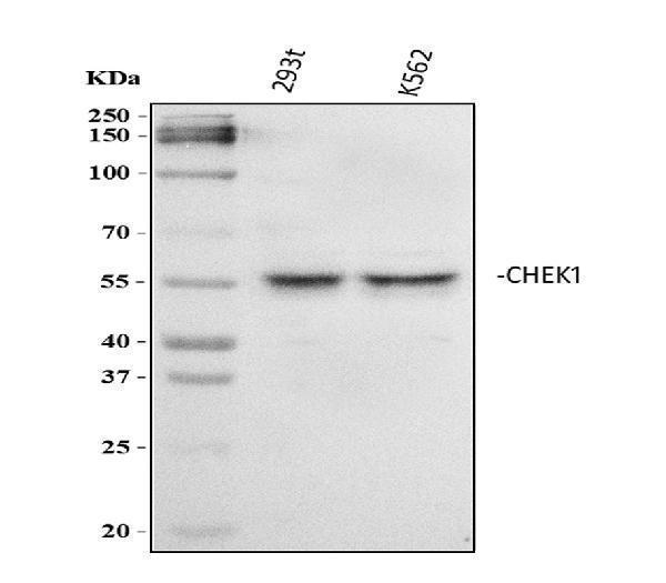
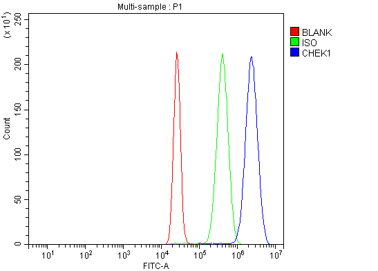
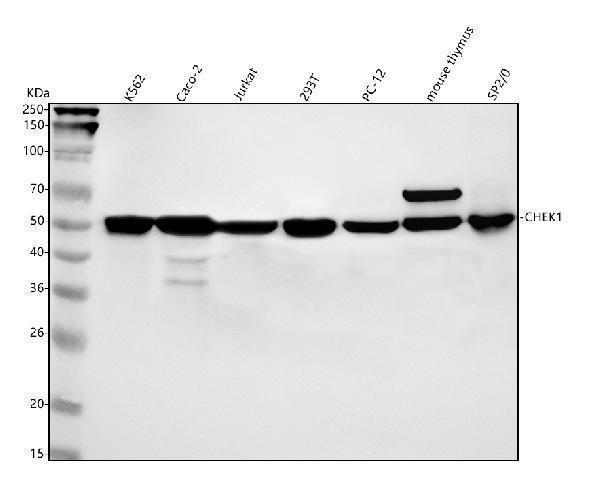
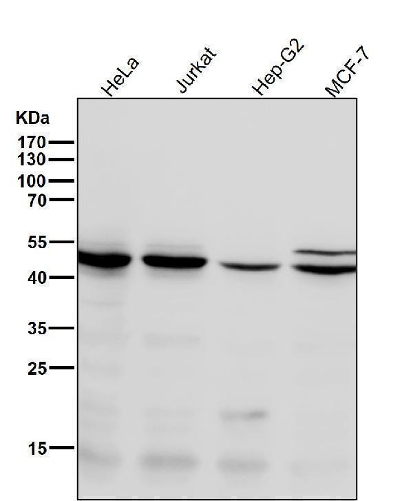
| Gene Name: | CHEK1 |
| Uniprot: | O14757 |
| Entrez: | 1111 |

| Belongs to: |
|---|
| protein kinase superfamily |

Checkpoint, S. pombe, homolog of, 1; CHEK1; CHK1 (checkpoint, S.pombe) homolog; CHK1 checkpoint homolog (S. pombe); Chk1; CHK1serine/threonine-protein kinase Chk1; EC 2.7.11; EC 2.7.11.1
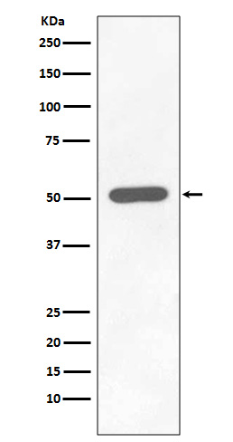
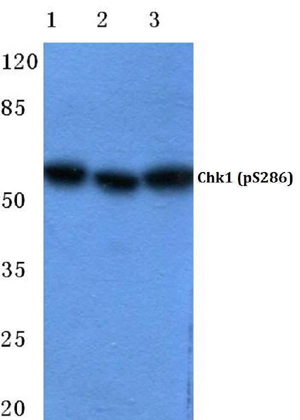
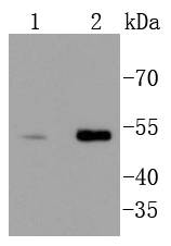
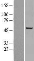
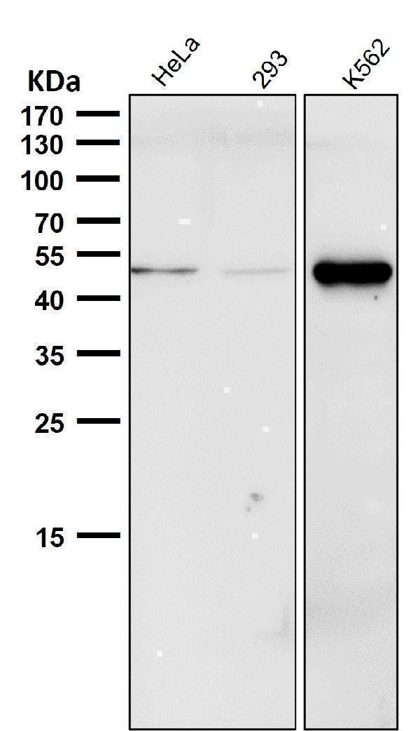
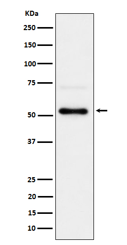
Mass (kDA):
54.434 kDA

| Human | |
|---|---|
| Location: | 11q24.2 |
| Sequence: | 11; NC_000011.10 (125624910..125676256) |
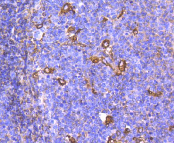
Expressed ubiquitously with the most abundant expression in thymus, testis, small intestine and colon.
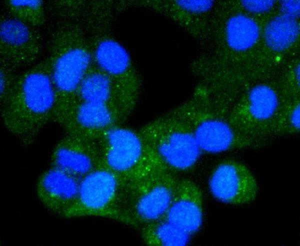
Nucleus. Cytoplasm. Cytoplasm, cytoskeleton, microtubule organizing center, centrosome. Nuclear export is mediated at least in part by XPO1/CRM1. Also localizes to the centrosome specifically during interphase, where it may protect centrosomal CDC2 kinase from inappropriate activation by cytoplasmic CDC25B.


PMID: 9278511 by Sanchez Y., et al. Conservation of the Chk1 checkpoint pathway in mammals: linkage of DNA damage to Cdk regulation through Cdc25.
PMID: 9382850 by Flaggs G., et al. Atm-dependent interactions of a mammalian chk1 homolog with meiotic chromosomes.