This website uses cookies to ensure you get the best experience on our website.
- Table of Contents
21 Citations 16 Q&As
45 Citations 16 Q&As
10 Citations 16 Q&As
4 Citations 5 Q&As
3 Citations 16 Q&As


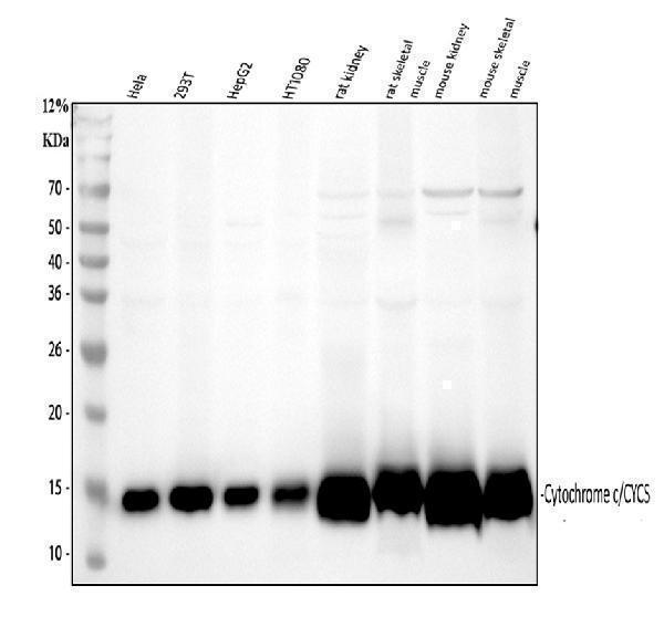
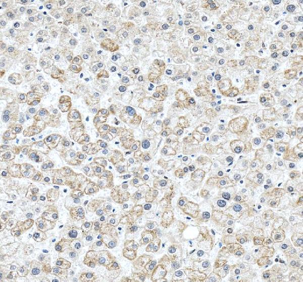
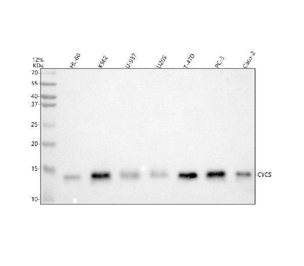
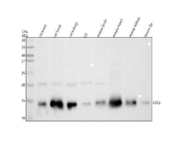
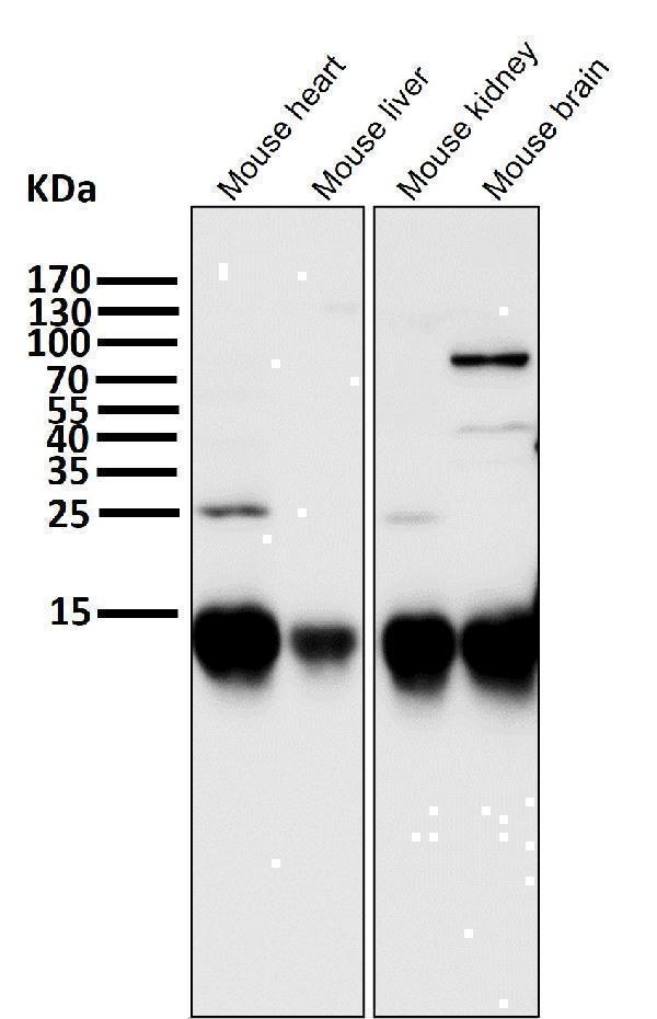
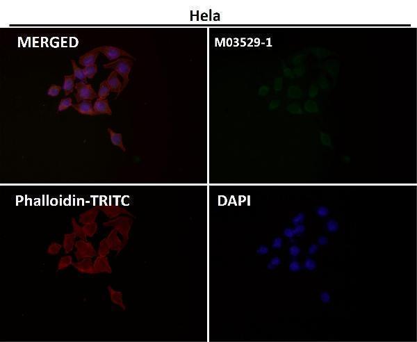
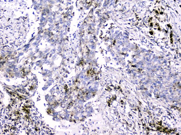
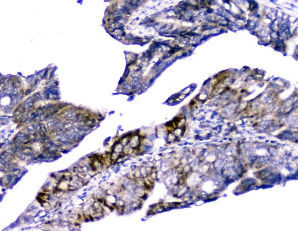
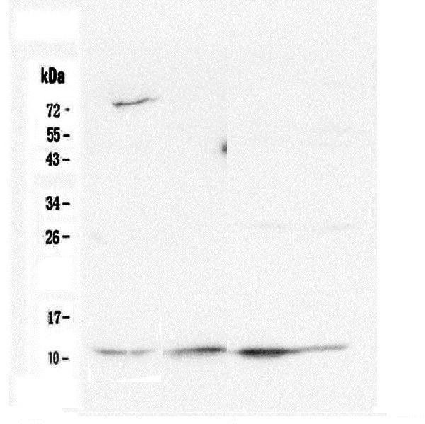
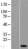
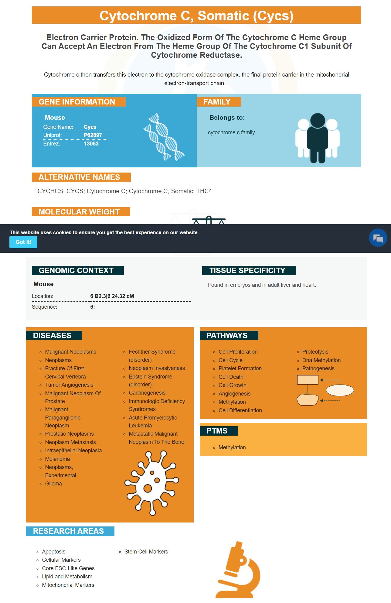
Facts about Cytochrome c, somatic.
Cytochrome c then transfers this electron to the cytochrome oxidase complex, the final protein carrier in the mitochondrial electron-transport chain. .
| Mouse | |
|---|---|
| Gene Name: | Cycs |
| Uniprot: | P62897 |
| Entrez: | 13063 |

| Belongs to: |
|---|
| cytochrome c family |

CYCHCS; CYCS; Cytochrome c; cytochrome c, somatic; THC4
Mass (kDA):
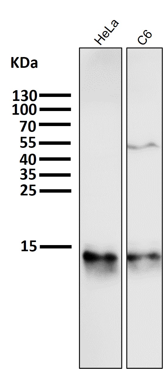
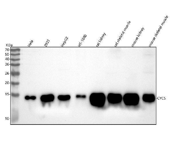
11.605 kDA

| Mouse | |
|---|---|
| Location: | 6 B2.3|6 24.32 cM |
| Sequence: | 6; |
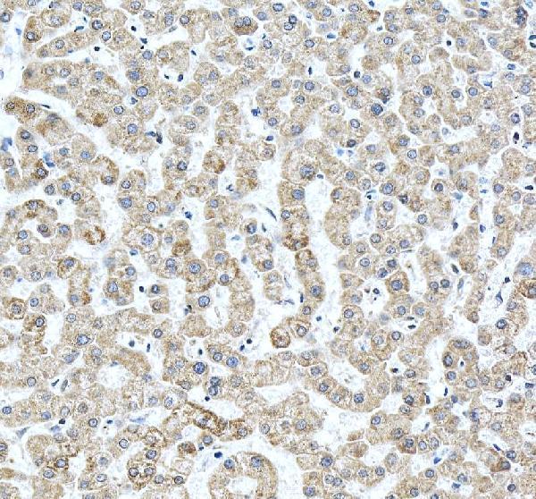
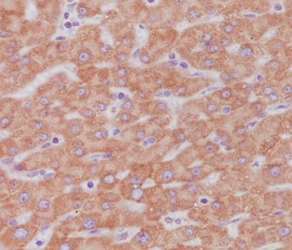
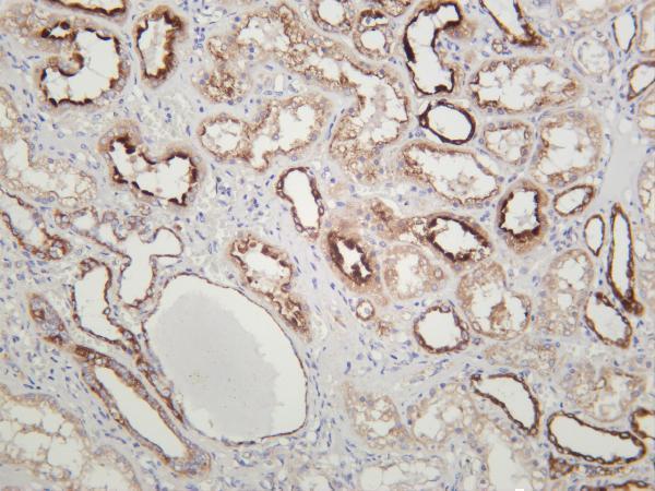
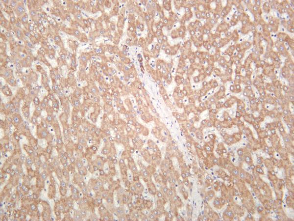
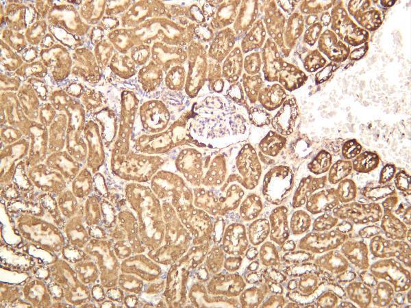
Found in embryos and in adult liver and heart.


PMID: 2987801 by Limbach K.J., et al. Characterization of a mouse somatic cytochrome c gene and three cytochrome c pseudogenes.
PMID: 191069 by Carlson S.S., et al. Primary structure of mouse, rat, and guinea pig cytochrome c.
*Showing only the more recent 20. More publications can be found for each product on its corresponding product page