This website uses cookies to ensure you get the best experience on our website.
- Table of Contents
1 Citations 17 Q&As
1 Citations 4 Q&As
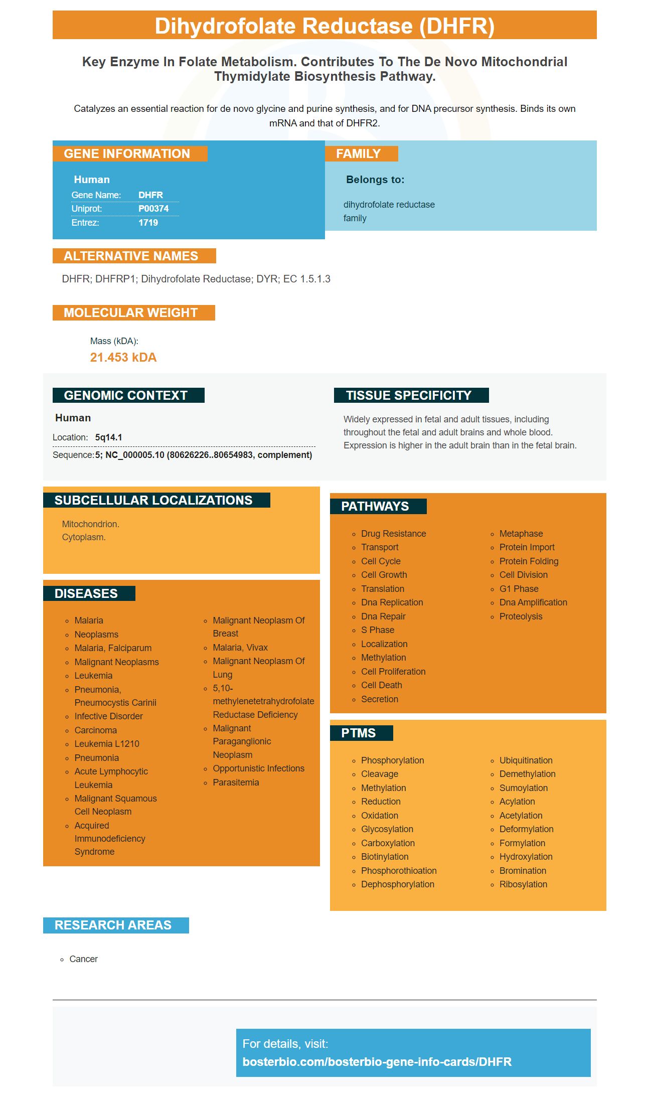
Facts about Dihydrofolate reductase.
Catalyzes an essential reaction for de novo glycine and purine synthesis, and for DNA precursor synthesis. Binds its own mRNA and that of DHFR2.
Human | |
---|---|
Gene Name: | DHFR |
Uniprot: | P00374 |
Entrez: | 1719 |
Belongs to: |
---|
dihydrofolate reductase family |
DHFR; DHFRP1; Dihydrofolate Reductase; DYR; EC 1.5.1.3
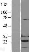
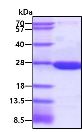
Mass (kDA):
21.453 kDA
Human | |
---|---|
Location: | 5q14.1 |
Sequence: | 5; NC_000005.10 (80626226..80654983, complement) |
Widely expressed in fetal and adult tissues, including throughout the fetal and adult brains and whole blood. Expression is higher in the adult brain than in the fetal brain.
Mitochondrion. Cytoplasm.
This article will review the DHFR Marker and its benefits. We will also talk about the sensitivity of this marker to inhibitors of DHFR and how this marker enhances the stability of the cell lines created by using it. These benefits will ultimately increase your research's success rate. We'll talk about the possibilities of the DHFR Marker within the lab in the following paragraphs.
The DHFR Marker in Boster Bio was created by an antibody manufacturer. It was designed for detecting rEg-DHFR protein in serum. It is HRP-conjugated which is used to determine the presence of rEg-DHFR in serum. Its detection limit minimum for serum was 0.4140 milliliters and its mean absorbance was 0.527.
The DHFR subunit's charge is different in Eg-DHFR and humans. Due to the different charge between the proteins, there is a difference in the size of the loop. This leads to the development of specific inhibitors. Specific charge-charge features can be added to DHFR inhibitors. Asn64 is positively charged, as are Trp24 and Trop26 positively charged. Thus, the DHFR subunits can have a wider hydrophobic binding pocket.
The subunits of DHFR are assigned to the backbone structure by spectroscopy. The dispersion curves of the principal DHFR residues were characterized by an overall rate of 555 +24 s-1 at 306.4 K. These residues were located in the FG or GH loops, which are near site of binding to the substrate. The DHFR subunits are also subjected to conformational exchange processes.
Boster Bio's DHFR marker can be used to screen for antibodies against a variety of pathogens and diseases. The DHFR gene has been extensively studied in a variety of organisms and is now employed to target drug therapy for the treatment of cancer as well as parasitic infection. The Eg DHFR gene isn't yet fully understood in tapeworms. However it has been identified in sheep, and an iELISA for CE was identified.
There are a lot of mutant alleles in human DNA from the DHFR gene. This gene is crucial in preventing HIV infection. The DHFR gene is present in nearly all mammals, including humans. There are two types of DHFR gene: DHFR and DHPS. Both contain high levels of DHFR mutations. Both DHFR mutations and DHPS mutations are involved in HIV prevention.
The Eg-DHFR gene has been successfully in pET28a (+). After induction, a purified protein recombinant was produced. Western blotting revealed a single band with a size that was comparable to the predicted 26kDa. Western Blotting revealed an extremely high degree of antigenicity and reactivity. It was also determined that rabbit anti-rEgDHFR IgG recognized the protein 22 kDa.
Boster Bio's DHFR inhibitors are tolerant because they are sensitive to specific antagonists. Boster Bio Anti Dihydrofolate Reductase V2 reagents are constructed from recombinant Human and Mouse DHFR proteins. Boster Bio DHFR inhibitor (V2) reacts with a subdiluted 1% of human, mouse or rats DHFR proteins.
The structure of the human DHFR differs from the Eg-DHFR. The main difference between these two enzymes is in the charge distribution of the residues. H. sapiens DHFR has negative charge whereas Eg-DHFR has a positive charge. This distinction allows the design of inhibitors that are sensitive to enzymes. For instance there is a negative charge that can be introduced at Asn64 or a non-charged amino acid could replace the random coil residue, and an amino acid that is polar can replace an unpolar residue.
Both Methotrexate as well as Aminopterin block the rEg-DHFR enzyme. They have different structures, however both share a Pteridine rings. They are very similar to glutamic acidwhich is the typical substrate for DHFR. The IC50 of antifolates can be determined by relying on their standard errors of mean. Utilizing the standard errors of means we can determine if the inhibitor is of more sensitivity to specific enzyme.
Metformin, Q54, and Q48 were effective against CLIC1-dependent tumors. Although Q54 and Q48 were more effective than metformin in fighting CLIC1-dependent tumors not show any moderate inhibitory effects. Additionally, these agents have a restricted access to intracellular compartments. Unlike metformin, Q54's sensitivity to DHFR was time-dependent and likely related to insufficient internalization.
A variety of parasitic helminths have been discovered to contain the recombinant protein Eg–DHFR. It was discovered in larvae, germinal layer and adult worms. The protein is present in the suckers and rostellum which are involved in adhesion of the small intestines to the absorption of nutrients. Therefore, the recombinant Eg-DHFR could be an effective DHFR inhibitor in the field of parasitic infections.
Stability of cell lines created with the DHFR Marker are essential for the creation and testing of therapies for a variety of human diseases. Gene therapy requires a vector that has an essential gene for the host cell's function. For instance, cells that are deficient in the enzyme dihydrofolate reductase are unable to survive without the presence of nucleosides. Transfected CHO cells carrying DHFR are capable of synthesizing the nutrient, making them suitable for production of nucleosides. Methotrexate exposure triggers gene amplification of DHFR genes, which results in multiple copies of the plasmid in the cells transfected.
Multiple rounds of selection are required in order to utilize the DHFR-based method. Each round requires a gradual increase in MSX concentration. The cell line might not survive if the selection marker is weakened too much. The stability of rCHO cell lines produced by the DHFR Marker hasn't been verified. A DHFR-based method of selection could improve productivity and optimized vectors for cell lines generation.
The DHFR-Marker is a tool to create stable cell lines. This is a wonderful way to increase throughput research to develop drugs or production of mAbs. While the DHFR marker is not able to confer the functional DHFR protein but it does permit cells to be created at high concentrations and yield greater quantities of therapeutic agents. The DHFR-/ lines could produce many grams of mAb simultaneously however the DHFR/ lines were unable to.
Cho DHFR-cells have been transfected with both the DI TPA vector and the standard tPA vector. The stability of the cell line that was created with the DHFR Marker is shown by the amount of stable colonies after selection. Moreover, the DI-cells produce 11-fold more tPA than the conventional clones. Thus, DHFR-based cell lineages are superior to those produced with the MTX-driven cells.
Intron of pRK vectors was inverted to insert cDNA with DHFR Marker. This vector is a source of an integrase that transforms the transgene cDNA into the host cell genome. An additional insertion of an EcoRV site into the BstXI site resulted in a 678-bp blunted-ended fragment that contains mouse DHFR DNA.
After the transgene was incorporated into the stable cell line showed strong transgene transcription. Recombinase-mediated exchange of cassettes is a method that involves a master line, assists in the integration of the reporter genes. Two distinct Flp recognition target locations protect the reporter gene which allow for high levels of transgene expression in the resulting cell lines.
Transfected HEK293 cells that contain DHFR Marker that have been altered to the Golgi apparatus exhibit high levels of deglycosylated glyproteins. This cell line was also used to show that protein deglycosylation occurs within the Golgi apparatus before the endoplasmic.reticulum. The endoplasmic regulator is responsible for the quality control of the secretory pathway. It would be possible for glycoproteins to crystallize in this environment without further deglycosylation.
PMID: 6323448 by Chen M.-J., et al. The functional human dihydrofolate reductase gene.
PMID: 6687716 by Masters J.N., et al. The nucleotide sequence of the cDNA coding for the human dihydrofolic acid reductase.
*More publications can be found for each product on its corresponding product page