GSK3B / Glycogen synthase kinase-3 beta · IHC design guide

Design Immunohistochemistry for GSK3B

Plan chromogenic GSK3B IHC in paraffin sections with the IHC-validated antibody A00791-3 (datasheet A00791-3). Use the predominantly cytoplasmic tissue pattern and abundant CNS staining as benchmarks (HPA tissue IHC), and check whether your antibody detects total GSK3B or phospho-Ser9 (datasheet M00791-1).

Evidence assembled Oct 2026 · For research use; verify linked source records and product datasheet before use
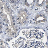
Immunohistochemistry protocol sheet for GSK3B (IHC for GSK3B): expected localisation Predominantly cytoplasmic tissue staining (HPA tissue IHC), antibody A00791-3, validated IHC image, and IHC protocol steps
Printable GSK3B IHC protocol sheet — expected localisation Predominantly cytoplasmic tissue staining (HPA tissue IHC), antibody A00791-3, controls and protocol steps. Open the full GSK3B IHC guide →

GSK3B Immunohistochemistry Experimental Design Guide

Expected localisation, validated protocols, controls and antibodies — the at-a-glance facts below, then the full design guide.

Must know before staining
Expected localisation Predominantly cytoplasmic tissue staining (HPA tissue IHC)
Staining pattern Cytoplasmic staining, most abundant in CNS and testis (HPA tissue IHC)
Antigen retrieval EDTA pH 8.0 HIER, heat-mediated (datasheet A00791-3)
Positive control ⓘ Caudate+4 more · see all
Negative control ⓘ Adipose tissue+4 more · see all
Important caveats
Reasons your staining may differ from the expected pattern.
Fixation Keep fixation consistent across sections (standard IHC practice; not target-specific)
Caveat Phospho-Ser9 staining may differ from total GSK3B (datasheet M00791-1)
Regulation AKT1 phosphorylates Ser9 (UniProt)
Isoform / epitope 2 isoforms; confirm epitope coverage (UniProt)
Section 1

Recommended GSK3B IHC & IF Protocols

The catalog antibody uses EDTA pH 8.0 retrieval (datasheet A00791-3). One published astrocytoma IHC protocol provides a pH 6.0 alternative (PMC10137203).

Recommended immunohistochemistry (IHC-P) protocol parameters
SampleParaffin-embedded human breast cancer tissue; fixative not specified (datasheet A00791-3)
FixationImage fixative and duration unreported (datasheet A00791-3); verify before use.
Sectioning4–5 µm sections on charged slides (standard)
DeparaffinisationXylene, graded ethanol series to water (standard)
Antigen retrievalHeat retrieval: EDTA pH 8.0 (datasheet A00791-3); 20 min, 95–100 °C (standard)
Peroxidase block3% H2O2, 10 min, room temperature (standard)
Blocking10% goat serum (datasheet A00791-3)
Primary antibodyRabbit anti-GSK3B, 2-5 μg/ml (datasheet A00791-3)
Primary incubationOvernight at 4 °C (datasheet A00791-3)
DetectionHRP-conjugated secondary, DAB chromogen (datasheet A00791-3)
CounterstainHematoxylin, blue, dehydrate and mount (standard)
Expected resultGSK3B-positive staining in neuronal cells of caudate (HPA tissue IHC: High). HPA tissue profile: Cytoplasmic expression in most tissues, most abundant in CNS and testis. No signal in the no-primary control.
💡Decision noteStart with heat-mediated EDTA pH 8.0 retrieval for the catalog antibody (datasheet A00791-3); evaluate the published pH 6.0 method if staining needs optimization (PMC10137203).
Section 2

What Is the Expected GSK3B Staining Pattern?

In paraffin section IHC, expect predominantly cytoplasmic GSK3B staining across many tissues, with strong staining in neuronal cells of the caudate, cerebral cortex and hippocampus and in cerebellar molecular layer cells (HPA: tissue IHC). GSK3B is also annotated in the nucleus and at the cell membrane and has no transmembrane segment (UniProt P49841: localization and topology). HPA rates its tissue IHC pattern Approved, with medium RNA agreement and external verification pending (HPA: reliability).

What am I looking at on my slide?
Strong cytoplasmic staining in caudate, cortical or hippocampal neuronal cells, or cerebellar molecular layer cells.This matches the reported high staining in those cells (HPA: tissue IHC). Judge the distribution within identifiable cells, rather than treating all cells in a positive section as equally positive; the reported result is cell specific (HPA: tissue IHC).
Signal appears only in vessel lumens, extracellular material or a uniform edge deposit, without convincing cellular staining.Those patterns do not match the reported cellular cytoplasmic tissue pattern (HPA: tissue IHC). Check section morphology, chromogen precipitate and detection controls (general IHC practice). Nuclear or membrane associated staining alone is not automatically artefactual because UniProt also lists those compartments (UniProt P49841: localization).
Strong staining appears in cardiomyocytes, adipocytes or skeletal myocytes while expected positive cells stain.HPA reports those cell types as not detected (HPA: tissue IHC). Investigate antibody cross reactivity and, with peroxidase detection, endogenous activity using appropriate controls (general IHC practice). A discrepant result warrants review; HPA's Approved tissue pattern still awaits external verification (HPA: reliability).
Weak, even colour covers cells and surrounding tissue with little separation between positive and negative structures.The pattern is hard to score as specific cellular staining (general IHC practice). Review blocking, antibody concentration, washing and detection controls (general IHC practice). Compare neuronal cells reported high with cell types reported not detected on appropriately processed sections (HPA: tissue IHC).
No signal is visible in neuronal cells of a caudate, cerebral cortex or hippocampus section.These are reported high staining cells, so an absent result calls for a run and specimen check (HPA: tissue IHC). Verify the IHC antibody's stated application, retrieval and detection steps, then inspect tissue preservation and controls (general IHC practice). Absence in one run does not establish biological loss.
💡Expected GSK3B appearanceCall the result positive when identifiable neuronal cells show strong, predominantly cytoplasmic chromogen staining (HPA: High in caudate, cortex and hippocampus); diffuse extracellular colour alone is not a convincing cellular result (general IHC practice).
How each factor affects the staining
Tissue and cell choiceHPA reports high staining in selected CNS cells, medium staining in appendix, breast and colon glandular cells, and no detection in adipocytes or cardiomyocytes (HPA: tissue IHC). Choose comparison cells by identity, not by organ name alone (general IHC practice).
Compartment and phosphorylationUniProt lists cytoplasm, nucleus and cell membrane; it specifically links the phosphorylated form to cytoplasm and membrane (UniProt P49841: localization). A total GSK3B IHC pattern cannot by itself assign phosphorylation state or kinase activity (general IHC interpretation).
Isoform and epitope coverageTwo isoforms and a kinase domain at residues 56–340 are annotated (UniProt P49841: isoforms and domains). The supplied record gives no catalog antibody epitope, so it cannot establish which isoforms that antibody detects; consult its validated application and epitope documentation.
IF/ICC Q: Must its pattern match tissue IHC?A: HPA describes mainly nucleoplasmic signal in ICC-IF, while tissue IHC is predominantly cytoplasmic (HPA: subcellular ICC-IF; HPA: tissue IHC). Assess each application against its own evidence; this section supplies no IF/ICC protocol.
Why is my staining missing, weak or wrong?
SituationLikely causeNext action
Known high staining neuronal cells are blank.A failed retrieval, primary incubation or detection step is possible (general IHC practice); the cells are reported high (HPA: tissue IHC).Check the catalog antibody's IHC-P instructions, retrieval settings and reagent controls, then repeat with an appropriate positive section (general IHC practice).
The whole section develops brown colour.Excess detection background or endogenous peroxidase activity may obscure cellular signal in chromogenic IHC (general IHC practice).Review peroxidase blocking, washes and a primary-omission detection control; score only discernible cellular staining (general IHC practice).
Cells reported not detected stain strongly.Cross reactivity or detection background is possible; HPA reports no detection in adipocytes, cardiomyocytes and skeletal myocytes (HPA: tissue IHC).Check cell identity and compare positive and negative controls; investigate specificity before interpreting the unexpected signal (general IHC practice).
Only extracellular deposits or section edges stain.Precipitate or uneven reagent coverage is possible (general IHC practice); this differs from HPA's cellular cytoplasmic pattern (HPA: tissue IHC).Inspect morphology and reagent coverage, review washes and repeat with controls before assigning a cellular score (general IHC practice).
Nuclear staining appears alongside cytoplasmic staining.Nuclear localization is annotated by UniProt, and HPA reports nucleoplasmic ICC-IF staining (UniProt P49841: localization; HPA: subcellular ICC-IF).Record compartments separately and verify the IHC signal with controls; do not infer phosphorylation from compartment alone (general IHC interpretation).

Sample controls for GSK3B IHC & IF

🧪Run caudate first and confirm staining in neuronal cells (HPA: High in caudate neuronal cells); use adipose tissue as the negative comparison and assess adipocytes (HPA: Not detected in adipocytes). On the caudate slide, unstained neighboring cells can serve as background comparators, but no cell type there is established as an internal GSK3B-negative population (HPA: caudate neuronal-cell staining).
Positive control tissue: Caudate (Neuronal cells, HPA High)
Negative control tissue: Adipose tissue (HPA Not detected)
ICC-IF cell lines (HPA subcellular resource): HPA ICC-IF images show GSK3B in A-431, U-251MG, U2OS, A-549, OE19, SK-MEL-30, SiHa, SuSa, hTERT-RPE1 (serum starved), with annotated localisation: Nucleoplasm (enhanced) (HPA subcellular).
Technical controls: Include a no-primary, secondary-only control, a concentration-matched rabbit IgG isotype control (caption: rabbit primary antibody), and GSK3B-knockout material as a biological specificity control. Quench endogenous peroxidase and inspect caudate pigment before interpreting DAB signal (caption: HRP/DAB detection; standard IHC practice).
⚠️Feasibility: A target-specific fixation window or fixation effect is unreported, and the selected A00791-3 tissue-IHC caption does not state its fixative (caption: fixative unreported). Heat retrieval in EDTA at pH 8.0 was used for the shown paraffin-section IHC, but retrieval dependence is unreported (caption: EDTA retrieval). Frozen-section feasibility and whether IF is easier are unreported; ICC-IF images are available for this protein, and neuronal pigment should be checked when interpreting caudate signal (HPA: ICC-IF images; standard microscopy practice).

HPA tissue IHC evidence for GSK3B

Comprehensive Human Protein Atlas IHC scoring per tissue (reliability: Approved — Medium consistency between antibody staining and RNA expression data. Pending external verification.). Rows are taken directly from the HPA tissue chart — click any row's HPA link to view the source.

Positive expression · recommended positive controls

TissueCell typeLevelEvidenceSource
Caudate Neuronal cells High Protein (IHC) HPA →
Cerebellum Cells in molecular layer High Protein (IHC) HPA →
Cerebral cortex Neuronal cells High Protein (IHC) HPA →
Hippocampus Neuronal cells High Protein (IHC) HPA →
Appendix Glandular cells Medium Protein (IHC) HPA →

Undetected expression · recommended negative controls

TissueCell typeLevelEvidenceSource
Adipose tissue Adipocytes Not detected Protein (IHC) HPA →
Bone marrow Hematopoietic cells Not detected Protein (IHC) HPA →
Heart muscle Cardiomyocytes Not detected Protein (IHC) HPA →
Lung Alveolar cells Not detected Protein (IHC) HPA →
Skeletal muscle Myocytes Not detected Protein (IHC) HPA →
Section 3

Advanced GSK3B IHC Tips

Troubleshoot GSK3B staining in paraffin sections by checking retrieval, compartment, controls and scoring before interpreting a chromogenic signal.

What retrieval should I try when GSK3B staining is weak in paraffin sections?
Start with heat-mediated retrieval in EDTA at pH 8.0 (datasheet A00791-3). The catalog antibody detected GSK3B in a paraffin-embedded human breast cancer section after this retrieval, followed by 2 μg/ml primary antibody overnight at 4°C (datasheet A00791-3). If staining remains weak, compare a shorter and longer heating exposure on adjacent sections while holding antibody concentration and detection constant (standard IHC practice). Check tissue integrity and background alongside signal, since excessive heating can damage morphology and increase nonspecific staining (standard IHC practice). Keep a known positive section in each retrieval run; neuronal cells in cerebral cortex show high tissue staining (HPA: cerebral cortex).
How can I troubleshoot fixation without a GSK3B-specific fixation recommendation?
Target-specific fixation sensitivity is unknown: the selected tissue caption identifies a paraffin-embedded section but does not state its fixative (datasheet A00791-3). Record the actual fixative and fixation duration for each specimen, and compare sections processed under matched conditions before changing retrieval or antibody concentration (standard IHC practice). Formalin fixation can mask epitopes, while inconsistent fixation can produce uneven staining across a section (standard IHC practice). Begin troubleshooting with the documented EDTA pH 8.0 retrieval and 2 μg/ml primary incubation overnight at 4°C (datasheet A00791-3). Do not attribute a weak result to a particular GSK3B residue without antibody epitope evidence (UniProt P49841: modified residues; datasheet A00791-3: epitope unreported).
Should GSK3B stain the cytoplasm, nucleus or cell membrane?
Assess cytoplasmic and nuclear staining separately: GSK3B is annotated in both compartments, as well as at the cell membrane (UniProt P49841: subcellular location). Tissue IHC is predominantly cytoplasmic, whereas the separate cell imaging dataset reports mainly nucleoplasmic localisation (HPA: tissue profile; HPA: subcellular summary). A phosphorylated form can localise to cytoplasm and cell membrane, so membrane-associated signal warrants comparison with a phosphorylation-specific assay rather than attribution from total staining alone (UniProt P49841: subcellular location). Check compartment boundaries against the counterstain and section morphology (standard IHC practice). Compare cells within the same section before treating a change in compartment as a biological shift (standard IHC practice).
Can this stain distinguish GSK3B isoforms or phosphorylation states?
GSK3B has 2 annotated isoforms, and its protein kinase domain spans residues 56–340 (UniProt P49841: isoforms; UniProt P49841: domains). The supplied tissue caption specifies the antibody concentration and retrieval, but gives no epitope or isoform recognition information (datasheet A00791-3). Therefore, treat chromogenic signal as GSK3B staining without assigning it to a particular isoform (datasheet A00791-3: epitope unreported; UniProt P49841: isoforms). Ser-9 and Tyr-216 are annotated modified residues, but total GSK3B staining does not measure either modification (UniProt P49841: modified residues; standard antibody interpretation). For state-specific questions, use an independently validated modification-specific assay and matched controls (standard IHC practice).
How should I extend a GSK3B experiment to multiplex IF?
Use IF as a separate validation workflow: the selected antibody evidence describes chromogenic staining of a paraffin section, while the HPA subcellular result comes from cell imaging (datasheet A00791-3; HPA: subcellular summary). Pair GSK3B with a validated marker for the expected cell type; neuronal cells show high tissue staining in cerebral cortex (HPA: cerebral cortex; standard IF practice). Choose spectrally separated fluorophores, considering a far-red channel when tissue autofluorescence obscures shorter wavelengths (standard IF practice). Because GSK3B is intracellular and has no transmembrane segment, include permeabilisation when testing cytoplasmic or nuclear epitopes (UniProt P49841: topology and subcellular location; standard IF practice). Optimise permeabilisation and single-stain controls before interpreting overlap (standard IF practice).
What should I check when the DAB signal is diffuse?
Separate background from cellular GSK3B signal by examining a section processed without primary antibody and an adjacent positive section (standard IHC practice). The documented workflow used 10% goat serum block, 2 μg/ml primary antibody overnight at 4°C, and DAB development (datasheet A00791-3). If the omission control stains, review the peroxidase block, secondary antibody and DAB development time before changing retrieval (standard chromogenic IHC practice). If background appears only with primary antibody, titrate its concentration and strengthen washing while preserving the same tissue controls (standard IHC practice). Score staining within intact cells; acellular deposits and tissue edges can accumulate misleading chromogen (standard IHC practice).
How should I score GSK3B IHC across sections? ⚠ ANSWER MARKED FOR VERIFICATION
Define the cell population and compartment before scoring, then report the percentage of positive cells and staining intensity separately or combine them in an H-score (standard IHC quantification practice). Where cell counts are unreliable, report positive-cell density per mm² of viable tissue and state the area measured (standard IHC quantification practice). Normalise comparisons to the same eligible cell type, viable tissue area and staining run, using identical image settings and thresholds (standard IHC quantification practice). Keep cytoplasmic and nuclear scores separate because both locations are annotated for GSK3B (UniProt P49841: subcellular location). Include a reference section across batches; cerebral cortex neuronal cells have high reported staining (HPA: cerebral cortex).
How do I distinguish convincing GSK3B staining from artefact?
Give greatest weight to staining within intact cells in an expected compartment: tissue IHC is mainly cytoplasmic, while GSK3B is also annotated in the nucleus (HPA: tissue profile; UniProt P49841: subcellular location). Compare cell types within the section; neuronal cells in cerebral cortex show high staining, whereas adipocytes were not detected in the HPA tissue survey (HPA: cerebral cortex; HPA: adipose tissue). Edge-only staining, necrotic debris and signal in a no-primary control favor processing or detection artefact (standard IHC practice). Investigate endogenous peroxidase if DAB persists without primary antibody (standard chromogenic IHC practice). Treat the HPA pattern as supporting context because its tissue reliability is Approved with medium RNA-staining consistency and pending external verification (HPA: reliability).
Boster reagents

Best GSK3B / Glycogen synthase kinase-3 beta IHC Antibodies

These GSK3B antibodies have IHC images from human paraffin sections; catalog IF/ICC applications include human samples, with mouse and rat reactivity listed for selected products (catalog applications, reactivity and image captions).

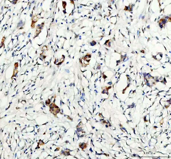
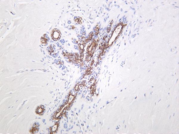
Real IHC data IHC analysis of GSK3 beta/GSK3B using anti-GSK3 beta/GSK3B antibody (A00791-3). GSK3 beta/GSK3B was detected in a paraffin-embedded section of human breast cancer tissue. Heat mediated antigen retrieval was performed in EDTA buffer (pH 8.0, epitope retrieval solution). The tissue section was blocked with 10% goat serum. The tissue section was then incubated with 2 μg/ml rabbit anti-GSK3 beta/GSK3B Antibody (A00791-3) overnight at 4°C. Peroxidase Conjugated Goat Anti-rabbit IgG was used as secondary antibody and incubated for 30 minutes at 37°C. The tissue section was developed using HRP Conjugated Rabbit IgG Super Vision Assay Kit (Catalog # SV0002) with DAB as the chromogen.
Anti-GSK3 beta/GSK3B Antibody ®
Cat # A00791-3
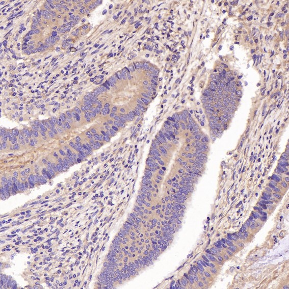
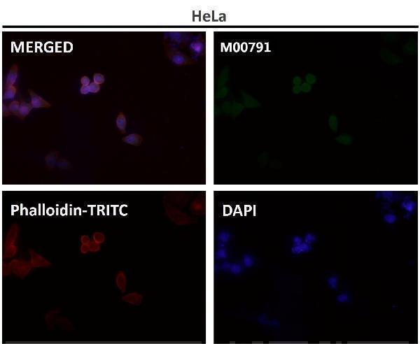
Real IHC data Immunohistochemical analysis of paraffin-embedded Human colon cancer, using the Antibody at 1:100 dilution.
Anti-GSK3 beta Rabbit Monoclonal Antibody
Cat # M00791
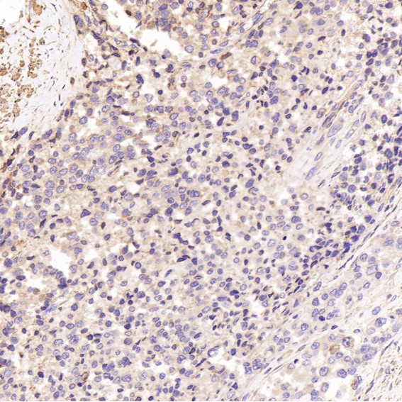
Real IHC data Human breast carcinoma was stained with Anti-GSK3 β (phospho Ser9) rabbit antibody
Anti-GSK3 β Rabbit Monoclonal Antibody
Cat # M00791-1
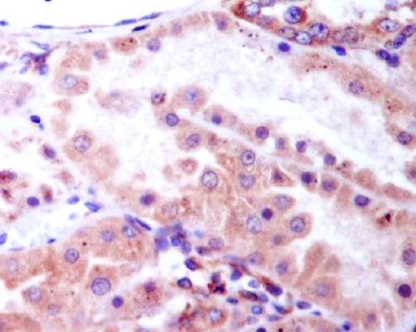
Real IHC data Immunohistochemical analysis of paraffin-embedded human kidney, using Phospho-GSK3 beta (Ser9) Antibody.
Anti-Phospho-GSK3 beta (Ser9) Rabbit Monoclonal Antibody
Cat # P00791-1

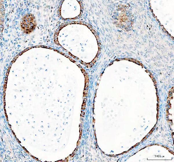
A00791-3 shows IHC staining in human breast cancer, ovarian cancer, endometrial adenocarcinoma and spleen; M00791 shows staining in human colon, prostate, small cell lung and pancreatic cancers (catalog IHC image captions). M00791-1 shows phospho-Ser9 staining in human breast carcinoma, while P00791-1 shows phospho-Ser9 staining in human kidney; both list IHC and IF applications (catalog IHC image captions and applications).

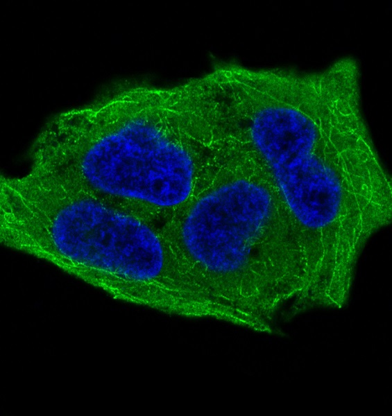
Which to pick: For total GSK3B tissue IHC, choose A00791-3 for its documented paraffin-section workflow with EDTA retrieval at pH 8.0 and 2 μg/ml primary antibody; its IHC caption does not report the fixative (A00791-3 IHC image caption). For IF/ICC, A00791-3 has a documented U2OS cell IF example, while M00791 lists both IF and ICC; choose a phospho-Ser9 antibody only when that modification is the target (A00791-3 IF image caption; M00791 applications; M00791-1 short description; P00791-1 title). For cross-species work, A00791-3 lists human, mouse and rat reactivity; M00791 and M00791-1 list human and mouse, while P00791-1 lists human only (catalog reactivity).

Each figure is that product's own IHC / IF validation image from its datasheet.

References

  1. UniProt Consortium. UniProt entry P49841 (GSK3B_HUMAN, Glycogen synthase kinase-3 beta).
  2. Human Protein Atlas. GSK3B tissue IHC expression (reliability: Approved).
  3. Human Protein Atlas. GSK3B subcellular location (ICC-IF): Mainly localized to the nucleoplasm..
  4. Human Protein Atlas. GSK3B antibody validation summary (8 antibodies).
  5. Galectin-3 Mediates Tumor Progression in Astrocytoma by Regulating Glycogen Synthase Kinase-3β Activity. Current issues in molecular biology 2023 — PMC10137203.
  6. Characterization of neuroendocrine regulation- and metabolism-associated molecular features and prognostic indicators with aid to clinical chemotherapy and immunotherapy of patients with pancreatic cancer. Frontiers in endocrinology 2022 — PMC9895410.
  7. GSK3B inhibition reduced cervical cancer cell proliferation and migration by modulating the PI3K/Akt signaling pathway and epithelial-to-mesenchymal transition. Brazilian journal of medical and biological research = Revista brasileira de pesquisas medicas e biologicas 2024 — PMC11338547.
  8. Construction and Validation of a Necroptosis-Related Gene Signature for Predicting Prognosis and Tumor Microenvironment of Pancreatic Cancer. Disease markers 2022 — PMC9214653.
  9. PubMed PMID:7980435 — UniProt-cited evidence.
  10. PubMed PMID:15489334 — UniProt-cited evidence.
  11. PubMed PMID:10486203 — UniProt-cited evidence.