This website uses cookies to ensure you get the best experience on our website.
- Table of Contents
1 Citations 5 Q&As
2 Citations 6 Q&As
16 Q&As
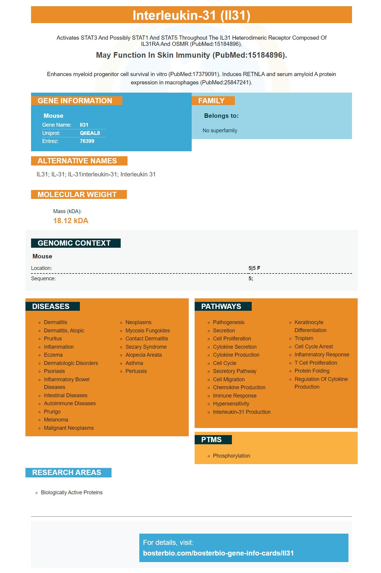
Facts about Interleukin-31.
Activates STAT3 and possibly STAT1 and STAT5 Throughout the IL31 heterodimeric receptor composed of IL31RA and OSMR (PubMed:15184896).
May function in skin immunity (PubMed:15184896).Enhances myeloid progenitor cell survival in vitro (PubMed:17379091). Induces RETNLA and serum amyloid A protein expression in macrophages (PubMed:25847241).
Mouse | |
---|---|
Gene Name: | Il31 |
Uniprot: | Q6EAL8 |
Entrez: | 76399 |
Belongs to: |
---|
No superfamily |
IL31; IL-31; IL-31interleukin-31; interleukin 31
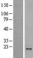
Mass (kDA):
18.12 kDA
Mouse | |
---|---|
Location: | 5|5 F |
Sequence: | 5; |
This article explores the role of Interleukin-31 and its potential for the treatment of allergies with a specific gene. The article also explains the biological mechanisms that allow interleukin 31 to be produced. This marker plays a major role in interleukin-31 production and is a potential treatment target. Boster Bio is a company that offers gene therapy solutions and ongoing clinical trials.
Biological assays often use antibodies to detect Interleukin 31. These antibodies can react with different proteins and may be monoclonal or polyclonal or both. Boster Bio has developed these antibodies using rabbit and mouse samples. Interleukin-31 serves many functions that include skin immunity, stimulating myeloid precursor cell survival and triggering serum amyloid A and RETNLA expression in macrophages. It is also involved in activating the STAT3/calcium Axis and is related with the neuropeptide beta endorphin.
The Interleukin-31 gene is a highly abundant protein in the human body. In a recent study researchers studied the expression of IL-31 during inflammatory skin disease, prurigo nodularis. The mRNA levels of IL-31 in patients with the disease were observed to be significantly higher than healthy controls. In addition, IL-31 levels are elevated in the skin tissues of patients with prurigo nodularis which is correlated with the itch-scratch-cycle.
IL-31 plays an important role in the homeostasis of progenitor cells from hematopoietic stem cells. It regulates osteoclasts, cells that specialize in bone resorption. Additionally, it promotes monocyte activation through the upregulation of proinflammatory genes. Proinflammatory effects can also be observed in activated human macrophages and monocytes.
Its binding complex includes intracellular motifs and phosphorylation sites. These sites attract STATs and the PI3K/AKT signaling pathways. Furthermore, IL-31 interacts with an adaptor protein known as Shc. This protein activates RAS/RAF/MEK/ERK and JNK pathways. This antibody is an essential part of IBD research.
The IL-31 marker can be detected in a variety of biological tests by using antibodies that are monoclonal or polyclonal as they are in nature. Boster Bio uses rabbits and mice as models to create antibodies against Interleukin-31. This protein is a receptor that plays a role in the immune system by enhancing the survival of myeloid precursor cells and increasing the expression of RETNLA and serum amyloid-A protein in macrophages. Researchers have also found that it is produced by T cells that have been activated.
Different studies have proven that IL-31 plays a significant role in the treatment of various inflammation-related skin conditions, such as Psoriasis. Treatments that target this protein may be beneficial in the treatment of various diseases. Authors acknowledge they took part in the creation of the manuscript and gave final approval. They also acknowledge that they are responsible for the content. Both authors have received grants from different companies and have received personal expenses from Maruho.
Recent research suggests that IL-31 may play a significant role in pruritus. Patients with pruritus had skin lesions that contained IL-31 and were higher in protein levels and gene expression. Normal skin did not have IL-31 levels. The condition is not fully understood. Therefore, drug development has been driven to IL-31 and the related pathways.
Allergen-specific immunotherapy with the target IL31 marker involves targeting the T cell receptor IL-31 which is expressed by a variety of allergens. T cells that produce IL-5, IL-10, stimulate B cell switching to the IgE class. They then activate allergic inflammatory cells which leads to the persistence of allergic inflammation.
Allergen-specific IgE levels increase after exposure to allergens, but decrease over time. Additionally the levels of specific IgA2 increase, but are not well-correlated with the clinical efficacy. This could be because specific IgA has a protective role on mucosal surfaces . Additionally, isotype IgA2 acts like blocking antibodies. Further studies are required to confirm these findings.
In a prior study, researchers found that anti-IL31 antigens can be a therapeutic and prophylactic therapy for murine allergic asthma. While they were effective in reducing allergen-specific IgE titers but did not affect symptoms or lung function, omalizumab or quilizumab weren't as effective. However, they did find that anti IL31 immunotherapy reduced the circulating IgE levels. These findings suggest that other compartments in the IgE memory play a vital role in the production of allergen-specific IgE.
Atopic dermatitis is treated with allergy-specific immunotherapy (ASIT). The IL31 marker, a key IL31-targeting drug, has helped to standardize treatments in this field. It works on the basis of allergen-specific antigens affecting peripheral T-cell tolerance. This assists the body to develop immunity to allergens. Allergen-specific immunotherapy is a method of activating regulatory T cells in the immune system. The patient is provided with an antigen-specific vaccination, which stimulates the production of T-helper1 cells (TH2 cells). These cells interact with B cells and release cytokines, which trigger the specific immune response.
There are several advantages of this treatment. AIT has shown significant improvements in the progression of disease in patients with atopic diseases. It has been successfully used in the treatment of human allergies for over 100 years. Noon et al., from Canada were the first to apply it to the treatment of hay fever. The IL31 marker is believed to work because it alters the immune response by blocking the binding of allergen-specific IgE antibody to IgG4-specific IgG4 antibodies. It is also a biomarker which can be used to detect AIT.
IL-31 is a promising therapeutic target, due to its role in several inflammatory skin diseases. In addition, many companies are working on anti-IL-31 treatments. Nemolizumab is a humanized monoclonal antigen, targets IL-31. This article will provide an overview of the current clinical knowledge concerning IL-31 in the treatment of pruritic diseases and also the most recent advances and future prospects in the development of drugs.
Electroporation into C57BL/6N ES cells was used to target the Il31 gene. Three distinct targeted clones were made to make male chimeric animals. After mating, Il31/ mice were genotyped by PCR. To identify mutant alleles, a primer was used that was specifically designed for IL-31 mRNA. The Il31-/ mice were fertile and did not show any phenotypic differences when kept in pathogen-free conditions.
ELISA assay was used to measure the expression of IL-31 in HMC-1 cells. In this study, IL-31, ERK1/2, and OX40L were identified and quantified. The method of immunoblotting was used to determine the expression levels of IL-31, ERK1/2, as well as p38. The experiments were performed in triplets, and each sample was examined at least twice.
The intracellular signaling pathways NFKB and the MAPK cascade control the activity of immune cells. These signaling pathways are thought to activate NF-kB which coordinates the expression of proinflammatory cytokines. Since IL-31 is linked to NFKB/p65 medications that target both these pathways can block the release of these inflammatory mediators. These studies show that IL-31 is produced by the MAPK/NF-kB signaling pathways.
PMID: 15184896 by Dillon S.R., et al. Interleukin 31, a cytokine produced by activated T cells, induces dermatitis in mice.
PMID: 17379091 by Broxmeyer H.E., et al. Regulation of myeloid progenitor cell proliferation/survival by IL-31 receptor and IL-31.
*More publications can be found for each product on its corresponding product page