This website uses cookies to ensure you get the best experience on our website.


- Table of Contents
Plan LMNA IHC in paraffin sections around the distinct nuclear membrane pattern reported across cell types (HPA tissue IHC). Use the staining pattern to assess localisation, and consider age-related prelamin-A/C accumulation when interpreting arterial smooth muscle staining (UniProt).

Expected localisation, validated protocols, controls and antibodies — the at-a-glance facts below, then the full design guide.
| Expected localisation | Nuclear membrane in tissue (HPA tissue IHC) | |
| Staining pattern | Distinct nuclear rim staining in all cell types (HPA tissue IHC) | |
| Antigen retrieval | EDTA pH 8.0 HIER, heat-mediated (datasheet M00438-6) | |
| Positive control | Adipose tissue+4 more · see all | |
| Negative control | None in HPA (detected in all 45 tissues); use no-primary + isotype controls |
| Fixation | Matched tissue-IHC evidence does not establish the fixation claim. Validate the specimen-specific method before use. (selected-SKU IHC image M00438-6) | |
| Caveat | Prelamin-A/C can accumulate in aged arterial VSMCs (UniProt) | |
| Regulation | Oxidative stress can lower ZMPSTE24 (UniProt) | |
| Isoform / epitope | 6 isoforms; C-terminal processing may change epitope coverage (UniProt) |
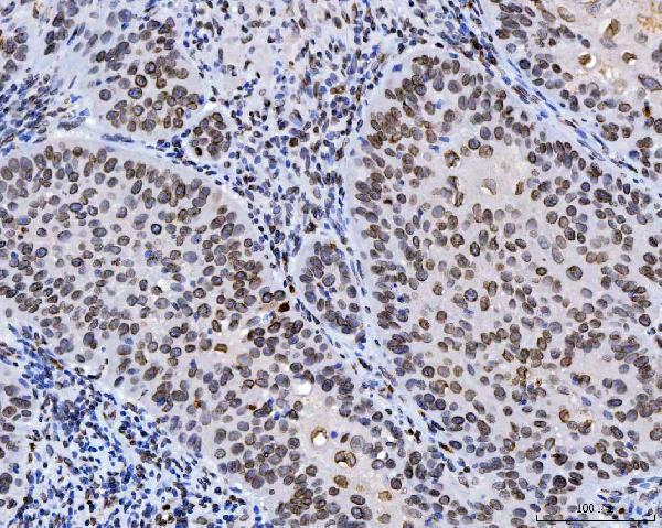
The catalog antibody’s IHC-P protocol (datasheet: M00438-6) is paired with a published chromogenic LMNA protocol for human motor nerve sections (PMC5159906).
| Sample | Paraffin-embedded human laryngeal squamous cell carcinoma tissue; fixative not specified (datasheet M00438-6) |
| Fixation | Image fixative and duration unreported (datasheet M00438-6); verify before use. |
| Sectioning | 4–5 µm sections on charged slides (standard) |
| Deparaffinisation | Xylene, graded ethanol series to water (standard) |
| Antigen retrieval | Heat retrieval: EDTA pH 8.0 (datasheet M00438-6); 20 min, 95–100 °C (standard) |
| Peroxidase block | 3% H2O2, 10 min, room temperature (standard) |
| Blocking | 10% goat serum (datasheet M00438-6) |
| Primary antibody | Mouse monoclonal (clone 5F3C12) anti-LMNA, 2-5 µg/ml (datasheet M00438-6) |
| Primary incubation | Overnight at 4 °C (datasheet M00438-6) |
| Detection | HRP-conjugated secondary, DAB chromogen (datasheet M00438-6) |
| Counterstain | Hematoxylin, blue, dehydrate and mount (standard) |
| Expected result | LMNA-positive staining in adipocytes of adipose tissue (HPA tissue IHC: High). HPA tissue profile: Distinct nuclear membrane expression in all cell types. No signal in the no-primary control. |
LMNA should outline the nuclear envelope in many cell types on paraffin section IHC; the HPA tissue profile describes distinct nuclear membrane staining across cell types and rates its tissue evidence Supported (HPA: tissue IHC). A rim around the nucleus fits its nuclear lamina location (UniProt P02545: subcellular location). LMNA has no transmembrane segment, so a nuclear rim should not be read as staining of the cell surface (UniProt P02545: topology).
| A crisp brown rim outlines nuclei in several cell populations, including adipocytes or respiratory epithelial cells. | This fits the expected nuclear membrane pattern; HPA reports High staining in adipocytes and bronchial respiratory epithelial cells (HPA: tissue IHC). Judge the rim against the counterstained nuclear boundary, since chromogen can obscure a thin outline (standard IHC practice). |
| Brown signal fills cytoplasm or traces the outside of whole cells while nuclear boundaries remain unstained. | That distribution conflicts with the nuclear lamina and envelope assignment (UniProt P02545: subcellular location). Check whether the apparent signal follows tissue edges, debris, or pigment before calling it LMNA; those appearances can mislead compartment scoring (standard IHC practice). |
| One cell population appears strongly positive while neighboring cell types show no nuclear rims. | An isolated population is not automatically a wrong cell type: HPA describes nuclear membrane expression across cell types, with differing observed levels (HPA: tissue IHC). If the signal also occupies the wrong compartment, consider cross-reactivity or endogenous chromogen activity; neither is established by the image alone (standard IHC practice). |
| Pale brown color covers tissue broadly without resolving individual nuclear outlines. | Diffuse background cannot establish LMNA localisation. Compare it with a no primary control and inspect washing, blocking, and detection conditions as general IHC checks (standard IHC practice). HPA's Supported rating describes agreement between staining and RNA data; it does not validate every stain in a new experiment (HPA: tissue IHC). |
| No nuclear rim is visible in a tissue expected to contain strongly stained cells. | First verify that the expected cells are present: HPA reports High staining in bone marrow hematopoietic cells and adrenal glandular cells, among other examples (HPA: tissue IHC). An absent signal warrants checks of antibody application, detection, retrieval, and tissue integrity; this record does not identify an LMNA specific failure mechanism (standard IHC practice). |
| Cell population and reference tissue | HPA lists several High populations but Low staining in spleen red pulp cells; its supplied profile lists no negative tissue (HPA: tissue IHC). Use a documented High population to assess assay performance, and avoid treating weak red pulp staining as an assay failure. |
| Isoforms and antibody epitope | UniProt lists six LMNA isoforms and precursor versus processed chains (UniProt P02545: isoforms, processing). The supplied evidence gives no epitope for the chosen antibody, so the pattern alone cannot establish which isoform or processing state it recognizes. |
| Prelamin A processing and age | Prelamin A undergoes cleavage during maturation; UniProt reports prelamin A accumulation in aged or diseased arterial vascular smooth muscle cells (UniProt P02545: processing, tissue specificity). Ordinary LMNA nuclear staining cannot by itself identify accumulated prelamin A. |
| IHC antibody validation | HPA marks tissue IHC for HPA006660 and CAB004022 as Supported, while the supplied IHC entry for HPA006523 is blank (HPA: antibodies). Match any validation claim to the antibody actually used; these entries do not establish a dilution or retrieval condition. |
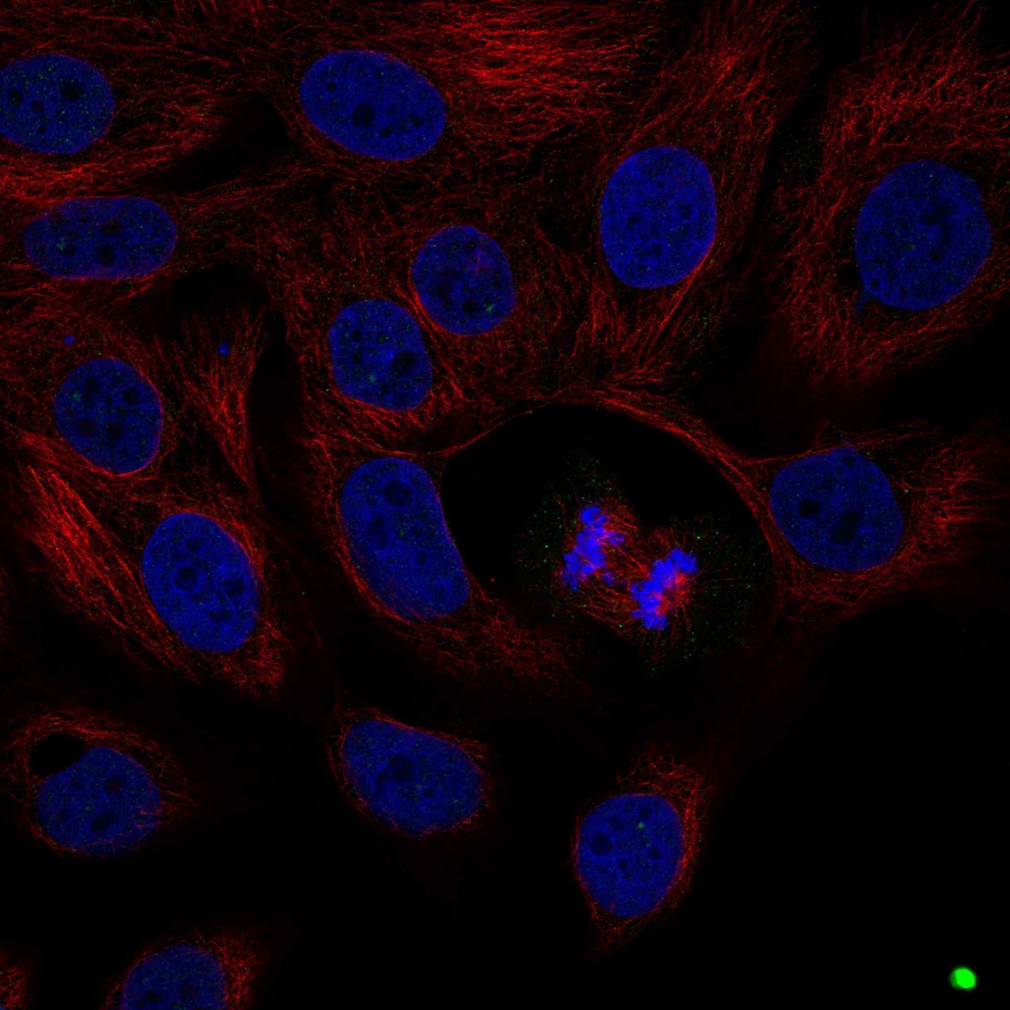
| IF/ICC Q: Should a speckled image override an IHC rim? | No. HPA's ICC IF summary reports supported nuclear speckles, whereas its tissue IHC profile reports nuclear membrane staining (HPA: subcellular ICC IF; HPA: tissue IHC). Interpret each assay against its own evidence and investigate a discordant compartment. |
| Situation | Likely cause | Next action |
|---|---|---|
| Known High population lacks a nuclear rim. | The stain or detection run may have failed, or the expected cells may be absent from the section (HPA: tissue IHC; standard IHC practice). | Confirm cell identity on the counterstain, then review the IHC validated antibody, retrieval, primary application, and detection controls; no LMNA specific retrieval setting is supplied (standard IHC practice). |
| Only spleen red pulp cells look weak. | Low staining is reported for this population, so weak signal can fit the reference (HPA: Low in spleen red pulp cells). | Assess a documented High population in the same run before changing conditions (HPA: tissue IHC; standard IHC practice). |
| Color follows tissue edges or fills cytoplasm. | That shape is inconsistent with the expected nuclear rim and may reflect nonspecific deposition (UniProt P02545: subcellular location; standard IHC practice). | Inspect the counterstained nuclei and a no primary control; reassess washing and detection before scoring positive cells (standard IHC practice). |
| Brown background appears in the no primary control. | Background can arise from endogenous detection activity or the detection reagents (standard IHC practice). | Review the appropriate endogenous activity block and detection controls for the chromogenic system in use (standard IHC practice). |
| Rims vary markedly between cell populations. | HPA reports different staining levels across sampled populations, including High examples and Low spleen red pulp cells (HPA: tissue IHC). | Score cell populations separately and compare each with its documented reference; avoid treating intensity alone as evidence of a different compartment (HPA: tissue IHC; standard IHC practice). |
| A nuclear rim is present, but the question concerns prelamin A accumulation. | The supplied antibody epitope is unspecified, and LMNA includes isoforms and processed forms (UniProt P02545: isoforms, processing). | Report the rim as LMNA pattern only; require evidence of antibody specificity for the relevant form before assigning prelamin A accumulation (UniProt P02545: processing; standard IHC practice). |






Comprehensive Human Protein Atlas IHC scoring per tissue (reliability: Supported — High consistency between antibody staining and RNA expression data.). Rows are taken directly from the HPA tissue chart — click any row's HPA link to view the source.
| Tissue | Cell type | Level | Evidence | Source |
|---|---|---|---|---|
| Adipose tissue | Adipocytes | High | Protein (IHC) | HPA → |
| Adrenal gland | Glandular cells | High | Protein (IHC) | HPA → |
| Appendix | Endocrine cells | High | Protein (IHC) | HPA → |
| Bone marrow | Hematopoietic cells | High | Protein (IHC) | HPA → |
| Breast | Adipocytes | High | Protein (IHC) | HPA → |
| Tissue | Cell type | Level | Evidence | Source |
|---|---|---|---|---|
| None in HPA: LMNA is detected in all 45 scored tissues. Use a no-primary (secondary-only) and an isotype control instead. | ||||
Troubleshoot LMNA staining in paraffin sections by checking nuclear localization, retrieval, antibody specificity, and scoring against matched tissue controls.
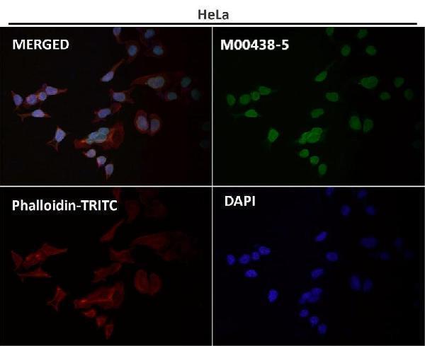
Anti-LMNA antibodies have IHC images from human, mouse and rat paraffin sections, plus IF images from cultured cells and an unspecified sample (catalog image captions).
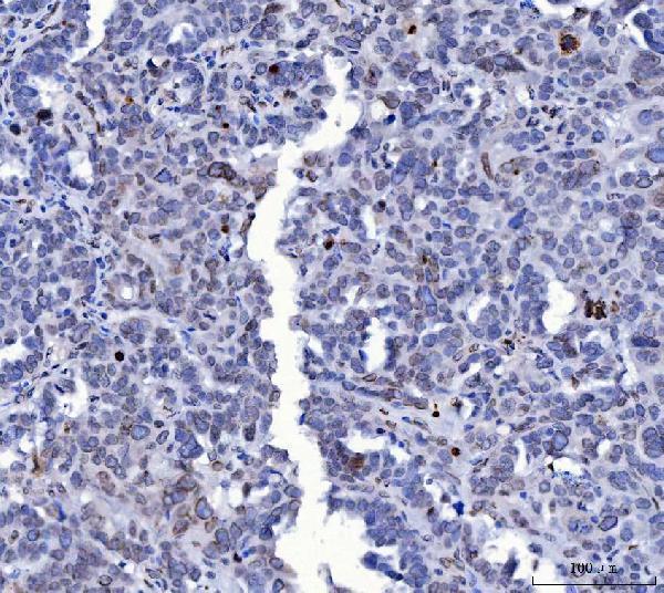
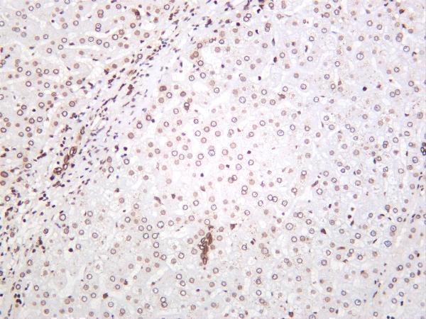
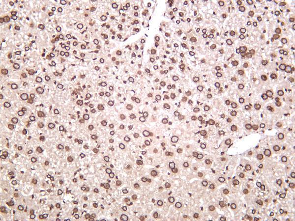
M00438-6 shows human laryngeal carcinoma IHC, PB9280 shows mouse intestine IHC, and M00438-8 shows human liver IHC (catalog image captions). PA1103 shows rat intestine IHC, while M00438-5 has an IF image whose sample is unspecified (catalog image captions).
Which to pick: For tissue IHC, choose M00438-6 for human paraffin sections or PB9280 for mouse paraffin sections; their captions report EDTA retrieval at pH 8.0, and neither reports a fixative (M00438-6 and PB9280 IHC image captions). For IF/ICC, PB9280 has documented A431 and NIH3T3 cell images at 2 µg/ml; M00438-5 is a rabbit monoclonal with an IF image at 1:50, but its sample is unspecified (PB9280 IF image captions; M00438-5 catalog and IF image caption). For cross-species tissue IHC, PB9280 has human, mouse and rat paraffin-section images and lists reactivity with all three species (PB9280 IHC image captions; PB9280 catalog).