This website uses cookies to ensure you get the best experience on our website.


- Table of Contents
Plan chromogenic IHC for MAP1LC3B in paraffin sections using the catalog antibody’s validated conditions (datasheet A01524-2). Compare cytoplasmic staining with the tissue profile, while accounting for the antibody specificity warning (HPA tissue IHC).

Expected localisation, validated protocols, controls and antibodies — the at-a-glance facts below, then the full design guide.
| Expected localisation | Cytoplasmic staining in several tissues (HPA tissue IHC) | |
| Staining pattern | Cytoplasmic in neuronal and glandular cells (HPA tissue IHC) | |
| Antigen retrieval | EDTA pH 8.0 HIER, heat-mediated (datasheet A01524-2) | |
| Positive control | Caudate+4 more · see all | |
| Negative control | Appendix+4 more · see all |
| Fixation | Keep fixation consistent across paraffin sections. (standard IHC practice; not target-specific) | |
| Caveat | Antibody may detect proteins from more than one gene (HPA tissue IHC) | |
| Regulation | Expression regulation not specified (UniProt) | |
| Isoform / epitope | No isoforms annotated; residues 121–125 are cleaved (UniProt) |
The catalog antibody’s IHC-P protocol is accompanied by 4 published MAP1LC3B IHC protocols spanning gastric, breast, and xenograft tumor tissue, plus a tissue staining review (PMC10883967; PMC8615094; PMC5045373; PMC5617963).
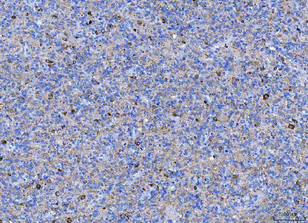
| Sample | Paraffin-embedded human glioma tissue; fixative not specified (datasheet A01524-2) |
| Fixation | Image fixative and duration unreported (datasheet A01524-2); verify before use. |
| Sectioning | 4–5 µm sections on charged slides (standard) |
| Deparaffinisation | Xylene, graded ethanol series to water (standard) |
| Antigen retrieval | Heat retrieval: EDTA pH 8.0 (datasheet A01524-2); 20 min, 95–100 °C (standard) |
| Peroxidase block | 3% H2O2, 10 min, room temperature (standard) |
| Blocking | 10% goat serum (datasheet A01524-2) |
| Primary antibody | Rabbit anti-MAP1LC3B, 1:50 recommended; image 1:100 (datasheet A01524-2) |
| Primary incubation | Overnight at 4 °C (datasheet A01524-2) |
| Detection | HRP-conjugated secondary, DAB chromogen (datasheet A01524-2) |
| Counterstain | Hematoxylin, blue, dehydrate and mount (standard) |
| Expected result | MAP1LC3B-positive staining in neuronal cells of caudate (HPA tissue IHC: High). HPA tissue profile: Cytoplasmic expression in several tissues. No signal in the no-primary control. |
MAP1LC3B is associated with cytoplasmic vesicles and autophagosome membranes; it has no transmembrane segment (UniProt Q9GZQ8). In paraffin-section IHC, expect cytoplasmic staining in selected cell populations, including caudate neuronal cells and thyroid glandular cells, both scored High by HPA (HPA: tissue IHC). Interpret these patterns cautiously: HPA rates its tissue IHC reliability Uncertain because the antibodies may target proteins from more than one gene (HPA: tissue IHC reliability).
| Cytoplasmic staining in caudate neuronal cells or thyroid glandular cells, with clearer signal in the listed positive cells than nearby unstained areas (HPA: High in both cell populations). | This agrees with HPA tissue IHC and with MAP1LC3B's cytoplasmic vesicle and autophagosome association (HPA: tissue IHC; UniProt Q9GZQ8 localization). It is a plausible positive pattern, but HPA's Uncertain reliability prevents attribution to MAP1LC3B alone (HPA: tissue IHC reliability). |
| Predominantly nuclear or extracellular chromogen, without a discernible cytoplasmic component (UniProt Q9GZQ8 localization). | That compartment conflicts with the supplied MAP1LC3B localization evidence and warrants an artefact check (UniProt Q9GZQ8 localization). Compare the slide with a control processed in parallel and inspect counterstain, precipitate, and tissue edges (general IHC practice). |
| Strong staining in a cell population listed as Not detected, such as appendix glandular cells or colon endothelial cells (HPA: tissue IHC). | Investigate antibody cross-reactivity or endogenous detection activity before calling it MAP1LC3B (HPA: Uncertain IHC reliability; general IHC practice). An HPA Not detected call is specific to the listed cells and assay; it does not prove that an entire organ lacks protein (HPA: tissue IHC). |
| Broad, similar chromogen across cells and tissue structures, obscuring cytoplasmic detail (HPA: cytoplasmic tissue profile). | This is poor evidence for a cell-specific positive result (HPA: tissue IHC profile). Check a control without primary antibody, blocking, reagent concentration, washing, and development time to distinguish detection background from interpretable staining (general IHC practice). |
| No cytoplasmic signal in caudate neuronal cells or thyroid glandular cells (HPA: High in those cells). | First check section quality and the staining run with a suitable control (general IHC practice). HPA's positive examples make these useful comparison tissues, but its Uncertain reliability means a negative result cannot by itself establish loss of MAP1LC3B (HPA: tissue IHC reliability). |
| Subcellular distribution (UniProt Q9GZQ8 localization). | LC3-II binds autophagic membranes, while the record also places MAP1LC3B in the cytoplasm and endomembrane system (UniProt Q9GZQ8). Thus a cytoplasmic IHC pattern is biologically coherent; a punctate appearance may be informative, but puncta alone do not establish autophagic flux (UniProt Q9GZQ8 function; general IHC interpretation). |
| Cell selection and apparent tissue differences (HPA: tissue IHC; UniProt Q9GZQ8 tissue specificity). | HPA scores caudate neuronal and thyroid glandular cells High, yet heart cardiomyocytes Not detected; UniProt describes abundant expression in heart (HPA: tissue IHC; UniProt Q9GZQ8 tissue specificity). Keep those observations separate when choosing controls; neither source resolves the discrepancy (HPA: Uncertain IHC reliability). |
| Antibody validation and processing (HPA: antibody validation; UniProt Q9GZQ8 processing). | The four listed HPA IHC antibodies have Uncertain validation, and the tissue profile warns of recognition of proteins from multiple genes (HPA: antibodies; HPA: tissue IHC reliability). UniProt records a 1–120 chain and 121–125 propeptide; epitope position for an unspecified antibody is unknown here, so processing effects cannot be predicted (UniProt Q9GZQ8 processing). |
| IF/ICC Q&A: What pattern should a separate IF experiment assess? (HPA: subcellular ICC-IF). | Look primarily for vesicles; HPA additionally reports cytosol and basal body, both with uncertain localization, and cautions that its antibodies may target proteins from multiple genes (HPA: subcellular ICC-IF). This ICC-IF observation does not establish the appearance of chromogenic paraffin-section IHC (HPA: subcellular ICC-IF; HPA: tissue IHC). |
| Situation | Likely cause | Next action |
|---|---|---|
| A positive reference section shows no interpretable cytoplasmic staining (HPA: High in caudate neurons and thyroid glandular cells). | A failed staining run, unsuitable antibody conditions, or an unsuitable section is possible (general IHC practice). | Review section integrity and run controls; check the catalog antibody's IHC-P instructions for its validated conditions before adjusting the assay (general IHC practice). |
| Chromogen appears mainly nuclear despite expected cytoplasmic distribution (UniProt Q9GZQ8 localization). | Non-specific signal, precipitate, or counterstain confusion may distort compartment assignment (general IHC practice). | Inspect morphology at higher magnification, compare a control without primary antibody, and score cytoplasmic signal separately (general IHC practice). |
| Appendix glandular cells stain strongly despite HPA's Not detected call (HPA: tissue IHC). | Cross-reactivity is plausible given HPA's multi-gene targeting warning; endogenous detection activity is another assay-level possibility (HPA: tissue IHC reliability; general IHC practice). | Compare an independent antibody when available and include a control without primary antibody and an appropriate detection control (general IHC practice). |
| Diffuse background masks differences between cell populations (HPA: cytoplasmic tissue profile). | Incomplete blocking or washing, excessive reagent concentration, or overdevelopment can raise general background (general IHC practice). | Check controls without primary antibody, optimize blocking and washing, and follow the validated IHC-P detection conditions (general IHC practice). |
| Heart cardiomyocytes are negative despite UniProt's high heart expression statement (HPA: Not detected in cardiomyocytes; UniProt Q9GZQ8 tissue specificity). | The supplied sources disagree at different levels of observation; the record does not identify an assay or fixation cause (HPA: tissue IHC; UniProt Q9GZQ8 tissue specificity). | Report the cardiomyocyte observation as measured, verify run controls, and avoid treating heart as an assured positive IHC control (HPA: Uncertain IHC reliability; general IHC practice). |
| Many small cytoplasmic dots are visible and are being scored as increased autophagy (UniProt Q9GZQ8 localization). | Membrane-associated LC3-II can produce a vesicular pattern, but a static stain does not measure formation and clearance rates (UniProt Q9GZQ8 localization; general autophagy interpretation). | Record puncta and diffuse cytoplasmic staining separately; limit the IHC conclusion to observed localization and seek an independent flux assay for a flux claim (general autophagy interpretation). |

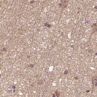


Comprehensive Human Protein Atlas IHC scoring per tissue (reliability: Uncertain — Caution, targets protein from more than one gene. Pending external verification.). Rows are taken directly from the HPA tissue chart — click any row's HPA link to view the source.
| Tissue | Cell type | Level | Evidence | Source |
|---|---|---|---|---|
| Caudate | Neuronal cells | High | Protein (IHC) | HPA → |
| Thyroid gland | Glandular cells | High | Protein (IHC) | HPA → |
| Adrenal gland | Glandular cells | Medium | Protein (IHC) | HPA → |
| Breast | Glandular cells | Medium | Protein (IHC) | HPA → |
| Bronchus | Respiratory epithelial cells | Medium | Protein (IHC) | HPA → |
| Tissue | Cell type | Level | Evidence | Source |
|---|---|---|---|---|
| Appendix | Glandular cells | Not detected | Protein (IHC) | HPA → |
| Colon | Endothelial cells | Not detected | Protein (IHC) | HPA → |
| Esophagus | Squamous epithelial cells | Not detected | Protein (IHC) | HPA → |
| Fallopian tube | Glandular cells | Not detected | Protein (IHC) | HPA → |
| Heart muscle | Cardiomyocytes | Not detected | Protein (IHC) | HPA → |
Use the documented paraffin section workflow as the starting point, then assess cytoplasmic staining and puncta with controls that account for antibody specificity.
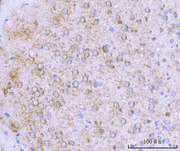
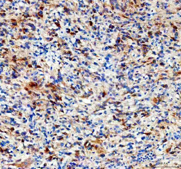
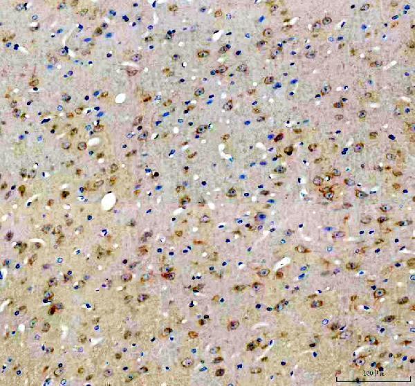
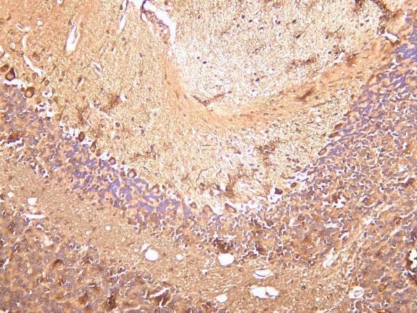
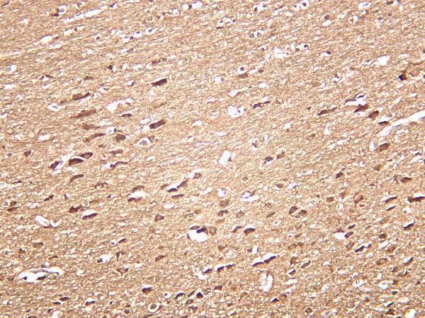
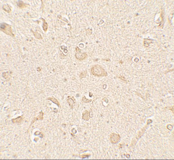
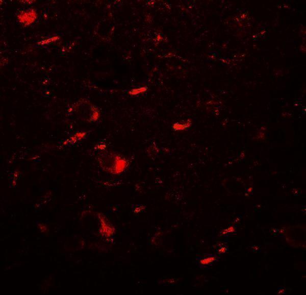
Anti-MAP1LC3B antibodies have IHC images from human tissue and mouse and rat brain, plus IF images from human brain tissue and HeLa cells (catalog image captions).
A01524 has IHC and IF images from human brain tissue; A01524-2 has paraffin-section IHC images from human glioma and mouse and rat brain (catalog image captions). M01524 has paraffin-section IHC images from human glioma and mouse and rat brain, plus IF images including HeLa cells; M01524-2 has IHC images from human, mouse and rat brain (catalog image captions).
Which to pick: For tissue IHC, choose A01524-2 when a documented paraffin-section workflow matters: its images describe EDTA retrieval at pH 8.0 and primary antibody at 1:100 overnight at 4°C; the fixative is unreported (A01524-2 IHC captions). For IF/ICC, M01524 lists both applications and has an IF image of HeLa cells, although IHC is absent from its application list despite its paraffin-section images (M01524 applications and image captions). For IHC across species, A01524-2 has separate paraffin-section images from human glioma and mouse and rat brain; the fixative is unreported (A01524-2 IHC captions).