This website uses cookies to ensure you get the best experience on our website.


- Table of Contents
Plan NFKB1 staining in paraffin sections around the generally cytoplasmic pattern and additional nuclear staining seen in some tissues (HPA tissue IHC). Use the guide to choose controls and interpret nuclear signal in light of NF-κB activation and p105 processing to p50 (UniProt).

Expected localisation, validated protocols, controls and antibodies — the at-a-glance facts below, then the full design guide.
| Expected localisation | Cytoplasmic; nuclear in several tissues (HPA tissue IHC) | |
| Staining pattern | Generally cytoplasmic, with nuclear staining in some tissues (HPA tissue IHC) | |
| Antigen retrieval | Tris-EDTA pH 9.0 HIER, 95–98 °C, 20 min (rule: nuclear antigen) | |
| Positive control | Bone marrow+4 more · see all | |
| Negative control | Adipose tissue+4 more · see all |
| Fixation | Keep fixation consistent across sections. (standard IHC practice; not target-specific) | |
| Caveat | Activation can shift staining into nuclei (UniProt) | |
| Regulation | IκB loss increases nuclear signal (UniProt) | |
| Isoform / epitope | 3 isoforms; p105 processing removes the p50 C terminus, so epitope matters (UniProt) |
The catalog antibody protocol is accompanied by published IHC workflows for bladder cancer tissue (PMC11618553), colorectal cancer tissue (PMC4574027), and tissue microarrays (PMC4724081).
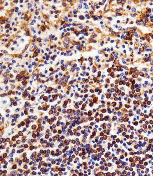
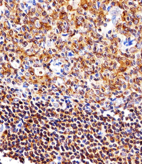
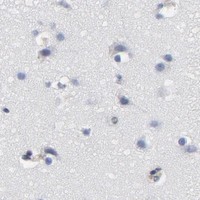
| Sample | Paraffin-embedded H. spleen tissue; fixative not specified (datasheet M00283-2) |
| Fixation | Image fixative and duration unreported (datasheet M00283-2); verify before use. |
| Sectioning | 4–5 µm sections on charged slides (standard) |
| Deparaffinisation | Xylene, graded ethanol series to water (standard) |
| Antigen retrieval | Heat-induced epitope retrieval in Tris-EDTA buffer, pH 9.0, 20 min at 95–98 °C (standard rule: nuclear antigen) |
| Peroxidase block | 3% H2O2, 10 min, room temperature (standard) |
| Blocking | 10% normal serum of the secondary host, 30 min, room temperature (standard) |
| Primary antibody | Mouse monoclonal (clone 1298CT792.105.117.133) anti-NFKB1, 1:25 (datasheet M00283-2) |
| Primary incubation | Overnight at 4 °C (standard) |
| Detection | HRP-polymer secondary, DAB chromogen 5–10 min (standard) |
| Counterstain | Hematoxylin, blue, dehydrate and mount (standard) |
| Expected result | NFKB1-positive staining in hematopoietic cells of bone marrow (HPA tissue IHC: High). HPA tissue profile: General cytoplasmic expression. Additional nuclear expression in several tissues. No signal in the no-primary control. |
NFKB1 is a cytoplasmic and nuclear protein with no transmembrane segment (UniProt P19838). In paraffin-section IHC, expect general cytoplasmic staining and additional nuclear staining in several tissues; high staining is reported in lung macrophages, bone-marrow hematopoietic cells, and lymph-node or tonsil germinal-center cells (HPA tissue IHC). HPA rates the tissue staining Enhanced, with medium consistency between antibody staining and RNA expression (HPA tissue IHC).
| Cytoplasmic staining appears in lung macrophages or germinal-center cells, with nuclear staining in some cells (HPA tissue IHC). | This fits HPA’s general cytoplasmic and additional nuclear tissue pattern. Nuclear signal can reflect NFKB1 localization, so record nuclear and cytoplasmic staining separately (HPA tissue IHC; UniProt P19838). |
| Staining outlines cell membranes or appears extracellular, without convincing cytoplasmic or nuclear signal (UniProt P19838). | Treat this as a possible localization artifact: NFKB1 has no transmembrane segment, and its supported compartments are cytoplasm and nucleus. Check morphology and detection controls before scoring it as positive (UniProt P19838; standard IHC practice). |
| Strong staining appears in cardiomyocytes or adipocytes while expected positive cells also stain (HPA tissue IHC). | HPA reports NFKB1 as not detected in those cell types. Consider antibody cross-reactivity or endogenous chromogenic activity, but do not treat a single HPA negative observation as proof of absolute absence (HPA tissue IHC; standard IHC practice). |
| Brown signal spreads across tissue, vessels, and cell-free areas without clear cell boundaries (standard chromogenic IHC practice). | Diffuse background prevents reliable compartment scoring. Consider nonspecific antibody binding, insufficient washing, or endogenous detection activity; compare with a primary-antibody-omission control (standard IHC practice). |
| No clear staining is seen in lung macrophages or tonsil germinal-center cells (HPA: High in both). | An assay failure is possible when an HPA high-staining cell population is negative. Confirm that the relevant cells are present, then review antibody dilution, retrieval, and detection performance; HPA supplies no NFKB1-specific retrieval requirement (HPA tissue IHC; standard IHC practice). |
| Compartment and activation state (UniProt P19838) | NFKBIA association retains inactive NFKB1 complexes in the cytoplasm; following NFKBIA degradation, they can move into the nucleus. Nuclear staining is therefore compatible with NFKB1 biology, though staining alone does not establish pathway activation (UniProt P19838). |
| Precursor processing and antibody epitope (UniProt P19838) | NFKB1 yields p105 (residues 1–968) and p50 (residues 1–433). An antibody against a shared region may detect both; one against the p105-only region may not detect p50. The supplied record does not identify the catalog antibody’s epitope (UniProt P19838). |
| Cell type selection for slide controls (HPA tissue IHC) | HPA reports high staining in bone-marrow hematopoietic cells, lung macrophages, germinal-center cells, spermatogonia, and urothelial cells. Choose a documented cell population within the section and score that population, rather than assigning one intensity to an entire tissue (HPA tissue IHC; standard IHC practice). |
| Reliability and apparent negatives (HPA tissue IHC) | HPA labels tissue IHC Enhanced but reports only medium agreement with RNA data. Its ‘not detected’ calls for adipocytes and cardiomyocytes are useful comparisons, not universal biological negatives; interpret unexpected signal with morphology and controls (HPA tissue IHC; standard IHC practice). |
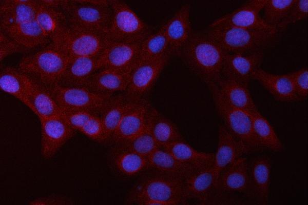
| IF/ICC Q: Should the same compartment pattern be expected? (HPA subcellular ICC-IF) | A: HPA ICC-IF mainly shows nucleoplasmic NFKB1 with additional cytosolic signal, whereas tissue IHC describes general cytoplasmic and additional nuclear expression. Compare each result with its own assay evidence; this section offers no IF/ICC protocol (HPA subcellular ICC-IF; HPA tissue IHC). |
| Situation | Likely cause | Next action |
|---|---|---|
| High-staining reference cells show no signal (HPA tissue IHC). | The run may have insufficient assay sensitivity, or the expected cell population may be absent from the section (standard IHC practice). | Check cell identity and slide integrity; review the antibody’s IHC dilution, retrieval, and chromogenic detection against its validated instructions (standard IHC practice). |
| Staining is diffuse across cells and empty spaces (standard IHC practice). | Nonspecific binding or inadequate washing can obscure NFKB1’s reported compartments (standard IHC practice; UniProt P19838). | Compare a primary-antibody-omission control, optimize blocking and washes, and score only cell-associated signal with interpretable morphology (standard IHC practice). |
| Brown precipitate persists without primary antibody (standard chromogenic IHC practice). | Endogenous enzyme activity or another detection-system background source is possible (standard IHC practice). | Review the appropriate endogenous-enzyme block and detection reagents; do not score control-positive precipitate as NFKB1 (standard IHC practice). |
| Apparent signal is predominantly membranous (UniProt P19838). | This conflicts with the reported cytoplasmic and nuclear localization and lack of a transmembrane segment (UniProt P19838). | Inspect cell boundaries and counterstain, compare controls, and re-evaluate antibody specificity before interpreting the membrane pattern (standard IHC practice). |
| Strong staining appears in HPA not-detected cardiomyocytes or adipocytes (HPA tissue IHC). | Cross-reactivity or detection background is possible; HPA’s observations alone cannot prove biological absence (HPA tissue IHC; standard IHC practice). | Compare the same run with HPA high-staining cells and a primary-antibody-omission control; document the discrepancy by cell type (HPA tissue IHC; standard IHC practice). |
| Nuclear and cytoplasmic scores differ between specimens (HPA tissue IHC). | NFKB1 can occupy both compartments, and nuclear translocation can follow inhibitor degradation; staining alone cannot assign a mechanism (UniProt P19838). | Score each compartment separately in matched cell populations and report the observed distribution without inferring activation from localization alone (UniProt P19838; standard IHC practice). |


Comprehensive Human Protein Atlas IHC scoring per tissue (reliability: Enhanced — Medium consistency between antibody staining and RNA expression data.). Rows are taken directly from the HPA tissue chart — click any row's HPA link to view the source.
| Tissue | Cell type | Level | Evidence | Source |
|---|---|---|---|---|
| Bone marrow | Hematopoietic cells | High | Protein (IHC) | HPA → |
| Lung | Macrophages | High | Protein (IHC) | HPA → |
| Lymph node | Germinal center cells | High | Protein (IHC) | HPA → |
| Testis | Spermatogonia cells | High | Protein (IHC) | HPA → |
| Tonsil | Germinal center cells | High | Protein (IHC) | HPA → |
| Tissue | Cell type | Level | Evidence | Source |
|---|---|---|---|---|
| Adipose tissue | Adipocytes | Not detected | Protein (IHC) | HPA → |
| Heart muscle | Cardiomyocytes | Not detected | Protein (IHC) | HPA → |
| Hippocampus | Glial cells | Not detected | Protein (IHC) | HPA → |
| Liver | Cholangiocytes | Not detected | Protein (IHC) | HPA → |
| Skeletal muscle | Myocytes | Not detected | Protein (IHC) | HPA → |
Troubleshoot NFKB1 staining by evaluating retrieval, antibody epitope, cell compartment and controls before interpreting nuclear signal as pathway activity.
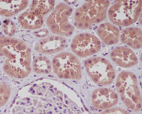
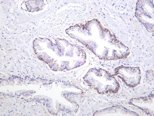
Anti-NFKB1 antibodies have IHC images from human spleen, tonsil, kidney and prostate carcinoma, plus an IF/ICC image from U2OS cells (catalog image captions). Catalog reactivity includes human, mouse and rat (catalog reactivity).
M00283-2 has paraffin-section IHC images from human spleen and tonsil; M00283 has a paraffin-section IHC image from human kidney (catalog image captions). M00283-3 has an IHC image from human prostate carcinoma, while PB9149 has an IF/ICC image from U2OS cells (catalog image captions).
Which to pick: For tissue IHC, choose M00283-2 for human paraffin sections at 1:25 or M00283 for paraffin-embedded human kidney at 1:50; neither IHC caption reports the fixative (M00283-2 and M00283 image captions; catalog IHC dilutions). For IF/ICC, choose PB9149 at 5 μg/ml, as shown in its U2OS cell image (PB9149 applications, dilution and image caption). For work across species, M00283-3 is a rabbit monoclonal listed for IHC and IF with human, mouse and rat reactivity; its IHC image shows human prostate carcinoma, but reports neither section processing nor fixative (M00283-3 catalog applications, reactivity and image caption).