Click image to see more details
-

-
-
-
-
+1
Product Info Summary
| SKU: | PB9149 |
|---|---|
| Size: | 100 μg/vial |
| Reactive Species: | Human |
| Host: | Rabbit |
| Application: | Flow Cytometry, IF, ICC, WB |
Customers Who Bought This Also Bought

Product info
Product Name
Anti-NFkB/NFKB1 p105/p50 Antibody Picoband®
SKU/Catalog Number
PB9149
Size
100 μg/vial
Form
Lyophilized
Description
Boster Bio Anti-NFkB/NFKB1 p105/p50 Antibody Picoband® catalog # PB9149. Tested in Flow Cytometry, IF, ICC, WB applications. This antibody reacts with Human. The brand Picoband indicates this is a premium antibody that guarantees superior quality, high affinity, and strong signals with minimal background in Western blot applications. Only our best-performing antibodies are designated as Picoband, ensuring unmatched performance.
Storage & Handling
Store at -20˚C for one year from date of receipt. After reconstitution, at 4˚C for one month. It can also be aliquotted and stored frozen at -20˚C for six months. Avoid repeated freeze-thaw cycles.
Cite This Product
Anti-NFkB/NFKB1 p105/p50 Antibody Picoband® (Boster Biological Technology, Pleasanton CA, USA, Catalog # PB9149)
Host
Rabbit
Contents
Each vial contains 4 mg Trehalose, 0.9 mg NaCl and 0.2 mg Na2HPO4.
Clonality
Polyclonal
Isotype
Rabbit IgG
Immunogen
E.coli-derived human NFkB/NFKB1 p105/p50 recombinant protein (Position: M1-Q360). Human NFkB p105/P50 shares 93% amino acid (aa) sequence identity with mouse NFkB p105/P50.
Cross-reactivity
No cross-reactivity with other proteins
Reactive Species
PB9149 is reactive to NFKB1 in Human
Observed Molecular Weight
50 kDa, 120 kDa
Calculated molecular weight
105.4 kDa
Background of NFKB1
Nuclear factor NF-kappa-B p105 subunit, also called EBP-1 is a protein that in humans is encoded by the NFKB1 gene. By fluorescence in situ hybridization, the gene was assigned to human chromosome 4q24. NFKB1 is a pleiotropic transcription factor present in almost all cell types and is the endpoint of a series of signal transduction events that are initiated by a vast array of stimuli related to many biological processes such as inflammation, immunity, differentiation, cell growth, tumorigenesis and apoptosis. NFKB1 appears to have dual functions such as cytoplasmic retention of attached NFKB1 proteins by p105 and generation of p50 by a cotranslational processing.
Antibody Validation
Boster validates all antibodies on WB, IHC, ICC, Immunofluorescence, and ELISA with known positive control and negative samples to ensure specificity and high affinity, including thorough antibody incubations.
Application & Images
Applications
PB9149 is guaranteed for Flow Cytometry, IF, ICC, WB Boster Guarantee
Recommend Dilution
| Application | Dilution | Species |
|---|---|---|
| Western blot | 0.1-0.5μg/ml | Human |
| Immunocytochemistry/Immunofluorescence | 5 μg/ml | Human |
| Flow Cytometry (Fixed) | 1-3 μg/1x106 cells | Human |
Tested application
Suggested blocking solution with 5% non-fat milk or BSA; (*)Recommended protein loading: 20-40 µg per lane
Validation Images & Assay Conditions
Click image to see more details
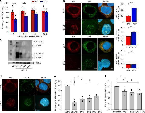
FLIP activates the canonical NF-κB pathway in myeloid cells. a Suppressive activity of THP1 cells transfected with c-FLIP RNA was measured by enumeration of an absolute number of human CD3 + T cells collected after in vitro co-culture. b Immunofluorescence confocal microscopy of p65 (red), p50 and p52 (green) nuclear translocation in transfected THP1 cells. c Nuclear c-FLIP protein expression by western blot in THP1 cells transfected with either GFP or c-FLIP RNA at different time points. d Immunofluorescence confocal microscopy of p50 (red) and c-FLIP (green) nuclear translocation in transfected THP1 cells. e CD11b + Ly6C + cells (M-MDSCs) were isolated from the spleen of MCA203 tumor-bearing, wild-type mice by flow sorter and transfected for 18 h with scramble, IKKα, IKKβ, or the combination IKKα plus IKKβ siRNAs. After transfection, cells were washed three times. M-MDSCs were co-incubated with peptide-stimulated CellTrace-labeled OT-I cells. Suppressive activity was measured by enumerating absolute numbers of CD8 + T cells collected after in vitro co-culture. f PD-L1 expression in transfected M-MDSCs. Data are presented either as mean ± s.e.m of three independent experiments ( a ) or as mean ± s.e.m of four independent experimental transfections ( e , f ) where each plot refers to CD11b + Ly6C + cells isolated from the pooled spleens of three tumor-bearing mice. Original images × 800 for all panels ( b , d ). * P < 0.05, ** P < 0.01; *** P < 0.001; n.s., not significant, by Mann–Whitney test ( a , b , e , f )
Index in PubMed under a CC BY license. PMID: 30518925

Click image to see more details
Western blot analysis of NFkB/NFKB1 p105/p50 using anti-NFkB/NFKB1 p105/p50 antibody (PB9149).
Electrophoresis was performed on a 10% SDS-PAGE gel at 80V (Stacking gel) / 120V (Resolving gel) for 2 hours. The sample well of each lane was loaded with 30 ug of sample under reducing conditions.
Lane 1: human U-87MG- WT whole cell lysates,
Lane 2: human U-87MG-NFKB1 KO whole cell lysates.
After electrophoresis, proteins were transferred to a nitrocellulose membrane at 150 mA for 50-90 minutes. Blocked the membrane with 5% non-fat milk/TBS for 1.5 hour at RT. The membrane was incubated with rabbit anti-NFkB/NFKB1 p105/p50 antigen affinity purified polyclonal antibody (PB9149) at 0.5 μg/mL overnight at 4°C, then washed with TBS-0.1%Tween 3 times with 5 minutes each and probed with a goat anti-rabbit IgG-HRP secondary antibody at a dilution of 1:5000 for 1.5 hour at RT. The signal is developed using an ECL Plus Western Blotting Substrate (Catalog # AR1196-200) with Tanon 5200 system. A specific band was detected for NFkB/NFKB1 p105/p50 at approximately 50/120 kDa. The expected band size for NFkB/NFKB1 p105/p50 is at 105 kDa.

Click image to see more details
Flow Cytometry analysis of Hela cells using anti-NFkB p105/p50/NFKB1 antibody (PB9149).
Overlay histogram showing Hela cells stained with PB9149 (Blue line). To facilitate intracellular staining, cells were fixed with 4% paraformaldehyde and permeabilized with permeabilization buffer. The cells were blocked with 10% normal goat serum. And then incubated with rabbit anti-NFkB p105/p50/NFKB1 Antibody (PB9149, 1 μg/1x106 cells) for 30 min at 20°C. DyLight®488 conjugated goat anti-rabbit IgG (BA1127, 5-10 μg/1x106 cells) was used as secondary antibody for 30 minutes at 20°C. Isotype control antibody (Green line) was rabbit IgG (1 μg/1x106) used under the same conditions. Unlabelled sample without incubation with primary antibody and secondary antibody (Red line) was used as a blank control.

Click image to see more details
Western blot analysis of NFkB p105/p50/NFKB1 using anti-NFkB p105/p50/NFKB1 antibody (PB9149).
Electrophoresis was performed on a 5-20% SDS-PAGE gel at 70V (Stacking gel) / 90V (Resolving gel) for 2-3 hours. The sample well of each lane was loaded with 30 ug of sample under reducing conditions.
Lane 1: human Raji whole cell lysates,
Lane 2: human SH-SY5Y whole cell lysates,
Lane 3: human Hela whole cell lysates,
Lane 4: human Jurkat whole cell lysates.
After electrophoresis, proteins were transferred to a nitrocellulose membrane at 150 mA for 50-90 minutes. Blocked the membrane with 5% non-fat milk/TBS for 1.5 hour at RT. The membrane was incubated with rabbit anti-NFkB p105/p50/NFKB1 antigen affinity purified polyclonal antibody (Catalog # PB9149) at 0.5 μg/mL overnight at 4°C, then washed with TBS-0.1%Tween 3 times with 5 minutes each and probed with a goat anti-rabbit IgG-HRP secondary antibody at a dilution of 1:5000 for 1.5 hour at RT. The signal is developed using an Enhanced Chemiluminescent detection (ECL) kit (Catalog # EK1002) with Tanon 5200 system. A specific band was detected for NFkB p105/p50/NFKB1 at approximately 50 kDa, 120 kDa. The expected band size for NFkB p105/p50/NFKB1 is at 105 kDa.

Click image to see more details
IF analysis of NFkB p105/p50/NFKB1 using anti-NFkB p105/p50/NFKB1 antibody (PB9149).
NFkB p105/p50/NFKB1 was detected in an immunocytochemical section of U2OS cells. Enzyme antigen retrieval was performed using IHC enzyme antigen retrieval reagent (AR0022) for 15 mins. The cells were blocked with 10% goat serum. And then incubated with 5 μg/mL rabbit anti-NFkB p105/p50/NFKB1 Antibody (PB9149) overnight at 4°C. Cy3 Conjugated Goat Anti-Rabbit IgG (BA1032) was used as secondary antibody at 1:500 dilution and incubated for 30 minutes at 37°C. The section was counterstained with DAPI. Visualize using a fluorescence microscope and filter sets appropriate for the label used.
Specific Publications For Anti-NFkB/NFKB1 p105/p50 Antibody Picoband® (PB9149)
Loading publications
Recommended Resources
Here are featured tools and databases that you might find useful.
- Boster's Pathways Library
- Protein Databases
- Bioscience Research Protocol Resources
- Data Processing & Analysis Software
- Photo Editing Software
- Scientific Literature Resources
- Research Paper Management Tools
- Molecular Biology Software
- Primer Design Tools
- Bioinformatics Tools
- Phylogenetic Tree Analysis
Customer Reviews
Have you used Anti-NFkB/NFKB1 p105/p50 Antibody Picoband®?
Share your experimental results or join a short interview to earn up to $1,000 in product credits or other rewards.
0 Reviews For Anti-NFkB/NFKB1 p105/p50 Antibody Picoband®
Customer Q&As
Have a question?
Find answers in Q&As, reviews.
Can't find your answer?
Submit your question
1 Customer Q&As for Anti-NFkB/NFKB1 p105/p50 Antibody Picoband®
Question
We are currently using anti-NFkB p105/p50/NFKB1 antibody PB9149 for rat tissue, and we are happy with the IHC results. The species of reactivity given in the datasheet says human, rat. Is it possible that the antibody can work on primate tissues as well?
R. Thomas
Verified customer
Asked: 2017-07-27
Answer
The anti-NFkB p105/p50/NFKB1 antibody (PB9149) has not been tested for cross reactivity specifically with primate tissues, though there is a good chance of cross reactivity. We have an innovator award program that if you test this antibody and show it works in primate you can get your next antibody for free. Please contact me if I can help you with anything.
Boster Scientific Support
Answered: 2017-07-27
