This website uses cookies to ensure you get the best experience on our website.


- Table of Contents
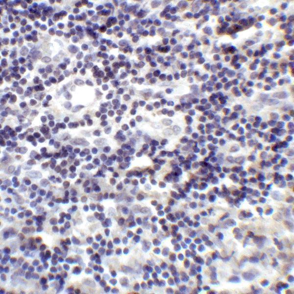
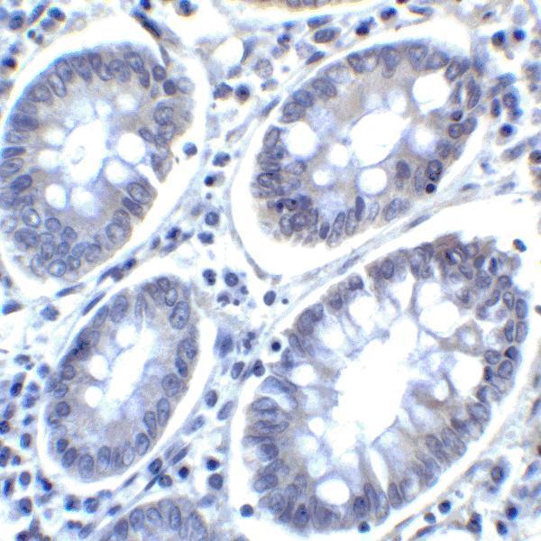
Plan human chromogenic IHC-P with M03295 at 2 μg/mL, as shown in tonsil and colon carcinoma images (datasheet M03295). Interpret variable cytoplasmic staining alongside the reported secreted variant (HPA tissue IHC).

Expected localisation, validated protocols, controls and antibodies — the at-a-glance facts below, then the full design guide.
| Expected localisation | Observed cytoplasm (HPA tissue IHC); membrane form (UniProt) | |
| Staining pattern | Variable cytoplasmic staining across tissue cell types (HPA tissue IHC) | |
| Antigen retrieval | Citrate pH 6 HIER, heat-mediated (datasheet A03295) | |
| Positive control | Appendix+4 more · see all | |
| Negative control | Adipose tissue |
| Fixation | Formaldehyde-fixed mouse sections are documented (datasheet A03295, IHC) (selected-SKU IHC image A03295) | |
| Caveat | A secreted variant may separate RNA and tissue staining (HPA tissue IHC) | |
| Regulation | Tissue-dependent staining intensity (HPA tissue IHC) | |
| Isoform / epitope | 3 isoforms; check epitope against extracellular and cytoplasmic regions (UniProt) |
Compare the catalog antibody’s IHC-P protocol with published PDCD1LG2 staining in colorectal carcinoma and lymphoma sections (PMC5668177; PMC6215508; PMC4994970).
| Sample | Formaldehyde-fixed, paraffin-embedded mouse brain tissue (datasheet A03295) |
| Fixation | Image formalin-fixed; duration unreported (datasheet A03295); verify before use. |
| Sectioning | 4–5 µm sections on charged slides (standard) |
| Deparaffinisation | Xylene, graded ethanol series to water (standard) |
| Antigen retrieval | Heat retrieval: Citrate pH 6 (datasheet A03295); 20 min, 95–100 °C (standard) |
| Peroxidase block | 3% H2O2, 10 min, room temperature (standard) |
| Blocking | 10% serum (datasheet A03295) |
| Primary antibody | Rabbit anti-PDCD1LG2, 2.5 μg/mL (datasheet A03295) |
| Primary incubation | Overnight at 4 °C (standard) |
| Detection | HRP-polymer secondary, DAB chromogen 5–10 min (standard) |
| Counterstain | Hematoxylin, blue, dehydrate and mount (standard) |
| Expected result | PDCD1LG2-positive staining in glandular cells of appendix (HPA tissue IHC: High). HPA tissue profile: Cytoplasmic expression at variable levels in all tissues. No signal in the no-primary control. |
PDCD1LG2 is a cell membrane, endomembrane and secreted protein with an extracellular domain and a cytoplasmic tail (UniProt Q9BQ51 topology). In paraffin-section IHC, HPA reports variable cytoplasmic staining across tissues, including high staining in selected epithelial cells, cardiomyocytes and kidney tubule cells (HPA tissue IHC). HPA rates its tissue IHC evidence Approved, while noting low consistency between antibody staining and RNA expression (HPA tissue IHC).
| Clear cytoplasmic staining in bronchial respiratory epithelial cells or kidney tubule cells. | This matches cell types scored High by HPA and supports an interpretable positive control (HPA tissue IHC). Compare the stain with tissue structure and the negative control before assigning a result (standard IHC practice). |
| Predominantly nuclear staining, especially without a cytoplasmic pattern. | A nuclear-only pattern is unsupported by the reported HPA tissue IHC profile and UniProt locations (HPA tissue IHC; UniProt Q9BQ51). Review control sections and detection background before treating it as PDCD1LG2 signal (standard IHC practice). |
| Strong staining confined to adipocytes in adipose tissue. | HPA reports PDCD1LG2 as Not detected in adipocytes (HPA tissue IHC). Check for cross-reactivity or endogenous detection activity; a stain in this cell type alone does not establish target expression (standard IHC practice). |
| Diffuse chromogen across tissue spaces, edges or many unrelated structures. | An unstructured deposit cannot establish the cell-associated cytoplasmic pattern reported by HPA (HPA tissue IHC). Compare a negative control and inspect blocking, washes and detection steps for nonspecific signal (standard IHC practice). |
| No visible stain in a high-staining reference tissue, such as bronchus or colon. | HPA scores bronchial respiratory epithelial cells and colonic glandular cells High (HPA tissue IHC). An absent reference signal makes a negative test section inconclusive; verify tissue integrity, antibody use and detection with controls (standard IHC practice). |
| Tissue and cell selection | HPA scores glandular cells in appendix, colon and duodenum, bronchial respiratory epithelium, cardiomyocytes and kidney tubule cells High; adipocytes are Not detected (HPA tissue IHC). Choose a reference by cell type, rather than assuming uniform staining throughout a section (standard IHC practice). |
| Protein topology and epitope | UniProt places residues 20–220 extracellularly, 221–241 across the membrane and 242–273 in the cytoplasmic tail (UniProt Q9BQ51 topology). The antibody epitope is unspecified here, so topology alone cannot predict its IHC compartment or retrieval response. |
| Processing and variants | UniProt lists a signal peptide at residues 1–19, 3 isoforms and a secreted location (UniProt Q9BQ51). HPA notes that at least one variant is secreted, complicating RNA-to-protein location comparisons; these records do not identify which variant produces a given stain (HPA tissue IHC). |
| Evidence strength | The listed HPA antibody, HPA013411, has Approved IHC status, while HPA reports low consistency between staining and RNA expression (HPA antibodies; HPA tissue IHC). Treat cell-level morphology and controls as essential to interpretation (standard IHC practice). |
| IF/ICC: what location is reported? | HPA reports cytosol as the main ICC-IF location but marks it uncertain; the listed antibody's ICC status is Uncertain (HPA subcellular; HPA antibodies). Use the separate IF/ICC guide for that application; the ICC observation does not establish an IHC protocol or override tissue IHC results. |
| General chromogenic workflow | Antigen retrieval, blocking, washes and detection controls can affect how an IHC slide reads (standard IHC practice). Neither supplied source establishes a PDCD1LG2-specific fixation or retrieval effect; judge any workflow change against a positive tissue and negative control. |
| Situation | Likely cause | Next action |
|---|---|---|
| A high-reference section has no stain. | A failed reference signal can reflect a section, antibody or detection problem; bronchial respiratory epithelium is scored High by HPA (HPA tissue IHC; standard IHC practice). | Check the reference tissue and cell identity, reagent setup and detection control; then review the catalog antibody's IHC-P instructions before interpreting test sections (standard IHC practice). |
| The test section is weak while its positive reference stains. | HPA reports variable cytoplasmic levels across tissues, so weak staining may reflect the sampled cells; the supplied sources give no target-specific fixation explanation (HPA tissue IHC). | Score the named cell population against its tissue-specific HPA level and the run controls; record weak staining without converting it into a categorical negative (HPA tissue IHC; standard IHC practice). |
| Most of the slide has diffuse color. | Diffuse deposit lacks the cell-associated cytoplasmic pattern described for tissue IHC and may reflect nonspecific background (HPA tissue IHC; standard IHC practice). | Compare the negative control, inspect washes and blocking, and assess whether color follows recognizable cell boundaries or cytoplasm (standard IHC practice). |
| Only nuclei stain. | Neither HPA's cytoplasmic tissue profile nor UniProt's membrane, endomembrane and secreted locations support a nuclear-only assignment (HPA tissue IHC; UniProt Q9BQ51). | Check negative controls and section morphology; withhold a PDCD1LG2-positive call unless a supported cell-associated pattern is demonstrable (standard IHC practice). |
| Adipocytes stain strongly while expected reference cells do not. | HPA scores adipocytes Not detected and several epithelial reference populations High, making this distribution discordant with its tissue IHC record (HPA tissue IHC). | Review cross-reactivity and endogenous detection activity with appropriate controls; verify the reference cell population before assigning target-specific staining (standard IHC practice). |
| RNA abundance seems inconsistent with the IHC result. | HPA explicitly reports low RNA–staining consistency and notes that a secreted variant can make RNA and protein tissue locations differ (HPA tissue IHC). | Report the observed staining by tissue, cell type and compartment alongside the RNA result; avoid using RNA alone to overrule a controlled IHC observation (HPA tissue IHC; standard IHC practice). |


Comprehensive Human Protein Atlas IHC scoring per tissue (reliability: Approved — Low consistency between antibody staining and RNA expression data. At least one protein variant secreted, tissue location of RNA and protein might differ and correlation is complex.). Rows are taken directly from the HPA tissue chart — click any row's HPA link to view the source.
| Tissue | Cell type | Level | Evidence | Source |
|---|---|---|---|---|
| Appendix | Glandular cells | High | Protein (IHC) | HPA → |
| Bronchus | Respiratory epithelial cells | High | Protein (IHC) | HPA → |
| Colon | Glandular cells | High | Protein (IHC) | HPA → |
| Duodenum | Glandular cells | High | Protein (IHC) | HPA → |
| Esophagus | Squamous epithelial cells | High | Protein (IHC) | HPA → |
| Tissue | Cell type | Level | Evidence | Source |
|---|---|---|---|---|
| Adipose tissue | Adipocytes | Not detected | Protein (IHC) | HPA → |
Troubleshoot PDCD1LG2 staining in paraffin sections by checking retrieval, compartment, cell identity and controls before comparing signal across samples (UniProt Q9BQ51; standard IHC practice).
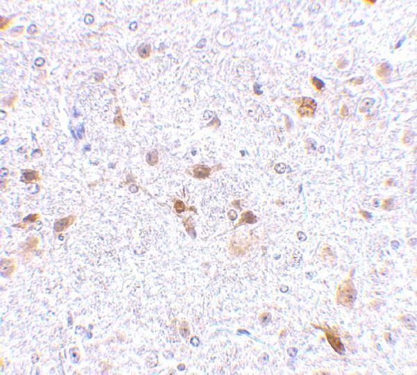
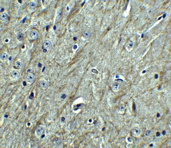
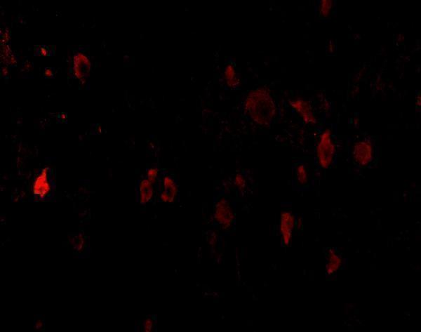
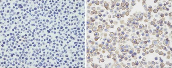
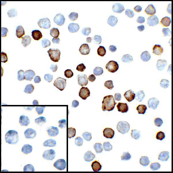
The catalog shows PDCD1LG2 IHC data from mouse brain, human tonsil, human colon carcinoma, and 293T cell sections, with IF data from mouse brain and human samples (catalog image captions).
A03295 has IHC data from formaldehyde-fixed paraffin mouse brain and IF data from mouse brain cells and tissue (A03295 image captions). M03295 has IHC data from human tonsil and colon carcinoma and IF/ICC data from those tissues and transfected HEK293 cells (M03295 image captions); M03295-1 has IHC data from untransfected and transfected 293T cell sections (M03295-1 image caption).
Which to pick: For human tissue IHC, choose M03295, a mouse monoclonal with tonsil and colon carcinoma IHC images; its IHC captions do not report a fixative (catalog M03295; M03295 IHC captions). For IF/ICC, choose M03295 for human samples (catalog M03295 applications and IF/ICC captions); M03295-1 is an IHC-P option supported by formaldehyde-fixed 293T cell sections retrieved with EDTA at pH 9.0 (M03295-1 image caption). For human and mouse reactivity, choose rabbit antibody A03295, whose IHC image shows formaldehyde-fixed paraffin mouse brain retrieved with citrate at pH 6; its catalog also lists IF, with mouse brain IF images (catalog A03295; A03295 image captions).