Click image to see more details
-

-
-
-
-
+6
Product Info Summary
| SKU: | A03295 |
|---|---|
| Size: | 0.1 mg |
| Reactive Species: | Human, Mouse |
| Host: | Rabbit |
| Application: | ELISA, IF, IHC-P, WB |
Customers Who Bought This Also Bought

Product info
Product Name
Anti-PD-L2 PDCD1LG2 Antibody
SKU/Catalog Number
A03295
Size
0.1 mg
Form
Liquid
Description
Boster Bio Anti-PD-L2 PDCD1LG2 Antibody (Catalog # A03295). Tested in ELISA, WB, IHC-P, IF applications. This antibody reacts with Human, Mouse.
Storage & Handling
PD-L2 antibody can be stored at 4°C for three months and -20°C, stable for up to one year. Avoid repeated freeze-thaw cycles. Antibodies should not be exposed to prolonged high temperatures.
Cite This Product
Anti-PD-L2 PDCD1LG2 Antibody (Boster Biological Technology, Pleasanton CA, USA, Catalog # A03295)
Host
Rabbit
Contents
PD-L2 Antibody is supplied in PBS containing 0.02% sodium azide.
Clonality
Polyclonal
Isotype
IgG
Immunogen
Anti-PD-L2 antibody was raised against a peptide corresponding to 16 amino acids near the center of human PD-L2. The immunogen is located within amino acids 140-190 of PD-L2.
Reactive Species
A03295 is reactive to PDCD1LG2 in Human, Mouse
Observed Molecular Weight
68 kDa
Calculated molecular weight
31.0 kDa
Background of PDCD1LG2
Cell-mediated immune responses are initiated by T lymphocytes that are themselves stimulated by co gnate peptides bound to MHC molecules on antigen-presenting cells (APC). T-cell activation is generally self-limited as activated T cells express receptors such as PD-1 (also known as PDCD-1) that mediate inhibitory signals from the APC. PD-1 can bind two different but related ligands, PD-L1 and PD-L2, both of which are thought act as a negative regulator of T cell activation. However, it has been suggested that PD-L2 can act to stimulate an immunogenic response through and alternative receptor from PD-1.
Antibody Validation
Boster validates all antibodies on WB, IHC, ICC, Immunofluorescence, and ELISA with known positive control and negative samples to ensure specificity and high affinity, including thorough antibody incubations.
Application & Images
Applications
A03295 is guaranteed for ELISA, IF, IHC-P, WB Boster Guarantee
Recommend Dilution
WB: 0.5-4 μg/mL; IHC: 2.5 μg/mL; IF: 20 μg/mL.
Antibody validated: Western Blot in human and mouse samples; Immunohistochemistry in mouse samples; Immunofluorescence in mouse samples. All other applications and species not yet tested. Optimal dilutions for each application should be determined by the researcher.
Validation Images & Assay Conditions
Click image to see more details
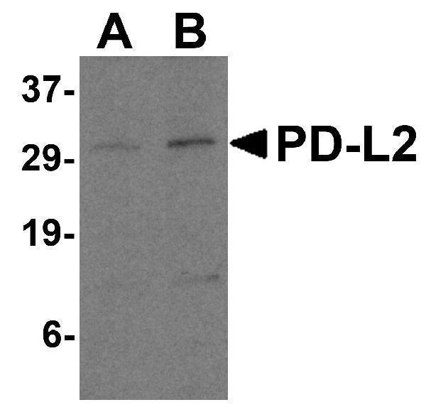
Western Blot Validation in Human Raji Cell Lysate
Loading: 15 μg of lysates per lane.
Antibodies: PD-L2 A03295 (A: 0.5 μg/mL and B: 1 μg/mL), 1h incubation at RT in 5% NFDM/TBST.
Secondary: Goat anti-rabbit IgG HRP conjugate at 1:10000 dilution.

Click image to see more details
Independent Antibody Validation (IAV) via Protein Expression Profile in Human and Mouse Cell Lines
Loading: 15 μg of lysates per lane.
Antibodies: PD-L2, A03295 (4 μg/mL), competitor antibody (4 μg/mL), and beta-actin (1 μg/mL), 1h incubation at RT in 5% NFDM/TBST.
Secondary: Goat anti-rabbit IgG HRP conjugate at 1:10000 dilution.

Click image to see more details
KO Validation in HeLa Cells
Loading: 15 μg of HeLa WT cell lysates or PD-L2 KO cell lysates. Antibodies: PD-L2, A03295 (4 μg/mL) and beta-actin 3779 (1 μg/mL), 1 h incubation at RT in 5% NFDM/TBST.
Secondary: Goat Anti-Rabbit IgG HRP conjugate at 1:10000 dilution.

Click image to see more details
KO Validation in HeLa Cells
Loading: 15 μg of HeLa WT cell lysates or PD-L2 KO cell lysates. Antibodies: PD-L2, A03295 (4 μg/mL) and beta-actin 3779 (1 μg/mL), 1 h incubation at RT in 5% NFDM/TBST.
Secondary: Goat Anti-Rabbit IgG HRP conjugate at 1:10000 dilution.

Click image to see more details
Immunohistochemistry Validation of PD-L2 in Mouse Brain Tissue
Immunohistochemical analysis of paraffin-embedded mouse brain tissue using anti-PD-L2 antibody (A03295) at 2.5 μg/ml. Tissue was fixed with formaldehyde and blocked with 10% serum for 1 h at RT; antigen retrieval was by heat mediation with a citrate buffer (pH6). Samples were incubated with primary antibody overnight at 4˚C. A goat anti-rabbit IgG H&L (HRP) at 1/250 was used as secondary. Counter stained with Hematoxylin.

Click image to see more details
Immunofluorescence Validation of PD-L2 in Mouse Brain Tissue
Immunofluorescent analysis of 4% paraformaldehyde-fixed mouse brain cells labeling PD-L2 with A03295 at 20 μg/mL, followed by goat anti-rabbit IgG secondary antibody at 1/500 dilution (red).

Click image to see more details
Immunohistochemistry Validation of PD-L2 in Mouse Brain Tissue
Immunohistochemical analysis of paraffin-embedded mouse brain tissue using anti-PD-L2 antibody (A03295) at 2.5 μg/ml. Tissue was fixed with formaldehyde and blocked with 10% serum for 1 h at RT; antigen retrieval was by heat mediation with a citrate buffer (pH6). Samples were incubated with primary antibody overnight at 4˚C. A goat anti-rabbit IgG H&L (HRP) at 1/250 was used as secondary. Counter stained with Hematoxylin.

Click image to see more details
Immunofluorescence Validation of PD-L2 in Mouse Brain Tissue
Immunofluorescent analysis of 4% paraformaldehyde-fixed mouse brain tissue labeling PD-L2 with A03295 at 20 μg/mL, followed by goat anti-rabbit IgG secondary antibody at 1/500 dilution (green) and DAPI staining (blue).

Click image to see more details
Regulated Expression Validation of PD-L2 in Mice with Melanoma Tumor (Knox et al., 2019)
Immunoblot analysis of PD-L2 expression with anti-PD-L2 (A03295) antibodies. PD-L2 expression was up-regulated by anti-PD1 antibody treatment whereas it was reduced by Next A alone or combination treatment (anti-PD1 antibody + NextA).

Click image to see more details
Immunohistochemistry Validation of PD-L2 in Lung Tumor of Mice (Kao et al., 2015)
Protein analysis for PD-L2 (E-H) by immunohistochemistry with anti-PD-L2 antibodies in mice lung tumors. hMUC1.Tg mice were induced with lung adenoma and then treated with concurrent or sequential cisplatin/radiotherapy. PD-L2 expression level at week 41 after treatment was similar in control and treatment groups.
Specific Publications For Anti-PD-L2 PDCD1LG2 Antibody (A03295)
Loading publications
Recommended Resources
Here are featured tools and databases that you might find useful.
- Boster's Pathways Library
- Protein Databases
- Bioscience Research Protocol Resources
- Data Processing & Analysis Software
- Photo Editing Software
- Scientific Literature Resources
- Research Paper Management Tools
- Molecular Biology Software
- Primer Design Tools
- Bioinformatics Tools
- Phylogenetic Tree Analysis
Customer Reviews
Have you used Anti-PD-L2 PDCD1LG2 Antibody?
Share your experimental results or join a short interview to earn up to $1,000 in product credits or other rewards.
0 Reviews For Anti-PD-L2 PDCD1LG2 Antibody
Customer Q&As
Have a question?
Find answers in Q&As, reviews.
Can't find your answer?
Submit your question
1 Customer Q&As for Anti-PD-L2 PDCD1LG2 Antibody
Question
Is 100ug enough for 100 patients for use in IHC-P?
Verified customer
Asked: 2020-12-28
Answer
For the Anti-PD-L2 PDCD1LG2 Antibody (A03295), our starting concentration is 2.5 ug/ml for use in IHC-P.
Boster Scientific Support
Answered: 2020-12-29
