Click image to see more details
-

-
-
-
-
+4
Product Info Summary
| SKU: | M03295 |
|---|---|
| Size: | 0.1 mg |
| Reactive Species: | Human |
| Host: | Mouse |
| Application: | ELISA, Flow Cytometry, IF, IHC-P, ICC, WB |
Customers Who Bought This Also Bought

Product info
Product Name
Anti-PDL2 PDCD1LG2 Monoclonal Antibody [4E10]
SKU/Catalog Number
M03295
Size
0.1 mg
Form
Liquid
Description
Boster Bio Anti-PDL2 PDCD1LG2 Monoclonal Antibody [4E10] (Catalog # M03295). Tested in ELISA, WB, IHC-P, ICC, IF, Flow Cytometry applications. This antibody reacts with Human.
Storage & Handling
PD-L2 antibody can be stored at 4°C for three months and -20°C, stable for up to one year. Avoid repeated freeze-thaw cycles. Antibodies should not be exposed to prolonged high temperatures.
Cite This Product
Anti-PDL2 PDCD1LG2 Monoclonal Antibody [4E10] (Boster Biological Technology, Pleasanton CA, USA, Catalog # M03295)
Host
Mouse
Contents
PD-L2 Antibody is supplied in PBS containing 0.02% sodium azide and 50% glycerol.
Clonality
Monoclonal
Clone Number
Clone: 4E10
Isotype
IgG1
Immunogen
PD-L2 antibody was raised against the extracellular domain of human PD-L2.
Reactive Species
M03295 is reactive to PDCD1LG2 in Human
Observed Molecular Weight
68 kDa
Background of PDCD1LG2
Cell-mediated immune responses are initiated by T lymphocytes that are themselves stimulated by co gnate peptides bound to MHC molecules on antigen-presenting cells (APC). T-cell activation is generally self-limited as activated T cells express receptors such as PD-1 (also known as PDCD-1) that mediate inhibitory signals from the APC. PD-1 can bind two different but related ligands, PD-L1 and PD-L2, both of which are thought act as a negative regulator of T cell activation. However, it has been suggested that PD-L2 can act to stimulate an immunogenic response through and alternative receptor from PD-1.
Antibody Validation
Boster validates all antibodies on WB, IHC, ICC, Immunofluorescence, and ELISA with known positive control and negative samples to ensure specificity and high affinity, including thorough antibody incubations.
Application & Images
Applications
M03295 is guaranteed for ELISA, Flow Cytometry, IF, IHC-P, ICC, WB Boster Guarantee
Recommend Dilution
PD-L2 antibody can be used for detection of PD-L2 by Western blot at 0.5 - 1 μg/mL. Antibody can also be used for immunohistochemistry starting at 2 μg/mL. For immunofluorescence start at 20 μg/mL.
Antibody validated: Western Blot in human samples; Immunohistochemistry in human samples; Immunocytochemistry in human samples; Immunofluorescence in human samples and Flow Cytometry in mouse samples. All other applications and species not yet tested. Optimal dilutions for each application should be determined by the researcher.
Validation Images & Assay Conditions
Click image to see more details
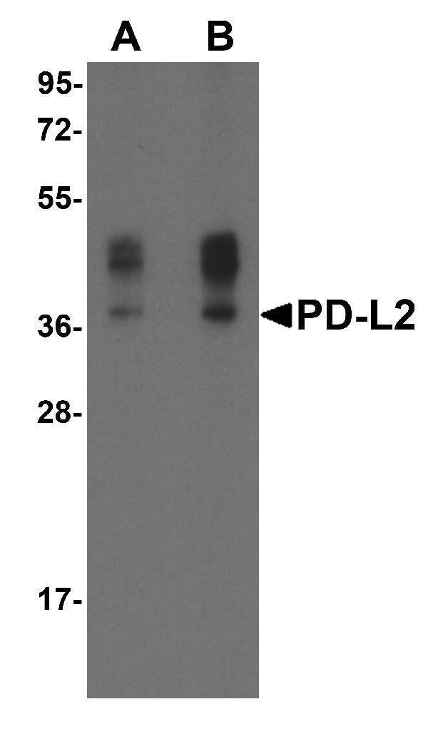
Western blot analysis of PD-L2 in overexpressing HEK293 cells PD-L2 antibody at 0.5 and 1 μg/ml

Click image to see more details
Immunocytochemistry of PD-L2 in transfected HEK293 cells with PD-L2 antibody at 5 μg/mL. Lower left: Immunocytochemistry in transfected HEK293 cells with control mouse IgG antibody at 5 μg/mL.

Click image to see more details
Immunofluorescence of PD-L2 in transfected HEK293 cells with PD-L2 antibody at 20 μg/mL.
Green: PDL2 Antibody [4E10] (M03295)
Blue: DAPI staining

Click image to see more details
Immunofluorescence of PD-L2 in human tonsil tissue with PD-L2 antibody at 20 μg/mL.
Green: PDL2 Antibody [4E10] (M03295)
Blue: DAPI staining

Click image to see more details
Immunofluorescence of PD-L2 in human colon carcinoma tissue with PD-L2 antibody at 20 μg/mL.
Green: PDL2 Antibody [4E10] (M03295)
Blue: DAPI staining

Click image to see more details
Immunohistochemistry of PD-L2 in human tonsil tissue with PD-L2 antibody at 2 μg/mL.

Click image to see more details
Immunohistochemistry of PD-L2 in human colon carcinoma tissue with PD-L2 antibody at 2 μg/mL.

Click image to see more details
Flow cytometry analysis of PD-L2 overexpressing HEK293 cells using PD-L2 antibody and control mouse IgG antibody at 10 μg/ml. Blue: Untransfected HEK293 cells. Yellow: PD-L2 overexpressing HEK293 cells.
Specific Publications For Anti-PDL2 PDCD1LG2 Monoclonal Antibody [4E10] (M03295)
Loading publications
Recommended Resources
Here are featured tools and databases that you might find useful.
- Boster's Pathways Library
- Protein Databases
- Bioscience Research Protocol Resources
- Data Processing & Analysis Software
- Photo Editing Software
- Scientific Literature Resources
- Research Paper Management Tools
- Molecular Biology Software
- Primer Design Tools
- Bioinformatics Tools
- Phylogenetic Tree Analysis
Customer Reviews
Have you used Anti-PDL2 PDCD1LG2 Monoclonal Antibody [4E10]?
Share your experimental results or join a short interview to earn up to $1,000 in product credits or other rewards.
0 Reviews For Anti-PDL2 PDCD1LG2 Monoclonal Antibody [4E10]
Customer Q&As
Have a question?
Find answers in Q&As, reviews.
Can't find your answer?
Submit your question
